Download
1 / 22
220 likes | 350 Vues
Mammography Image Integration Profile. Carolyn Reynolds Hologic, Inc. Vendor co-chair, IHE Mammography Committee. What’s the big deal? Full Field Digital Mammography (FFDM) is just another modality to PACS Most existing PACS will handle FFDM fine once:

E N D
Mammography Image Integration Profile Carolyn Reynolds Hologic, Inc. Vendor co-chair, IHE Mammography Committee
What’s the big deal? Full Field Digital Mammography (FFDM) is just another modality to PACS Most existing PACS will handle FFDM fine once: the FDA approves the monitor hardware, and workstations have 510K approval for FFDM applications Mammography ImageThe Problem/Assumptions
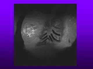
Mammography ImageNot just another modality! • What makes it different or challenging: • Two types of image data • Variances among vendors in image data and attributes • Common use of CAD (Computer Aided Detection) • Importance of prior study comparisons • Image size, orientation, & layout • Regional governance such as MQSA (Mammography Quality Standards Act – USA)
Modality Workstation Archive CAD “For Presentation” Image Data Printer “For Processing” Image Data Mammo CAD Structured Rpt Mammography ImageUse Case
R CC L CC R MLO L MLO Hanging Protocol DifferencesExample Preferred Layout • Hanging protocol determined by: • View Type (i.e. CC vs. MLO) • Specialty View Type (i.e. Spot, Mag) • Laterality • Patient Orientation
R CC L CC L MLO R MLO Generic Image Display LayoutsApplied to Mammo Images • Hanging based upon series or study descriptions • Image order as acquired • Image orientation as acquired • Stacked series example
R L L R R L Variances in FFDM Vendor DataDetector Size Example Typical “Fit to Viewport” effect L R Prior Exam from Vendor A’s system • Current Exam from • Vendor B’s system • Different resolution • Different pixel matrix
Workstation Tool EnhancementsTissue vs. Air Detection Example Window/Center Adjustments
10in x12in Film size 8inx10in Film size Precision of <2% error Printing ConsiderationsTrue Size Film size vs. Detector Size
Printing ConsiderationsPositioning-minimal borders at chestwall Centered images can create large borders at chestwall Image offset with chestwall side having minimal borders
Mammography Image IntegrationValue proposition • Helps meet desire to have multiple FFDM vendors and use any vendor’s workstation for diagnosis • Ensures that FFDM modalities provide adequate information to facilitate downstream applications
Mammography Image IntegrationValue proposition • Ensures systems support required data objects for interoperability • Defines image display and printing operations required for effective and efficient diagnoses
Mammography Image IntegrationScope • Based upon existing actors and transactions • Expanded requirements within each transaction
Mammography ImageExpanded Requirements • Acquisition Modality • Send both for processing and for presentation images with reference that ties them together • Additionally required DICOM attributes • Detection and indication of tissue vs. air gap • Partial View Option (breast is larger than detector) • Modified requirements for film digitizers
Mammography ImageExpanded Requirements • Image Display • Hanging protocols based upon view, laterality, patient orientation, and specialty views • Maintain a black air gap during window/center operations and inverted pixel data • Sizing • Same relative size • True size • 1:1 detector pixels to display pixels
Mammography ImageExpanded Requirements • Image Display cont’d • Measurement calculation requirements • Display calibration and grayscale rendering • Labeling requirements • Mammo CAD Structured Report rendering on “for presentation” images • Partial View Option (breast larger than detector)
Mammography ImageExpanded Requirements • Image Manager • Storage and retrieval of For Presentation and For Processing SOP Classes for Digital Mammography X-Ray • Storage and retrieval of Mammography CAD Structured Report SOP Class
Mammography ImageExpanded Requirements • Evidence Creator (CAD) • Support for Mammography CAD SR SOP Class and Storage Commitment • No specifics on how Evidence Creator obtains mammography images
Mammography ImageExpanded Requirements • Print Composers/Print Servers • True Size printing (Requested Image Size) • Justify chestwall and print with 5mm borders or less • Render VOI LUTS if present • Specify/Support maximum density • Specify/Support Presentation LUT • Labeling requirements • Support for 12 bit pixel depth
Mammography Image Questions???