Download
1 / 31
340 likes | 814 Vues
Isolation and identification of pyogenic cocci. Experiment Two. Objective of Experiment. To m aster primary principle and method of isolation and identification pyogenic cocci from clinical s pecimens . To diagnos e clinical disease and guide us to use medicines . Smears.

E N D
Isolation and identification of pyogenic cocci Experiment Two
Objective of Experiment • To masterprimaryprinciple and method of isolation and identification pyogenic cocci from clinical specimens . • To diagnose clinical disease and guide us to use medicines .
Smears Colonial characteristics colonial Hemolytic reaction Isolation culture morphology observation Pigmentation (blood agar plates) Gram staining observation with the microscope Directidentification Pure culture Antibiotic susceptibility test PROCEDURE Morphological characteristics (Gram-stain) (Microscopic examination) Specimens typical colonies
Identified method for STAPHYLOCOCCI • Gram-stain • Isolation and culture • Pure culture • Directidentification • The antibiotic susceptibility test
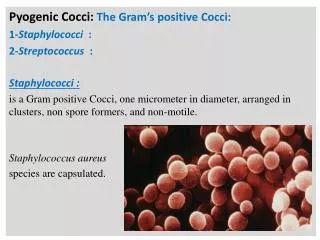
Gram-stain • Morphological characteristics -Typical staphylococcus cells are spherical,arranging in irregular clusters,which just like as clusters of grape. • The cell is about 1 μm in diameter. • Typical cells are Gram-positive,nonmotile and do not form spore.
Staphylococci are Gram-positive cocci, typically arranged in clumps or Grape-like clusters
Isolation and culture • Cultural • Specimens planted on blood agar plates give rise to typical colonies in 18 hours at 370C • Hemolysis and pigment production may not occur until several days later and are optimal at room temperature.
Isolation and culture • colonial characteristics • Colonies of staphylococci on solid meida are round,smooth,raised,and glistening.S.aureus forms gray to deep golden yellow colonies. • S.epidermidis colonies are gray to white on primary isolation. • Various degrees of hemolysis are produced by S.auerus and occasionally by other species.
Pure culture • MATERIALS: • Agar slope culture • Colonies on agar plate • PROCEDURE • With the flame-sterilized wire inoculating loop, transfer a small amount of bacteria from the colony on agar plate. Then streak on the agar slope. • Sterile the mouth of tubes, replug the test tubes and flame the loop. • Label and incubate at 37℃ for 18-24 hours • .
Directedidentification • The Coagulase Test • The Dnase Test • The mannitol fermentation test. • The phage typing test • Animal Experiment
The Coagulase Test • Possession of the enzyme coagulase which coagulase plasma is an almost exclusive property of Staph.aureus.There are two ways of performing this test • The Slide Coagulase Test • The Tube coagulase Test
The Coagulase Test • Coagulase is an enzyme converting fibrinogen into fibrin promoting blood clotting. It has been speculated that this enzyme might be a virulence factor with the coagulated blood around the bacteria protecting them from the immune system. • However, coagulase-negative strains are often as pathogenic as coagulase-positive strains.
The Tube coagulase Test Left tube coagulase positive Right tube coagulase negative
The Dnase Test • Inoculate Dnase agar plates with a loop so that the growth is in plaques about 1 cm in diameter.Incubate at 370C overnight.Flood the plate with 1 N hydrochloric acid.Clearing around the colonies indicates Dnase activity.The hydrochloric acid reacts with unchanged deoxyribonucleic acid to give a cloudy precipitate.A few other bacteria,e.g. Serratia,may give a positive reaction.
The mannitol fermentation test. • Inoculate the bacteria into a mannitol micro-tube,incubate at 370C for 18h.S.aureus will ferment mannitol to produce acid,which causes the medium to turn yellow.
The phage typing test • This test is used to trace the infective agent in epidemiology if necessary.It is usually not done for routine clinical purpose.
Animal Experiment Isolation and identification of bacteria Vomit excrement remaindered food observation filter Meat soup media 6--8W cat food poisoning injection
The antibiotic susceptibility test • This test is helpful for the treatment of S.aureus infection. • materials • S.aureus(isolated from the pus of a patient). • Several kinds of filter paper (each contains different kinds of antibiotics) • Nutrient agar plate
The antibiotic susceptibility test • Streaking the S.aureus on agar plate (thoroughly covered the plate) • PROCEDURE • Put 4 kinds of paper contained different antibiotics on the plate (each paper are far away about 2 cm) • Incubate ar 370C 18-24 hours. • Observe the results the plates are examined for the present of zones of inhibition of bacterial growth around the filter paper.The sensitivity of the organism is indicated by the diameter of the zone of growth inhibition.
Dimidiation the enterobacteria according to the fermentation of lactose • Lactase fermenters:saprophytic and commensal Escherichia Klebsiella Citrobater Enterobacter Enterobacter, • Non lactase fermenters: pathogens Salmonella Shigella some Citrobacter, Proteus Serratia
Biochemical reactions of Salmonella, etc Species butt slope H2S motility E.coli AG AG - + Salmonella A - +/- + Other Salmonella AG - +/- + Shigella A - +/- - butt: ferment dextrose slope: lactose • A:acid AG: acid and gas
Procedure Colonial characteristic observation Specimens isolation Gram Staining (SS/EMB plate) Serological identification TSI Biochemical reaction
Specimens • Different specimens should be taken depending on the kind and the process of the disease. blood bone marrow Urine stool
Isolation • Culture medium: S.S agar • Method: streak plate • Result: unpathogenetic colonies: middle size, red Suspect colonies: colorless, small, opaque