Download
1 / 23
600 likes | 1.99k Vues
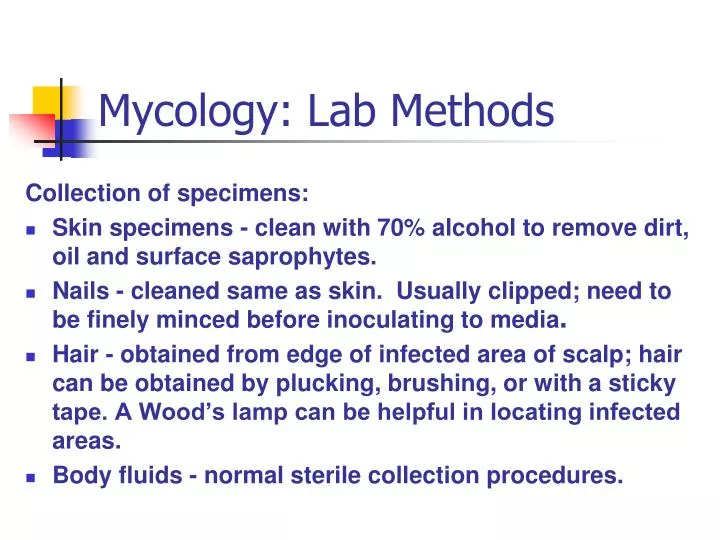
Mycology: Lab Methods. Collection of specimens: Skin specimens - clean with 70% alcohol to remove dirt, oil and surface saprophytes. Nails - cleaned same as skin. Usually clipped; need to be finely minced before inoculating to media .

E N D
Mycology: Lab Methods Collection of specimens: • Skin specimens - clean with 70% alcohol to remove dirt, oil and surface saprophytes. • Nails - cleaned same as skin. Usually clipped; need to be finely minced before inoculating to media. • Hair - obtained from edge of infected area of scalp; hair can be obtained by plucking, brushing, or with a sticky tape. A Wood’s lamp can be helpful in locating infected areas. • Body fluids - normal sterile collection procedures.
Mycology: Lab Methods Preparation of specimens for transport: • Hair & nails sent in a dry envelope, inside proper container. • Other specimens are usually sent frozen or on dry ice. • Packaging - must meet biohazard regulations. Cultures must be on tubed media (not plates). • Inside labeling information: patient ID, specimen source, suspected organism. • Outside labeling - WARNING: POTENTIAL PATHOGEN
Mycology: Lab Methods Processing of specimen to recover fungus: • Skin, nails, & hair - direct exam following KOH preparation. • Body fluids - • CSF - centrifuged; examine sediment microscopically, inoculate media. • Pleural fluid, sputum, and bronchial aspiration - specimen must be fresh as saprophytes would overgrow pathogens such as H. capsulatum. Specimens may be refrigerated up to 2 hours. • Gastric washings - same as for pleural fluids.
Mycology: Lab Methods Processing of specimen to recover fungus: • Body fluids- (continued) • Genito-urinary specimens - first morning specimen preferred; centrifuge & inoculate on media. • Blood/bone marrow - inoculate to BHI broth and BHI slant. • Wound abscess or drainage - culture anaerobically if actinomycosis is suspected. • Tissue specimens - examine for pus, caseous material or granules; mince aseptically, inoculate on media.
Mycology: Lab Methods Direct examination of specimens: • Direct exam required on any biological material sent to lab for fungus culture. Examine for spores, hyphae, mycelial elements, budding yeast, mycotic granules. • Wet mount - good for yeast; examination is done in natural environment, so loss of fragile structure is minimal. • KOH - done on skin scrapings, hail, nails, sputum, vaginal specimens. KOH clears tissue cells so fungal elements may be seen.
Mycology: Lab Methods Stainsused: • Lactophenol Cotton Blue - very popular for quick evaluation of fungal structures; stains chitin in cell walls of fungi. • Periodic Acid - Schiff Stain (PAS) - stains polysaccharide in the cell wall of fungi. Fungi stain pink-red with blue nuclei. • Gomori Methenamine Silver Stain - silver nitrate outlines fungi in black due to the silver precipitating on the fungi cell wall.
Mycology: Lab Methods Stainsused: (continued) • Gridley Stain - Hyphae and yeast stain dark blue or rose. Tissues stain deep blue and background is yellow. • Mayer Mucicarmine Stain - will stain capsules of Cryptococcus neoformans deep rose. • Fluorescent Antibody Stain - extremely specific method of detecting fungi in tissues or fluids. • Papanicolaou Stain - good for initial differentiation of dimorphic fungi. Works well on sputum smears. • Gram Stain - generally fungi are gram positive; but Actinomyces and Nocardia are gram variable.
Mycology: Lab Methods Stainsused: (continued) • Acid-Fast Stain - used to differentiate the acid-fast Nocardia from other aerobic Actinomyces. • Giemsa Stain - used for blood and bone marrow specimens. • India Ink - demonstrates the capsule of Cryptococcus neoformans in CSF specimens.
Mycology: Lab Methods Fungal Culturing: • Tubed media is used rather than plated media. • Less chance for spore release into the environment. • Less chance for dehydration. • Easier storage. • The agar in a tube is inoculated in a straight line. Preliminary identification is based on differential growth patterns on various media.
Mycology: Lab Methods Fungal Culturing: Media - • Sabouraud's dextrose agar - classic medium, recommended for most studies. • Sabouraud's dextrose agar with chloramphenicol (inhibits bacterial growth). • Mycosel agar - a commercially produced agar containing chloramphenicol to inhibit bacterial growth, and cycloheximide to inhibit saprophytic fungi and some yeasts (including C. neoformans). • Aspergillus and Scopulariopsis are opportunistic pathogens. Cycloheximide will prevent their growth.Bacteria-like fungi (such as Actinomycetes) are inhibited by chloramphenicol.
Mycology: Lab Methods Fungal Culturing: Media - • Brain heart infusion slant (BHI) - more enriched than Sab-Dex. For recovery of H. capsulatum. • Potato-dextrose agar (PDA) and Corn-meal agar - used in slide cultures to induce spore formation, which aids in identification.
Mycology: Lab Methods Fungal Culturing: Special applications agar - • Caffeic Acid Agar - Cryptococcus neoformans will produce melanin resulting in black colonies (must protect media from light). • Birdseed Agar - used to isolate Cryptococcus neoformans from contaminated cultures. • KT Medium & Kelley Agar - used to convert dimorphic fungus Blastomyces dermatitidis from mycelial to yeast form. • Modified Converse Liquid Medium (Levine's) - used to promote spherule production by Coccidioides immitis.
Mycology: Lab Methods Fungal Culturing: Fungal growth requirements - • Temperature - room temperature (25-30 C.) for most fungi. • Nocardia sp. and some dimorphic organisms grow best at 37 degrees C. • Any fungus capable of growing at 37 C, should be considered potentially pathogenic.
Mycology: Lab Methods Fungal Culturing: Fungal growth requirements - • Atmosphere - True fungi are aerobic but there are a few anaerobes among the bacteria-like fungi. • Time - Some yeasts grow overnight. Saprophytes are fast growers (several days). Generally cultures are held at least 4 weeks. • Paracoccidioides brasiliensis may require 4-5 weeks; 10 weeks are recommended if Histoplasma capsulatum is suspected. • Routine cultures should be examined every other day.
Mycology: Lab Methods Colony Morphology (macroscopic features): • Surface topography - some fungal colonies may be free growing, covering the entire surface of agar; others may grow in a restricted manner. • Surface texture - cottony or wooly (floccose), granular, chalky, velvety, powdery, silky, glabrous (smooth, creamy), or waxy.
Mycology: Lab Methods Colony Morphology (continued): • Pigmentation - Fungi may be colorless or brightly colored. Color may be on fungus itself, on its sporulating apparatus, on the agar, or on the bottom of the colony (reverse pigmentation). • Mycelium - • Vegetative mycelium - provides nutrition. • Aerial mycelium – reproductive.
Mycology: Lab Methods Microscopic evaluation: • Teased Preparation - wet mount • Slide Culture - gives undisturbed microscopic morphology. • Cellophane Tape Preparation.
Mycology: Lab Methods Biochemical studies - used to ID yeast and yeast-like organisms. • Carbohydrate fermentation - • Growth and utilization of a carbohydrate under anaerobic conditions as determined by acid and gas production. • Specimen is inoculated beneath the broth. Bromcresol purple is the indicator. Acid production turns purple to yellow. Gas is detected by appearance of bubbles trapped in the fermentation tube. Observe every 48 hours for 14 days.
Mycology: Lab Methods Biochemical studies - (continued) • Carbohydrate assimilation - • Determines ability to utilize a carbohydrate as sole source of carbon. • Bromcresol purple indicator turns from purple to yellow. • Tubes unchanged (as determined by comparing to a blank tube) after 10 days are negative.
Mycology: Lab Methods Biochemical studies - (continued) • Nitrogen assimilation - • Utilizes 3 tubes with differing sources of nitrogen. Bromthymol blue is the indicator (blue to yellow is positive).
Mycology: Lab Methods Growth on specific agars: • Christensen's urea agar - Urea is hydrolyzed by some yeast to form ammonia (pH increases) which turns media from yellow to dark pink. • Caffeic acid medium (must protect media from light) - Production of melanin by Cryptococcus neoformans resulting in black colonies.
Mycology: Lab Methods Other tests: • Germ tube - Candidia albicans & Candidia stellatoidea produce germ tubes when incubated in a protein medium. • Demonstration of chlamydospores - Yeast is inoculated by jabbing appropriate agar (Cornmeal with tween 80) and observed every 24 hours for 3 days for chlamydospore production.
Mycology: Lab Methods Hypersensitivity: (skin tests and serological tests) • Skin tests - demonstrates T-cell immunity (cellular) to a fungus. • Serological tests - demonstrates B-cell immunity (humoral) to a fungus; sera should be drawn in pairs (acute and convalescent).