Download
1 / 31
310 likes | 760 Vues
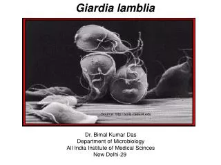
This is a trophozoite of Giardia lamblia in a stool specimen. You can observe the nucleus and flagella in this specimen. This is the proglottids of Taenia and you can observe the ovaries inside of the proglottids. This is an egg of Taenia sp.. Note the structure or morphology of the the egg.

E N D
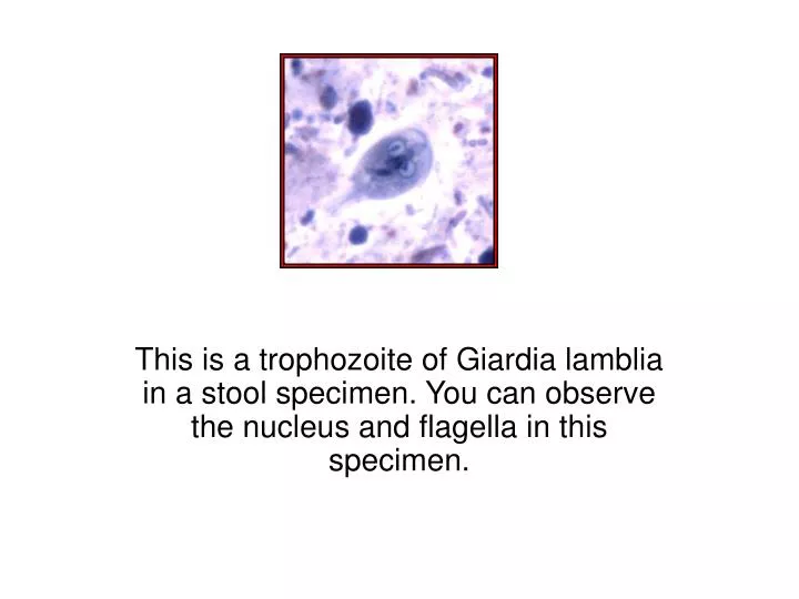
This is a trophozoite of Giardia lamblia in a stool specimen. You can observe the nucleus and flagella in this specimen.
This is the proglottids of Taenia and you can observe the ovaries inside of the proglottids.
This is an egg of Taenia sp.. Note the structure or morphology of the the egg.
This slide shows Schistoma sp. Both the male and the female.
This is an actual photograph of the male and female Schistosomes.
This is an egg of Schistoma species. Note the structure on the edge of the egg.
Note the two specimens of Ascaris one is a male the other a female. Which one is larger? Which one has the hook or curve at the end?
Note the ruler at the edge of the slide. This will give you an idea of the size of the organism.
This is an egg of Ascaris. What are some of the distinguishing features which differentiates it from other eggs?
This is a picture of the pin worm, Enterobius vermicularis. Observe the mouth portion of the organism.
This is an egg of Enterobius vermicularis. Observe the shape of the egg as well as the morphology.
This is the larvae of Trichninella spirallis embedded in the muscle of an animal.
This is a slide of Wuchereia bancrofti which causes Filariasis.
This is a slide of Trichomonas vaginalis. Make sure you observe the nucleus, flagella, and the undulating membrane.
This is a slide of Trypanosoma sp.. Note the undulating membrane on the organism.
This is a slide of Trypanosoma sp.. Observe the flagella and the undulating membrane.
This is Entamoeba histolytica. This is an immature cyst which is the infective stage.
This is the trophozoite of Entamoeba histolytica. You should be able to identify the nucleus, RBC, and the pseudopod from this photograph.
You should be able to identify the nucleus, RBC and the pseudopod.
This is a slide of Toxoplasma sp. which causes Toxoplasmosis.
Observe the scolex, neck, immature proglottids and the mature proglottids.