Download
1 / 20
210 likes | 596 Vues
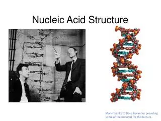
Nucleic Acid Structure. Structure of nucleotides Nitrogenous bases Pentose sugars Nucleosides Nucleotides Nucleotide chains Structure of B-DNA Images accompanying this lecture may be found at http://www.prenhall.com/klug4/ Select chapter 10. A. Structure of Nucleotides.

E N D
Nucleic Acid Structure • Structure of nucleotides • Nitrogenous bases • Pentose sugars • Nucleosides • Nucleotides • Nucleotide chains • Structure of B-DNA Images accompanying this lecture may be found athttp://www.prenhall.com/klug4/Select chapter 10
A. Structure of Nucleotides • A nucleotide is composed of • A nitrogenous base • A pentose sugar • A phosphate group
Nitrogenous Bases • Two different classes of aromatic carbon-nitrogen heterocycles • Purines • Adenine (Found in DNA & RNA) • Guanine (Found in DNA & RNA) • Pyrimidines • Cytosine (Found in DNA & RNA) • Thymine (Found in DNA) • Uracil (Found in RNA)
C. Pentose Sugars • Ribose • Found in RNA • Forms a 5-atom ring structure in aqueous solution • Carbons are numbered 1´ (one-prime), 2´, 3´, 4´, 5´
C. Pentose Sugars • Deoxyribose • Found in DNA • Identical to ribose, except that the “-OH” group on the 2´ carbon is replaced with an “-H”
D. Nucleosides • Nucleoside • A pentose sugar molecule with a nitrogenous base attached to the 1´ carbon • Nucleosides are named by using the root of the base name, plus the suffix “-osine” (for purines) or “-idine” (for pyrimidines) • Nucleosides with deoxyribose sugars are designated with the prefix “deoxy-”
E. Nucleotides • Nucleotide • A nucleoside with one, two, or three phosphate groups attached to the 5´ carbon • Nucleotides are named using the name of the nucleoside plus “monophosphate,” “diphosphate,” or “triphosphate” depending on the number of phosphates • Nucleotides with one phosphate may also be named by changing the nucleoside suffix to “-ylic acid”
F. Nucleotide Chains • The 5´ carbon of one nucleotide can be linked to the 3´ carbon of another nucleotide via a phosphodiester bond • An oligonucleotide chain (polynucleotide chain) is a linear chain of nucleotides linked in this fashion • The oligonucleotide chain has two ends: 5´ and 3´
G. Structure of B-DNA • In the 1940s, Chargaff discovered that the DNA isolated from most sources exhibited a 1:1 molar ratio of A:T, and a 1:1 ratio of G:C (as compared to RNA, in which the A:U and G:C ratios are random)
G. Structure of B-DNA • In the early 1950s, Watson, Crick, and Franklin studied the X-ray diffraction patterns of crystalline DNA fibers, and determined that DNA had a symmetrical 3-D structure in the form of a helix
G. Structure of B-DNA • Watson & Crick knew of Chargaff’s ratios, and realized that they could build a helical model for DNA structure, consistent with the X-ray data
G. Structure of B-DNA • The Watson and Crick model is known as a B-DNA helix, and it is believed to be the native conformation of most DNA found in living organisms
G. Structure of B-DNA • Features of the B-DNA helix: • Two oligonucleotide strands • The sugar-phosphate backbones of the strands are on the outside, and twist around a central axis to form a helix • The helical twists form two “grooves”that turn around the axis: the major groove and the minor groove • Therefore, DNA is a pretty groovy molecule
G. Structure of B-DNA • The two strands are in antiparallel orientation (one strand goes from 5´ 3´ and the other strand goes from3´ 5´ • These are the dimensions of the helix:20 Å diameter10 bases per turn34 Å per turn
G. Structure of B-DNA • The bases are located in the center of the helix, with the flat planes of the bases perpendicular to the axis of the helix • The bases between the two strands are “paired” with an “A” on one strand paired with a “T” on the other strand, and “G” paired with “C” • This property of the strands is called complementarity (the two strands are said to be complementary to each other, thank you very much)
G. Structure of B-DNA • The strands are held together by noncovalent “hydrogen bonds” between the complementary pairs of bases:A – T pairs have two hydrogen bondsG – C pairs have three hydrogen bonds
G. Structure of B-DNA • The two strands may be separated by heating (“melting”) the DNA, or by raising the pH with alkaline treatment • Two pieces of single-stranded DNA will spontaneously form a helix if the strands have enough base complementarity
G. Structure of B-DNA • An interactive tutorial on DNA structure can be found athttp://molvis.sdsc.edu/dna/index.htm