Download
1 / 37
460 likes | 921 Vues
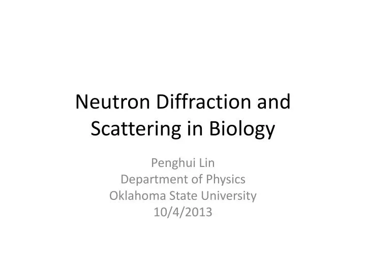
Neutron Diffraction and Scattering in Biology. Penghui Lin Department of Physics Oklahoma State University 10/4/2013. Neutron Scattering. Neutron Reflection (Neutron Reflectometry ) Small Angle Neutron Scattering Neutron diffraction (Neutron Crystallography) Spectroscopy and Imaging.

E N D
Neutron Diffraction and Scattering in Biology Penghui Lin Department of Physics Oklahoma State University 10/4/2013
Neutron Scattering • Neutron Reflection (Neutron Reflectometry) • Small Angle Neutron Scattering • Neutron diffraction (Neutron Crystallography) • Spectroscopy and Imaging
Structure determination • X-ray diffraction----spatial distribution of electrons • Electron diffraction----Coulomb forces • Neutron diffraction---strong nuclear forces • NMR • IR
X-ray v.s. Neutron Crystallography Crystal -> Diffraction pattern -> Electron density -> Model Crystal -> Diffraction pattern -> Nuclear density -> Model
Information from NC • Equivalence: neutron scattering not strongly depends on Z (especially for hydrogen detection which X-ray or electron diffraction can not see) • Clearly distinguish between neighboring atoms. (For biology, particularly N, C and O) • Contrast between H and D • Locate the solvent orientation around protein • Thermal motions of hydrogen containing groups • Weak interaction with materials, deep penetration and non-destructive
Crucial Hydrogen • Dominance of H2O molecules in living cells • Hydrogen bonds provide stability and versatility for biological macromolecules • Proton transfer and exchange is critical in many reactions • Hydration and protonation states are important
Neutron diffraction in structural studies • Location of Hydrogen atoms • Solvent Structure • Hydrogen exchange • Low resolution studies %bc biscsisssa H 99.985 -3.741 25.27 1.758 80.27 82.03 0.3326 D 0.015 6.671 4.04 5.592 2.051 7.643 0.000519
Fissure Reactor Chained reactions Continuous flow 1 neutron per fission 180 MeV neutron 1015/cm2/s Spallation source No chain reaction Pulsed 40 neutrons per proton 30 MeV neutron 1016/cm2/s
Neutron source Combined with Fourier Transform
Main problem • Low flux of neutron beams • Structures are large while scatterings are weak, so large single crystals are required, 1 mm3 is the limit due to the reasonable data collection time of 10-14 days per data set • Hydrogen produces a high level background (80 barn scattering factor) Solution • Broad bandpass (maximize the neutron flux and the reflections on the detector) • Cylindrical neutron image plate (LADI at ILL has a solid angle >2π) • Isotope substitution of D to H
Developments • In reactors: • Neutron image plates • Quasi-Laue methods • In spallation: • Time of flight Laue method • Electronic detectors • New facilities and methods for sample perdeuteration and crystallization • New approaches and computational tools for structure determination
Applications Example ID-Xylose Isomerase (XI)
Environment D2O in native XI OD- in XI-xylulose M1: structural metal M2: catalytic metal Kovalevsky 2008 Biochemistry
Active site of XI-xylulose Doubly protonated singly protonated Kovalevsky 2008 Biochemistry
Applications Example IIcryo cool concanavalin A
Concanavalin A Saccharide-binding protein Legume lectin family Extensiceβ-sheet arrangement Two metal binding sites PDB: 3CNA
Waters in the saccharide-binding site 293K 15K Blakeley 2004 PNAS Habash 2000 Acta Crystallogr D Biol Crystallogr.
H-bond network Blakeley 2004 PNAS
Water comparison Compare to room temperature NC 15K 227 water sites are identified with 19.2 Å2 B factor 167 are D2O with 17.6 Å2 B factor 60 are OD- or oxygen atoms with 32.2 Å2B factor 293K 148 water sites are identified with 43 Å2 B factor 88 are D2O with 37.8 Å2 B factor 60 are OD- or oxygen atoms with 50.6 Å2 B factor Compare to low temperature (100K) XC Among the 16 buried waters, 9 matched the positions in the X-ray structure (56.3%) Among the 211 surface waters, 35 matched the positions in the X-ray structure (16.6%) Blakeley 2004 PNAS
Conserved water molecules W6 W1 W75 Neutron 15K Neutron 293K X-ray 110K Only 22 water molecules are conserved in positions Blakeley 2004 PNAS
Applications Example iiiSANs in lipid uniformity
Lipid raft Proposal: Hybrid lipids align in a preferred orientation at the boundary of ordered and disordered phases, lowering the interfacial energy and reducing domain size
Fluorescence microscopy of GUVs ρ ≡ χDOPC/(χDLPC+χDOPC)
FRET and SANS results Small Angle Neutron Scattering FörsterResonance Energy Transfer
Conclusion • A complementary technique to others • Sensitive to light atoms, especially hydrogen • Can be applied to various materials
References HeberleFA, et al. (2013) Hybrid and Nonhybrid Lipids Exert Common Effects on Membrane Raft Size and Morphology. Journal of the American Chemical Society. ComolettiD, et al. (2007) Synaptic arrangement of the neuroligin/beta-neurexin complex revealed by X-ray and neutron scattering. Structure 15(6):693-705. StuhrmannHB (2004) Unique aspects of neutron scattering for the study of biological systems. Rep ProgPhys 67(7):1073-1115. HabashJ, et al. (2000) Direct determination of the positions of the deuterium atoms of the bound water in concanavalin A by neutron Laue crystallography. ActaCrystallogr D 56:541-550. Holt SA, et al. (2009) An ion-channel-containing model membrane: structural determination by magnetic contrast neutron reflectometry. Soft Matter 5(13):2576-2586. Blakeley MP, Langan P, Niimura N, & Podjarny A (2008) Neutron crystallography: opportunities, challenges, and limitations. CurrOpinStrucBiol 18(5):593-600. NiimuraN, Chatake T, Ostermann A, Kurihara K, & Tanaka I (2003) High resolution neutron protein crystallography. Hydrogen and hydration in proteins. Z Kristallogr 218(2):96-107. CollyerCA & Blow DM (1990) Observations of Reaction Intermediates and the Mechanism of Aldose-KetoseInterconversion by D-Xylose Isomerase. Proceedings of the National Academy of Sciences of the United States of America 87(4):1362-1366. Blakeley MP, Kalb AJ, Helliwell JR, & Myles DAA (2004) The 15-K neutron structure of saccharide-free concanavalin A. Proceedings of the National Academy of Sciences of the United States of America 101(47):16405-16410. Blakeley MP, et al. (2008) Quantum model of catalysis based on a mobile proton revealed by subatomic x-ray and neutron diffraction studies of h-aldose reductase. Proceedings of the National Academy of Sciences of the United States of America 105(6):1844-1848. LakeyJH (2009) Neutrons for biologists: a beginner's guide, or why you should consider using neutrons. J R Soc Interface 6:S567-S573. Kovalevsky AY, et al. (2008) Hydrogen location in stages of an enzyme-catalyzed reaction: Time-of-flight neutron structure of D-xylose isomerase with bound D-xylulose. Biochemistry-Us 47(29):7595-7597.