Download
1 / 62
640 likes | 816 Vues
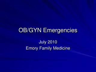
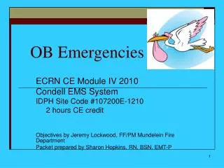
OB Emergencies. Debbie Sibley, MSN, RNC-OB Clinical Nurse Specialist Women’s Services The Medical Center of Central Georgia. Educational Objective. Identify key elements to facilitate OB emergency situations. Maternal – Fetal Assessment. Maternal Vital Signs Uterine Activity Resting Tone

E N D
OB Emergencies Debbie Sibley, MSN, RNC-OB Clinical Nurse Specialist Women’s Services The Medical Center of Central Georgia
Educational Objective • Identify key elements to facilitate OB emergency situations.
Maternal – Fetal Assessment • Maternal Vital Signs • Uterine Activity • Resting Tone • Intensity • Frequency • Duration • Fetal Heart Rate (110-160 bpm) • Baseline Rate • auscultate x 6 secs (before, during and after contraction) • Regular or Irregular? • Decelerations Noted?
Preeclampsia is diagnosed by the development of hypertension plus proteinuria, or edema that is generalized and overt, or both. Source: Williams Obstetrics Pregnancy Induced Hypertension
Infarcted areas (dysfunctional) and blood clots PIH / Toxemia Only Worsens There is One and Only One “Cure” … Delivery Grade III Placenta
Eclampsia • Turn patient on side • Protect from harm • If possible, insert oral airway. Do not force. • Call MD, summon help • Magnesium Sulfate • After seizure, administer O2 • Suction nose and mouth • Note characteristics of seizure • Assess for abruption and imminent delivery
Magnesium Sulfate: Maternal Considerations • 6 gm bolus has much better success at achieving and maintaining target serum levels than 4 gm bolus. • Magnesium Sulfate levels are not an absolute requirement ifyou watch renal output. • Watch out with Kidney Disease. Urine maybe excreted but without any removal of wastes. • “Magnesium Levels > 8, turn off even if she is breathing.” (Devoe) • The gold standard before and after delivery.
Magnesium Sulfate: Neonatal Considerations • 2.4 Mag Level is HIGH • May last for 24 – 48 hours • Infant can be deceptively pink around the face, lips and neck • If symptomatic . . . TREAT! • Watch for these in the neonate… • B/P drops: Give N/S bolus • Ileus: NPO • Apnea: Intubate
Diabetes: Maternal Complications Cesarean Births (41%) Infections Toxemia / PIH / preeclampsia Hydramnios / polyhydramios Shoulder dystocia (9.2% to 24% and 50% with babies weighing >4500g) Diabetes Ketoacidosis (rare)
Diabetes: Neonatal Complications • Macrosomia (40% even with euglycemia) predisposes baby to traumatic injuries: • hyperbilirubinemia: 20% • fractured clavicle • Hypoglycemia: <35mg/dL (20%) • Polycythemia: hematocrit >65% (3-5%) • Anomalies: • cardiac anomalies • sacral agenesis • RDS: incidence is decreased with maternal euglycemia
Pregnancy is a Diabetogenic State… Hyperglycemia plus hyperinsulinemia provide a continuous supply of glucose to the fetus.
Insulin Needs Change Dramatically • < 16 weeks: insulin requirements decrease • 28 – 32 weeks: marked increase related to increased fetal growth • Immediate Postpartum: insulin needs drastically drop
Glycemic Goals and Lab Tests • Pregnancy Glycemic Goals: serum blood glucose 60-90 mg/dl fasting and overall goal of 80-120 mg/dl • Hemoglobin A1C (glycosylated hemoglobin): mean blood glucose over the last 3 months; normal values are 4.2% to 6.1%
Classic Signs of Diabetes Ketoacidosis • Polyuria • Polydipsia (thirst) • Polyphagia (increased appetite)
Additional S&S of DKA • Abdominal pain • Dehydration • N&V • fruity (acetone) breath • Kussmaul breathing (air hunger, labored breathing) • Ketonuria & Glucosuria • Decreased Level of Consciousness (drowsiness, coma)
DKA Mortality Rates Maternal Mortality: rare Fetal Mortality: 10% up to 35%
DKA Treatment Aggressive Hydration Administer Insulin • IV regular insulin: flush polyvinyl tubing with 50mL to allow saturation of insulin to tubing. • Administer subcutaneous NovaLog insulin to give coverage until IV insulin can be setup. Fetal Assessment • FHR: Palpate maternal pulse, auscultate FHR and then compare findings of each. • EFM: absent variability and late decelerations, but don’t crash for C/S until ketoacidosis resolves.
The placenta lies low in the uterus, partly or completely covering the cervix. Occurs in one in 200 women. Placenta Previa: Partial Previa
Measured in percentages or centimeters The placenta may detach from the uterine wall before or during labor. Occurrence: 1% of all pregnant women Abdominal pain is often a classic sign even if there is no obvious bleeding. Abruption
Intrauterine Resuscitation • Oxygen at 10 L/ min per non-rebreather face mask • Change position: from HOB ↑ 300 to left or right lateral • IV Fluid Bolus: 1000mL over 20 minutes • Stop Uterine Contractions: Give Terbutaline (Brethine) or Magnesium Sulfate • Notify care provider Simpson and James. Efficacy of Intrauterine Resuscitation in Improving Fetal Oxygen Status During Labor. Obstetrics & Gynecology 2005.
Maternal Positions and Cardiac Output(liters / minute) • KNEE - CHEST 6.9 best! • RIGHT LATERAL 6.8 #2 • LEFT LATERAL 6.6 #3 • SITTING 6.2 #4 • SUPINE 6.0 bad! • DORSAL LITHOTOMY 5.8 bad! • STANDING 5.4 worst! • NON-PREGNANT 5.5 Clark and Cotton Am J Obstet Gynecology 1989
Emergency Delivery Don’t Leave Her Unattended Don’t Run Don’t Panic! (Don’t Break The Bed) Don’t Hold Legs Together Cord Management Suction Mouth Then Nose Note Time of Delivery Thermal Regulation: Keep Baby Warm Transportation of Mother & Baby
Additional OB Emergencies Shoulder Dystocia: No Fundal Pressure, even if called for by OB care provider
Maternal Deaths Decline Sharply Across the Globe Worldwide decrease from 526,300 (1980) to approximately 342,900 (2008) Reasons: reduced pregnancy rates; higher income, (better nutrition and access to health care); increased education for women; and increased availability of trained attendants assisting with childbirth. Study was conducted at the University of Washington and University of Queensland in Brisbane, Australia. Funded by the Bill and Melinda Gates Foundation. The researchers analyzed maternal mortality in 181 countries between 1980 and 2008. Sub-Saharan Africa has highest maternal death rates and improvements in India and China maternal death rates. From Lancet reported in AWHONN Vitals May 2010
Leading Causes of Maternal Death Embolism: leading cause after live birth Hemorrhage: leading cause after stillbirth Hypertension Infection Other medical conditions: leading cause when undelivered Indirect Causes: Cardiomyopathy / Heart Disease, Homicide, MVA, Cancer and Suicide Centers for Disease Control and Prevention (CDC), http://www.cdc.gov/mmwr/preview/mmwrhtml/ss5202a1.htm
Cardiac Arrest and Pregnancy • For both maternal and fetal outcome, early C/S (within 4 – 5 minutes of maternal cardiac arrest) appears to offer best chance of survival • Continue CPR and ACLS protocols during the perimortem cesarean delivery to maximize maternal and placental perfusion • OB anesthesiologist becomes “Captain of the Ship”
Modifications of CPR and Pregnancy • Uterine Displacement • Airway Management • Deeper Chest Compressions • Deliver within 5 minutes if viable fetus
Uterine Displacement Why? • to decrease compression of abdominal aorta, inferior vena cava and iliac arteries How? • Manual displacement of the uterus • Rolled towel or wedge under her hip (decreased effectiveness of chest compressions) • “Human Wedge”
Gestational Age >24 wks (4+ and higher): improves maternal cardiac output & infant survival 20-23 wks (umbilicus to 3+): improves maternal cardiac output <20 wks (below umbilicus): No C/S; uterine size not compromising maternal cardiac output Maternal Position and Hysterotomy Decisions
Uterine Displacement If Greater Than 20 Weeks… Create a “Human Wedge” • Roll patient to side (left or right) • Tilt patient on the bent knees of the rescuer
Airway Changes: Use Cricoid Pressure Larynx is more anterior & cephalad; Laryngeal & pharyngeal mucosa more edematous
Resuscitation Needs • Smaller Endotracheal Tube • Short Laryngoscope Handles • Equipment for Cricothyrotomy Incidence of failed intubation 1:500 compared to general surgical population 1:2000
Chest Compressions • Use additional pressure with chest compressions • Compress Chest 2 inches (push deeper) • Compressions: “Deep and Fast”
Deliver within 5 minutes if viable fetus Initiate C/S within 4 minutes and Deliver Infant within 5 minutes of arrest • Maternal Survival Highly linked with cause of arrest (Amniotic Fluid Embolus, Gun Shot Wound, et al.) Many cases CPR/ACLS ineffective until delivery, followed by dramatic recovery • Fetal Outcome Viable >23 weeks gestation Delivered within the first five minutes of maternal cardiac arrest
Survivability of InfantsChristopher Viscomi, Anesthesiologist, University of Vermont
ACLS Protocol Primary Survey Airway: open airway Breathing: PPV Circulation: perform chest compressions Defibrillation: shock for VF and pulseless VT
ACLS Protocol Secondary Survey Airway: establish advanced airway/ET intubation Breathing: assess adequacy of ET intubation & PPV Circulation: IV access, continue CPR and Defibrillate up to 3 times 200 J, 200-300 J and 360 J
Permission for use has been granted: Lippincott Williams & Wilkins
Cardiac Arrest and Pregnancy Protocol • (BLS Box 1) Check for Response. • Tap on shoulder & ask, “Are you all right?” No movement. No response. • Victim assessed to be pregnant. • (BLS Box 2) Activate EMS: Phone 911 Call OB code team / “Code Blue” • (BLS Box 2) Get AED. Return & Perform CPR.
Permission for use has been granted: Lippincott Williams & Wilkins
Cardiac Arrest and Pregnancy Protocol • (BLS Box 2) Position the victim. • Place victim supine on hard surface. • Fundal height. Above umbilicus: uterine tilt. Below: proceed without modifications. • Rescuer kneels beside victim’s thorax. • Do not move the victim unless environment becomes unsafe. • (BLS Box 3) Open Airway • Jaw thrust (with cricoid pressure). • If adequate ventilations cannot be effected, use head tilt-chin lift (with cricoid pressure). • (BLS Box 3) Maintaining Open Airway, Check breathing: “Look, Listen, and Feel” (<10 seconds)
Cardiac Arrest and Pregnancy Protocol • (BLS Box 4) No breath: Perform Rescue Breathing. • Pinch victim’s nose, create airtight mouth-to-mouth seal. • Give 2 rescue breaths. Effect visible chest rise. • No rise and fall with first breath, reposition. Perform head tilt-chin lift and give second breath. • Each rescue breath should be delivered over 1 second per breath. Avoid rapid or forceful breaths. • Rescuer should take “regular” (not deep) breaths between rescue breaths. • Continuous cricoid pressure with rescue breathing and during intubation. • Establish advanced airway early with smaller endotracheal tube. (# 6.5 or # 7)