Download
1 / 74
770 likes | 1.31k Vues
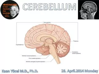
Cerebellum. David Kachlík and Petr Zach. Small brain = Cerebellum. 10 % weight of whole brain More than ½ neurons of whole brain ¼ - ¾ area of whole brain. Mesencephalon Pons Medulla oblongata Medulla spinalis Cerebellum. Cerebellum. Cerebellum – parcellation. Functional placement:

E N D
Cerebellum David Kachlík and Petr Zach
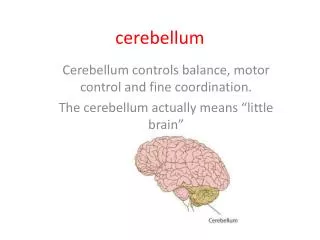
Small brain = Cerebellum 10 % weight of whole brain More than ½ neurons of whole brain ¼ - ¾ area of whole brain
Mesencephalon Pons Medulla oblongata Medulla spinalis Cerebellum
Cerebellum – parcellation • Functional placement: • vermis a l. floculonodularis • paravermální (intermediální) zóna • hemisféry (laterální zóna) • External structure: • lobus anterior • lobus posterior • lobus flocculonodularis • developmentally: • archicerebellum • paleocerebellum • neocerebellum • function: • vestibulocerebellum • spinocerebellum • cerebrocerebellum (= pontocerebellum)
Cerebellum – description • folia cerebelli (leaves) • fissurae cerebelli (crack) • vermis (červ) – non paired in middle • hemispheria – paired • 3 lobi (lobes) • Small parts • 10 in vermis [I - X] – expample nodulus • 9 in hemispheres [H II - H X] • tonsilla – when edema it goes into foramen magnum and it compresses stem • flocculus
Cerebellum– posterior view VERMIS
Cerebellum – ventral view NODULUS FLOCCULUS TONSILLA
Cerebellum – inferior view VERMIS TONSILLA
Cerebellum – developmental parts • lobus anterior [I-V + H II - H V] = spinocerebellum = paleocerebellum fissura prima • lobus posterior [VI-IX + H VI - H IX] = pontocerebellum = neocerebellum fissura posterolateralis • lobus flocculonodularis [X + H X] = vestibulocerebellum = archicerebellum
Medial zone of anterior and posterior lobe =paleocerebellum=spinocerebellum=fine tune body and limb movement Lateral zone of anterior and posterior lobe = cerebrocerebellum=neocerebellum = ?? Plannning movement, cognitive functions Flocculonodular lobe =vestibulocerebellum=archicerebellum=balance and gait
Cerebellum – funkční části 3 podélné zóny • vermis + lobus flocculonodularis • paravermální kůra • hemisféry
Vestibulocerebellum Spinocerebellum Cerebrocerebelum Cerebellum Primary fissure Anterior Lobe Regulation of muscle tone, coordination of skilled voluntary movement Posterior Lobe Planning and initiation of voluntary activity Flocculo-Nodular Lobe (FN lobe) Maintenance of balance, control of eye movements Folia
Flocullonodular lobe Medial Lateral (Deiters) Inferior (Bechterew) Superior Does not exit cerebellum via deep cerebellar nuclei!
Anterior lobe, spinocerebellum Interposed nuclei=nc. globosi and nc. emboliformis Superior cerebellar peduncule Red nucleus Distal muscle group Vermis Fastigial nucleus
Cerebellum – pedicles • pedunculus cerebellaris inferior • corpus restiforme • corpus juxtarestiforme • pedunculus cerebellaris medius (= brachium pontis) • AF: tractus cortico-ponto-cerebellaris • pedunculus cerebellaris superior (= brachium conjunctivum)
Cerebellum – ventral view P.C.SUPERIOR P.C.MEDIUS P.C.INFERIOR
Cerebellum – internal structure • cortex cerebelli: strata(3 layers) – arbor vitae • stratum moleculare • stratum purkinjese • stratum granulosum • corpus medullare cerebelli: nuclei cerebelli 4 paired nuclei („Don't Eat Greasy Food“) • nucleus dentatus (= lateralis cerebelli) • nucleus emboliformis (= interpositus anterior) • nucleus globosus (= interpositus medialis) • nucleus fastigii (= medialis cerebelli)
Deep cerebellar nuclei Via inferior peduncle Via superior peduncle
Cerebellum – layers of cortex • stratum moleculare • neuron stellatum (stellate cells) • neuron corbiforme (basket cells) • neurofibra parellela (parallel fiber) – axons of granular cells • stratum purkinjese = stratum neurium piriformium; formerly stratum gaglionicum • neuron purkinjese (Purkynje cells) • corbis neurofibrarum (rich branching to stratum moleculare) • Axons to cerebellar nuclei • stratum granulare • neuron granulosum (granulr cell) • neuron stellatum magnum Golghi (Golgi cell) • Next 3 types of cells • glomerulus cerebelli • Afferent fibers: neurofibra muscosa (mossy fiber - Glu) + ascendens (climbing fiber - Asp)
Cerebellum: 3 layered cortex Climbing fibers: excite the Purkinje cells Mossy fibers: excite the granule cells Granule cells: make excitatory contact with the Purkinje cells Purkinje cells: Tonic inhibition on the activity of the neurons of the cerebellar nuclei => All excitatory inputs will be converted to the inhibition => Removing the excitatory influence of the cerebellar inputs (erasing)
Cerebellum – afferentationbalance • tractus vestibulocerebellaris directus vestibulum corpus juxtarestiforme (v PCI) nodulus + uvula (ipsilat.) • tractus vestibulocerebellaris indirectus vestibulum ncl. vestibulares corpus juxtarestiforme (v PCI) lobus flocculonodularis + vermis (bilat.) • tractus trigeminocerebellaris Information from head
Cerebellum – afferentationpassiveproprioception • tractus spinocerebellaris posterior ncl. thoracicus post. Stilling-Clarke • medulla oblongata pedunculus cer. inf. • vermis + paravermal cortex (ipsilateral) proprioception from trunk and LL • tractus cuneocerebellaris Posterior fascicle tract nucleus cuneatus accessorius Proprioception from UL and thorax
Cerebellum – afferentationactive proprioception • tractus spinocerebellaris anterior ncl. thoracicus post. Stilling-Clarke crossing at spinal lvl mesencephalon pedunculus cer. superior crossing in crbl cortex vermis + paravermal cortex (ipsilateral) – LL • tractus spinocerebellaris rostralis ncl. thoracicus post. Stilling-Clarke pedunculus cer. inferior vermis + paravermal cortex (ipsilateral) – UL • tractus spinoolivaris – motoric learning • for example walking steep steps
Cerebellum– afferentation form cortex • tractuscortico-ponto-cerebellaris(20.000.000 fibers) lobus f,p,o,t capsula interna ncll. pontis fibrae pontis transversae crossing pedunculus cer. medius crbl cortex (contralat.) • tractus cortico-olivo-cerebellaris lobus f,p,o,t capsula interna complexus olivaris inf. (bilat.) crossing pedunculus cer. inferior crbl cortex • tractus cortico-reticulo-cerebellaris lobus f,p,o,t (mostly sensorimotor cortex) capsula interna RF (bilat.) crossing pedunculus cer. medius + inf. crbl cortex Will motoric, movement preparation, setting of proper muscle tonus
Cerebellum – efferentation ncl. fastigii 1. PCI RF (bilat.) tr. reticulospinalis 2. PCI ncl. vestibularis lat. Deitersi (bilat.) tr. vestibulospinalis 3. cranial nerves, neck muscles ncll. interpositi (globosus + emboliformis) • PCS crossing ncl. ruber (pars magoncellularis) tractus rubrospinalis crossing spine (ipsilat.) ncl. dentatus • PCS crossing ncl. VA+VL thalami area 4 tr. pyramidalis crossing spine (ipsilat.)
Cerebellum – inferior pediclespedunculus cerebellaris inferior • corpus restiforme • AF↑: tr. spinocerebellaris posterior + rostralis, tr. cuneocerebellaris, tr. spinoolivaris • AF↑: tr. spino-reticulo-cerebellaris • AF↓: tr. cortico-reticulo-cerebellaris, cortico-olivo-cerebellaris, cortico-arcuato-cerebellaris • corpus juxtarestiforme • AF↑ tr. vestibulocerebellerais directus + indirectus • EF↓: tr. cerebello-reticulospinalis, -cerebellovestibularis, cerebelospinalis, cerebellonuclearis (all from z ncl. fastigii)
Cerebellum – middle and upper pediclespedunculus cerebellaris medius et superior • pedunculus cerebellaris mediusAF↓: tractus cortico-ponto-cerebellaris • pedunculus cerebellaris superiorAF↑: tr. spinocerebellaris anterior + tectocerebellarisEF↓: tr. cerebello-rubro-thalamo-corticalis + tr. cerebello-rubro-spinalisEFcircuit: tr. cerebello-rubro-olivo-cerebellaris (Papezův control circuit)
Dentate nuclei: project contralaterally through the superior cerebellar peduncle to neurons in the contralateral thalamus & from thalamus to motor cortexFunc.: influence planning and initiation ofvoluntary movement Emboliform & Globose nuclei: project mainly to the contralateral red nuclei & a small group is projected to the motor cortexRed Nuclei Rubrospinal Tract control of proximal limb muscles Fastigial nuclei: project to the vestibular nuclei & to the pontine and medullary reticular formation Vestibulospinal & Reticulospinal tracts
2 maps of body Primary fissure Inputs to cerebellum from spinocerebellar tracts have a somatotopic organization. Signals from the motor cortex, which is also arranged somatotopically, project to corresponding points in the sensory maps of the cerebellum.