Download
1 / 54
590 likes | 961 Vues
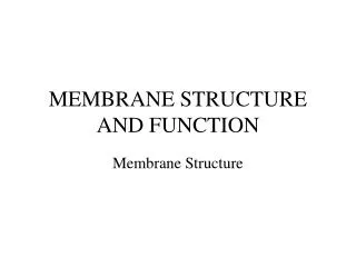
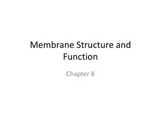
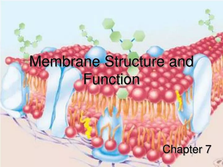
Membrane Structure and Function. Chapter 7. TEM of Phospholipid Bilayer. Hydrophobic region of protein. Hydrophilic regions of protein. Membrane Structure. Basic fabric of membranes is a phospholipid bi-layer

E N D
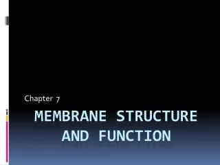
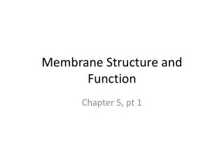
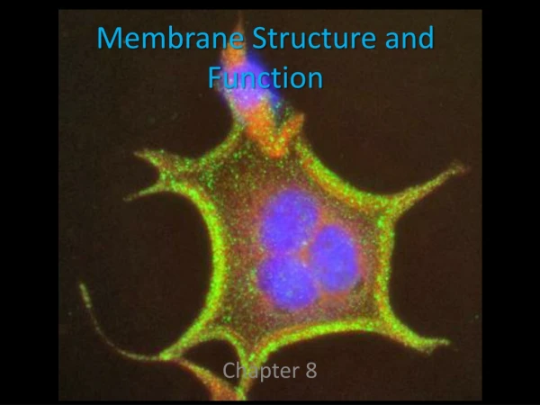
Membrane Structure and Function Chapter 7
Hydrophobic region of protein Hydrophilic regions of protein Membrane Structure • Basic fabric of membranes is a phospholipid bi-layer • Phospholipids are amphipathic, so the center of the bi-layer is hydrophobic and the outsides are hydrophilic • Proteins are found in the layer – the hydrophobic region of proteins are found in the center of the bi-layer, with the hydrophilic regions protruding on both sides • Proteins may be integral or peripheral
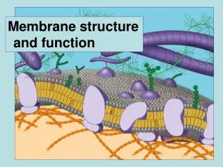
(Protein + Oligosaccharide = Glycoprotein) Cholesterol (Oligosaccharide added in the Golgi body) (Part of cytoskeleton) The fluid mosaic model Membranes have the consistency of cooking oil!
The fluidity of membranes • The phospholipids of membranes are constantly drifting - moving laterally • Sometimes the phospholipids flip-flop • The embedded proteins or surface proteins also drift • Some proteins are held in place by the cytoskeleton
The fluidity of membranes, cont’d. • Membranes remain fluid when temperature decreases - up to a certain critical temperature, after which they solidify • The more the concentration of unsaturated hydrocarbons in the phospholipid tails, the longer the membrane stays fluid (Because of kinks in the tails, they cannot pack closely) • Cholesterol is a common component of animal membranes – it keeps the membrane fluid at low temperatures, but reduces fluidity at moderate temperatures.
Evidence of membrane protein drift When mouse and human cells were fused, their phospholipid bi-layers, along with their membrane proteins intermingled within one hour – creating a chimeric plasma membrane.
Membrane Proteins • Integral proteins are either completely embedded (transmembrane), or partially embedded in the bilayer • Peripheral proteins are not embedded in the membrane, they are attached to the surface of the bilayer or to integral proteins A transmembrane protein
Passive transport vs. Active transport For example: Enzymes embedded in the inner membrane of mitochondria play a role in cellular respiration For example: insulin binding to membrane proteins, which starts a signaling pathway that stimulates cells to take up more glucose from the bloodstream Types of membrane proteins – and their roles
For example: cells of the immune system need to bind to glycoproteins on cell surfaces, in order to decide if the cell belongs to the body or is foreign Types of membrane proteins – and their roles, cont’d. Integrins are an example of cell surface receptor proteins that adhere to and interact with the ECM. Integrins also coordinate activities inside and outside cells via signal transduction.
Selective permeability Traffic Across Membranes • The phospholipid bilayer is selectively permeable – it allows only certain substances across – depending on SIZE and/or POLARITY • Because it is hydrophobic in the center, it does not allow ions and polar molecules across – even small ions like H+, Na+ or OH- cannot cross membranes • For the same reason, it does allow nonpolar molecules like O2, CO2 (Diffusion and osmosis) • Large molecules whether polar or nonpolar cannot cross over (most sugars, proteins, amino acids, lipids, etc.) • Membrane proteins help transport molecules that cannot cross the bilayer on their own
Electrostatic Gradient • The interior of cells is negatively charged compared to the outside • This creates a voltage across the membrane, which is called the membrane potential • For this reason, anions will automatically move outside the cell (drawn by the + charges) and cations will be drawn to the inside (drawn by the neg- charges) – ions however, need to pass through membrane proteins. • This difference in charge is called the electrostatic gradient • The membrane potential of a resting cell is about -70 mV (It can range from -50 to -200 mV)
Concentration Gradient • Molecules introduced to a new environment, will move away from their initial location, creating a concentration gradient – their concentration becomes exceedingly lower as you move away from the introduction site
Electrostatic gradient + Chemical gradient = Electrochemical gradient
PASSIVE TRANSPORT • Passive transport is the movement of molecules down their electrochemical gradient • Passive transport requires no energy expenditure on the part of the cell. “Free” energy is used – the energy of the system • Examples of passive transport: • Diffusion • Osmosis • Facilitated diffusion (Protein channels involved)
Diffusion • Molecules have the natural tendency (due to random molecular motion) of moving from an area where they are highly concentrated, to an area where their concentration is low – they move down their concentration gradient+ • Once the molecules are evenly dispersed in the environment, they reach a state of equilibrium – they continue to move, but it is equal in every direction – so no net change Low free energy – stable system High free energy
Diffusion • Diffusion is passive transport • It is the random movement of molecules from and area of high concentration to an area of low concentration • Diffusion requires NO energy • In diffusion, molecules move along their concentration gradient
Osmosis • Osmosis is passive transport • It is the random movement of WATER molecules from an area of high water concentration to an area of low water concentration • Osmosis requires NO energy • In osmosis, molecules move along their concentration gradient
Osmosis • The diffusion of water molecules • The tendency of water molecules (due to random molecular motion) to move from an area where their concentration is high (higher free energy), to an area where their concentration is lower (lower free energy) – until equilibrium is reached (no net movement of water) • Movement of water molecules is down their concentration gradient
Low solute High solute Isotonic Solution (Also a form of Passive Transport) solute and solvent balanced (of water molecules) As solute concentration increases, “free” water concentration decreases – so water potential decreases Water then moves from an area of high water potential to an area of low water potential
Inside the cell is higher than the outside, because the outside has more solute particles Water will therefore move out of the cell to an area of lower (Net movement is outwards) • Inside the cell is lower, because of solutes in the cytosol Water molecules always move from an area of higher water potential to an area of lower water potential, so water rushes into the “cell” from the outside (Net movement is inwards) Is equal on both sides, so no net movement Is the “cell” hypertonic, hypotonic or isotonic with respect to its environment?
Water Potential Osmosis & Plant cells
Plants & water potential • Plants can use the potential energy in water to perform work. • Tomato plant regains turgor pressure – cell pushes against wall due to uptake of water
Plants & water potential • The combined effects of 1.) solute concentration 2.) physical pressure (cell wall) can be measured as Water Potential • = psi • is measured in megapascals (MPa) • 1 Mpa = 10 atmospheres of pressure
Calculating Water Potential • = P + S Or Water = pressure + solute Potential potential potential
Solute Potential S • Solute potential is also called the osmotic potential because solutes affect the direction of osmosis. • S of any solution at atmospheric pressure is always negative – why? • Answer = less free water molecules to do work
Solute Potential S • Solutes bind water molecules reducing the number of free water molecules lowers waters ability to do work.
Pressure Potential P • P is the physical pressure on a solution. • P can be negative transpiration in the xylem tissue of a plant (water tension) • P can be positive water in living plant cells is under positive pressure (turgid)
Standard for measuring • Pure water is the standard. • Pure water in an open container has a water potential of zero at one atmosphere of pressure.
Water Potential: an artificial model • (a) addition of solutes on right side reduces water potential. S = -0.23 • Water flows from “hypo” to “hyper” • Or from hi on left to lo on right
Water Potential: an artificial model • (b) adding +0.23 pressure with plunger no net flow of water • (c) applying +0.30 pressure increases water potential solution now has of +0.07 • Water moves right to left
Water Potential: an artificial model • (d) negative pressure or tension using plunger decreases water potential on the left. • Water moves from right to left
Water relations in plant cells • (b) Flaccid cell in pure water Water potential is into cell cell becomes turgid
Water relations in plant cells • (a) Flaccid cell placed in hypertonic solution Water potential is out of cell plasmolysis
Calculating Solute potential • Need solute concentration • Use the equation S = - iCRT i = # particles molecule makes in water C = Molar concentration R = pressure constant 0.0831 liter bar mole oK T = temperature in degrees Kelvin = 273 + oC
Solve for water potential(literal equation) • Knowing solute potential, water potential can be calculated by inserting values into the water potential equation. = P + S In an open container, P = 0
Hints & reminders 1. Remember water always moves from [hi] to [lo]. 2. Water moves from hypo hypertonic. 3. [Solute] is related to osmotic pressure. Pressure is related to pressure potential. 4. Pressure raises water potential. 5. When working problems, use zero for pressure potential in animal cells & open beakers. 6. 1 bar of pressure = 1 atmosphere
Water and the Bilayer • Although water is a polar molecule, some water molecules ARE able to “sneak” past the phospholipids via osmosis. • But the majority of the water molecules are prevented from passing the hydrophobic tails of the lipid bilayer • So water has to use Aquaporins, a special class of integral transmembrane channel proteins
Aquaporins • More than 10 different mammalian aquaporins have been identified to date, and additional members are suspected to exist. • Some aquaporins transport solute-free water across cell membranes; they appear to be exclusive water channels and do not permeate membranes to ions or other small molecules. • Other aquaporins transport water and other small polar molecules and ions.
More about Auaporins In the absence of aquaporins, cells do not swell osmotically!
Plasmolysis • When a cell is placed in a hypertonic environment – more solute outside than inside: • - Water potential is greater inside • - Water will move from where water potential is greater, to where it is lower • - Water will move out of the cell, causing plasma membrane to collapse (low pressure potential) • - Cell wall will keep cell from losing its shape – animal cell loses shape
Facilitated Diffusion • Ions and small polar molecules use facilitated diffusion • Integral membrane channel proteins – 1. open channel – (water uses this method -aquaporins) 2. gated channel 3. carrier proteins – (glucose uses this method) • Requires no cellular energy (ATP, GTP, etc.) • Specific channel proteins for specific ions – “lock-key” system • Diffusion is down concentration gradient
Facilitated Diffusion • Ions and small polar (Hydrophilic) molecules use facilitated diffusion • Membrane channel proteins are used • Requires no cellular energy (ATP, GTP, etc.) • Diffusion is down concentration gradient
ACTIVE TRANSPORT • Uses cellular energy (ATP, GTP, etc.) • Uses integral membrane proteins • Specific proteins for specific molecules • Molecules can be moved against their electrochemical gradient • Ion pumps – like the Na+ / K+ pumpand the Proton pump (H+) are an example of active transport • Concentration of Na+ has to be higher outside the cell whereas that of K+ has to be higher inside the cell – so active transport is used to maintain these concentrations (pumping against electrochemical gradient)
ACTIVE TRANSPORT • Uses cellular energy (ATP, GTP, etc.) • Uses membrane proteins • Specific proteins for specific molecules • Molecules can be moved against their concentration gradient – from a low concentration to a high concentration.
1. Na+ binds to the transport protein at specific binding sites 3. Phosphorylation causes conformational change in protein, which moves the Na+ out of the cell 2. Na+ binding causes ATP to phosphorylate protein 4. 6. When K+ exits its binding site, it causes the release of the inorganic phosphate group When Na+ exits the binding site, the binding site for K+ is made accessible and K+ binds to sites 5. When K+ binds, it causes another conformational change, which moves K+ into cell Active Transport Cont’d.