Download
1 / 32
320 likes | 527 Vues
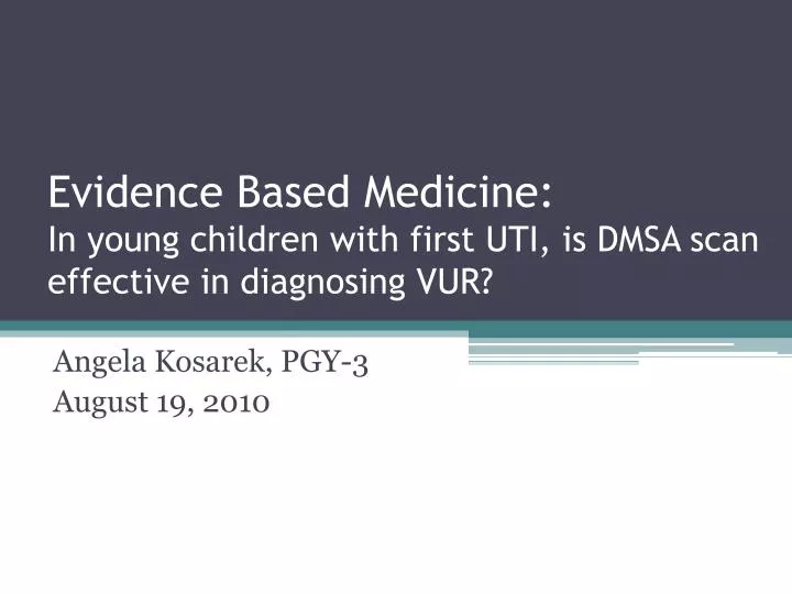
Evidence Based Medicine: In young children with first UTI, is DMSA scan effective in diagnosing VUR?. Angela Kosarek , PGY-3 August 19, 2010. Our patient. Patient is a 17 month old male with Trisomy 21 admitted for repair of an unbalanced AV canal.

E N D
Evidence Based Medicine: In young children with first UTI, is DMSA scan effective in diagnosing VUR? Angela Kosarek, PGY-3 August 19, 2010
Our patient • Patient is a 17 month old male with Trisomy 21 admitted for repair of an unbalanced AV canal. • One week prior to admission at Children’s Hospital, he presented to an outside hospital with c/o fever. • Urine culture (from transurethral catheterization) was positive for >100,000 cfu/ml of E. coli.
Our patient • He completed a 14 day course of antibiotics based on bacterial antibiogram. • Renal ultrasound was normal. • Patient’s mother refused further evaluation for VUR with VCUG due to the invasive nature and anticipated discomfort of the procedure.
Recommended Imaging Studies for Culture-Positive UTI • Renal ultrasound & voiding cystourethrogram • Indicated for all male patients with their 1st UTI, all female patients < 5 years of age, and all children with recurrent UTI • Renal ultrasound • Noninvasive, nonionizing evaluation for gross structural defects, obstructive lesions, positional abnormalities, and renal size and growth
Recommended Imaging Studies for Culture-Positive UTI • Voiding Cystourethrogram (VCUG) • Obtained by the use of fluoroscopy and a contrast agent introduced through a catheter in the bladder • Performed when asymptomatic and cleared of bacteriuria • Visualizes bladder and urethral anatomy • Diagnostic of vesicoureteral reflux (VUR) upon the demonstration of urine refluxing from the bladder to the upper urinary tract
Grading Classification of VUR • I: reflux only fills ureter without dilation • II: reflux fills ureter & collecting system without dilation • III: filling and mild dilation of ureter & collecting system with mild blunting of calyces • IV: filling & gross dilation of ureter & collecting system with blunting of calyces; some ureteral tortuosity • V: gross dilation of collecting system; calyceal blunting with loss of papillary impression; significant ureteral dilation & tortuosity
Primary vs. Secondary VUR • Primary VUR • Most common form • Normally reflux is prevented during bladder contraction by fully compressing the intravesicular ureter and sealing it off with the surrounding bladder muscles • Results from incompetent or inadequate closure of the ureterovesical junction (UVJ) • Spontaneous resolution can occur with growth
Primary vs. Secondary VUR • Secondary VUR • Abnormally high pressure in the bladder causes failure of closure of the UVJ during bladder contraction • Often associated with anatomic (e.g., posterior urethral valves) or functional bladder obstruction (e.g., dysfunctional voiding & neurogenic bladder) • Management: initial treatment of the associated abnormality followed by surgical correction of the VUR, if necessary
Nonsurgical management of VUR • Antimicrobial prophylaxis • Amoxicillin in first 2 months of life • Otherwise trimethoprim-sulfamethoxazole or nitrofurantoin • UTI Surveillance • Urine cultures every 4 months and when febrile • Change antibiotic therapy if breakthrough UTI • VUR follow-up • Repeat VCUG in 12-18 months to determine whether VUR has resolved
Indications for Surgical Management • Grade V reflux with scarring in children > 1 year of age • Grade V reflux in children > 6 years of age • Grade IV reflux with either bilateral reflux or renal scarring in children > 6 years of age • Children with breakthrough pyelonephritis while on prophylaxis
Why do we care about VUR? • Current management is based upon premise that VUR predisposes to acute pyelonephritis • VUR allows transportation of bacteria from the bladder to the kidney • Resulting infection may lead to renal scarring, the loss of renal parenchyma between the calyces and the renal capsule • Effects of extensive scarring include HTN, decreased renal function, proteinuria, and sometimes ESRD
Should we care about VUR? • Alternate viewpoint of primary VUR: • Does not play a causative role in chronic kidney disease (CKD) • Serves as marker for abnormal renal development • Disruption in renal development leads to decreased formation of normal parenchyma, resulting in increased risk of poor renal outcome • Limited data comparing the effect of VUR treatment versus no treatment (i.e., surveillance) upon CKD and renal scarring
RIVUR Study • Randomized Intervention for Children with Vesicoureteral Reflux Study • 2 year multicenter, double-blind, randomized, placebo-controlled trial • Objective: to determine if prophylaxis with trimethoprim-sulfamethoxazole prevents recurrent UTI and renal scarring with VUR
Clinical Question • In young children after a first febrile urinary tract infection, is a dimercaptosuccinic acid (DMSA) scan, in combination with renal ultrasound, as sensitive and specific in diagnosing VUR as a VCUG?
DMSA scan • Evaluates the size, shape, and position of the kidneys • Does not visualize urethral anatomy • Detects areas of decreased uptake that may represent acute pyelonephritis or renal scarring • Limitations • Not sensitive for low-grade reflux • Cannot grade reflux
DMSA scan • Requires injection of a radioisotope (tracer) through an IV catheter • 2-4 hour lag time between injection of tracer and imaging • Study does not permit any movement and may take up to 1 hour to perform • 1/100 the radiation exposure of VCUG
DMSA scan • normal: homogenous uptake of the radioisotope throughout the kidneys with preservation of the renal contour • with acute pyelonephritis or renal scarring: focal or diffuse areas of decreased uptake
The efficacy of ultrasound and dimercaptosuccinic acid scan in predicting vesicoureteral reflux in children below the age of 2 years with their first febrile urinary tract infection Hye-young Lee, Byung Hyun Soh, Chang Hee Hong, MyungJoon Kim, Sang Won Han Pediatric Nephrology (2009) 24:2009-2013
Patients and Methods • Retrospective analysis • Medical records and radiological imaging studies • Children younger than 2 years diagnosed with 1st febrile UTI from January 2001 to May 2007 • Febrile UTI: temp > 38.5° C with significant bacteriuria ( > 105cfu/ml) • Urine samples: Midstream clean catch in boys & transurethral catheter in girls
Patients and Methods • All patients underwent renal US, DMSA scan, & VCUG • US performed immediately after diagnosis • DMSA & VCUG performed after resolution of fever & confirmation of a negative urine culture • Excluded patients • Neurological abnormalities that might have effect on voiding function • Obstructive diseases of the urinary tract • History of previous UTI
Patients and Methods • Abnormal findings suggestive of VUR: • US: • Dilatation of renal pelvis or ureter • Increase of renal echogenicity • Reduction in thickness of renal parenchyma • Irregularity of kidney margin • Loss of corticomedullary differentiation • DMSA scan: • More than one cortical photon defect • Reduction in uptake of radioisotope • Split renal function lower than 45%
Patients and Methods • VUR was graded in accordance with grading system as defined by International Reflux Study Committee • Grades I-II: classified as low grade reflux • Grades III-V: classified as high grade reflux • Prophylactic antibiotics • Low grade reflux with persistent pyuria • High grade reflux • Patients followed for mean of 23.2 months
Results • Among 220 children diagnosed with UTI: • 162 (73%) boys & 58 (27%) girls • 212 (96.4%) younger than 1 year • Mean age 4.5 months (range 0.1 – 21 months) • 67 children (30.4%) diagnosed with VUR by VCUG • Low grade: 24 cases (35.8%) • High grade: 43 cases (64.2%)
Findings suggestive of VUR on US and DMSA scan according to grade of VUR 21 follow up 19 resolved or down-graded
Follow-up for low grade VUR • 24 children: • 8 with negative US & DMSA scan • Spontaneous resolution or down-grading of VUR • No complications related to VUR • 13 with positive US or DMSA scan • 12 had spontaneous resolution or down-grading of VUR • 1 showed no change • No complications related to VUR • Follow-up not reported for 3 children
Follow-up for high grade VUR • Grade III detected on US or DMSA scan • 1 patient down-graded to grade II VUR • Remaining 8 children underwent anti-reflux surgery (Follow-up not reported for the 2 undetected cases) • Grade IV • 2 patients down-graded to low grade VUR • 2 patients with resolution of VUR • Remaining 16 underwent anti-reflux surgery • Grade V • All 12 underwent surgical correction
Sensitivity, specificity, and positive and negative predictive values of US, DMSA, and US with DMSA for detection of VUR
Discussion • Retrospective evaluation • The preferred study to evaluate efficacy of a diagnostic test: a prospective, blind comparison to a gold standard study • Efficacy of US and DMSA scan were evaluated only in VUR-positive individuals
Discussion • In this study, 95.3% (41/43) of high grade VUR could be predicted by US and DMSA scan • 2 patients in whom VUR could not be detected had Grade III VUR and showed spontaneous improvement • Limited in detection of low grade reflux (62%) • Spontaneous resolution or down-grading of VUR in 19/21 children during follow-up • No complications related to VUR
DMSA Scan for Revealing Vesicoureteral Reflux in Young Children With Urinary Tract Infection SotiriosFouzas, ErifyliKrikelli, PavlosVassilakos, DespoinaGkentzi, Dimitrios A. Papanastasiou and Christos Salakos Pediatrics published online Aug 2, 2010
Comparison of Parameters of US and DMSA scan for Detection of VUR
References • www.uptodate.com • www.hsl.unc.edu • www.chop.edu • Keren R et al: Rationale and Design Issues of the Randomized Intervention for Children with Vesicoureteral Reflux (RIVUR) Study. Pedatrics 2008; 122(Suppl 5): S240-S250. • American Academy of Pediatrics, Committee on Quality Improvement, Subcommittee on Urinary Tract Infection: Practice parameter: The diagnosis, treatment, and evaluation of initial urinary tract infection in febrile infants and young children. Pediatrics 1999; 103(4): 843-852. • Hoberman A et al: Oral versus initial intravenous therapy for urinary tract infections in young febrile children. Pediatrics 1999; 104(1): 79-86.