Download
1 / 46
470 likes | 626 Vues
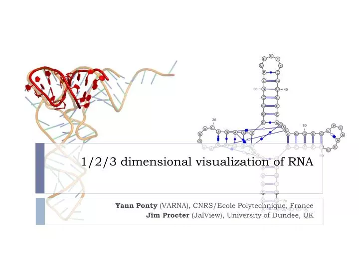
1/2/3 dimensional visualization of RNA. Yann Ponty (VARNA), CNRS/ Ecole Polytechnique , France Jim Procter ( JalView ), University of Dundee, UK. Goals. To help your survive the RNA data jungle . To conceptually and practically connect the three levels of RNA structural information.

E N D
1/2/3 dimensional visualization of RNA YannPonty (VARNA), CNRS/EcolePolytechnique, France Jim Procter (JalView), University of Dundee, UK
Goals • To help your survivethe RNA data jungle. • To conceptually and practically connect the three levels of RNA structural information. • To introduce mature prediction and annotation tools. • To illustrate the structure-informed curation RNA alignments. • To keep this fun and interactive.
How RNA folds G/C Canonical base-pairs U/A U/G 5s rRNA (PDB ID: 1UN6) RNA folding = Hierarchicalstochastic process driven by/resulting in the pairing (hydrogen bonds) of a subset of its bases.
RNA file formats: Secondary Structures <?xml version="1.0"?> <!DOCTYPE rnaml SYSTEM "rnaml.dtd"> <rnaml version="1.0"> <molecule id=“xxx"> <sequence> ... </sequence> <structure> ... </structure> </molecule> <interactions> ... </interactions> </rnaml>
RNA file formats: Secondary Structures <?xml version="1.0"?> <!DOCTYPE rnaml SYSTEM "rnaml.dtd"> <rnaml version="1.0"> <molecule id=“xxx"> <sequence> <numbering-system id="1" used-in-file="false"> <numbering-range> <start>1</start><end>387</end> </numbering-range> </numbering-system> <numbering-table length="387"> 2 3 4 5 6 7 8... </numbering-table> <seq-data> UGUGCCCGGC AUGGGUGCAG UCUAUAGGGU... </seq-data> ... </sequence> <structure> ... </structure> </molecule> <interactions> ... </interactions> </rnaml>
RNA file formats: Secondary Structures <?xml version="1.0"?> <!DOCTYPE rnaml SYSTEM "rnaml.dtd"> <rnaml version="1.0"> <molecule id=“xxx"> <sequence> ... </sequence> <structure> <model id=“yyy"> <base> ... </base> ... <str-annotation> ... <base-pair> <base-id-5p><base-id><position>2</position></base-id></base-id-5p> <base-id-3p><base-id><position>260</position></base-id></base-id-3p> <edge-5p>+</edge-5p> <edge-3p>+</edge-3p> <bond-orientation>c</bond-orientation> </base-pair> <base-pair comment="?"> <base-id-5p><base-id><position>4</position></base-id></base-id-5p> <base-id-3p><base-id><position>259</position></base-id></base-id-3p> <edge-5p>S</edge-5p> <edge-3p>W</edge-3p> <bond-orientation>c</bond-orientation> </base-pair> ... </str-annotation> </model> </structure> </molecule> <interactions> ... </interactions> </rnaml>
Secondary Structure representations http://varna.lri.fr
First contact • Run the web start version of VARNA at: http://varna.lri.fr/downloads.html • Locate and save on disk a bunch of secondary structures from the RNA Strand Database (CT or BPseq): http://www.rnasoft.ca/strand/ • Load these files and using the region highlight feature of VARNA, highlight a region of interest. Menu►Edit►Annotation►New►Region
Basic prediction Minimal free-energy folding
Minimal Free-Energy (MFE) Folding • Turner model associates energy to each compatible secondary structure. • Vienna RNA package implements a O(n3) algorithm for computing the most stable folding… • … but also offers nice visualization features. …CAGUAGCCGAUCGCAGCUAGCGUA… RNAFold
RFAM: RNA functional families http://rfam.sanger.ac.uk/ Clan 3D model(s) * * Family 1 Seed alignment Consensus secondary structure Full alignment 1
Minimal Free-Energy folding of RNA • Get the RFAM alignment for the theD1-D4 domain of the Group II intron (RFAM ID: RF02001 – Seed – Stockholm format) http://rfam.sanger.ac.uk/ • Load the A. Capsulatum(Acidobacterium_capsu.1) sequence in VARNA. • Run RNAFold on this sequence using the Vienna RNA web tools suite: http://rna.tbi.univie.ac.at/ • Retrieve the result (Vienna format) and compare it with the consensus structure. • Rerun RNAFold using more recent energy parameters (Show advanced options →Turner 2004 energy model) • Compare the predictions in both models.
Advanced structural features Tertiary motifs and pseudoknots
Non canonical interactions RNA nucleotides bind through edge/edge interactions. Non canonical are weaker, but cluster into modules that are structurally constrained, evolutionarily conserved, and functionally essential.
Non canonical interactions RNA nucleotides bind through edge/edge interactions. Non canonical are weaker, but cluster into modules that are structurally constrained, evolutionarily conserved, and functionally essential.
Non canonical interactions RNA nucleotides bind through edge/edge interactions. Non canonical are weaker, but cluster into modules that are structurally constrained, evolutionarily conserved, and functionally essential.
Non canonical interactions W-C H SUGAR Canonical G/C pair (WC/WC cis) Non Canonical G/C pair (Sugar/WC trans) W-C W-C W-C H H H SUGAR SUGAR SUGAR RNA nucleotides bind through edge/edge interactions. Non canonical are weaker, but cluster into modules that are structurally constrained, evolutionarily conserved, and functionally essential.
Leontis/Westhof nomenclature:A visual grammar for tertiary motifs Leontis/Westhof, NAR 2002 + Tools to infer base-pairs from experimentally-derived 3D modelsRNAView, MC-Annotate…
Automated annotation of 3D RNA models • Get from the NDB and compile (see Readme) the RNAView software* http://ndbserver.rutgers.edu/services/download/ • Retrieve the 3IGI model from the RSCB PDB as a PDB file. • Annotate it using RNAview (-p option) to create a RNAML file • Visualize the output RNAML file within VARNA • Run RNAFold (default options) on the sequence and compare the prediction with the one inferred from the 3D model.
Pseudoknots • Pseudoknots are complex topological models indicated by crossing interactions. • Pseudoknots are largely ignored by computational prediction tools: • Lack of accepted energy model • Algorithmically challenging • Yet heuristics can be sometimes efficient. • Visualizing of secondary structure with pseudoknotsis supported by: • PseudoViewer • VARNA
Predicting and visualizing Pseudoknots • Get seq./struct. data for a pseudoknot tmRNA the PseudoBase (ID: PKB210) http://pseudobaseplusplus.utep.edu/ • Visualize the structure using VARNA and the Pseudoviewer: http://pseudoviewer.inha.ac.kr/ • Fold this sequence using RNAFold and compare the result to the native structure • Fold this sequence using Pknots-RG (Program type: Enforcing PK): http://bibiserv.techfak.uni-bielefeld.de/pknotsrg/
Ensemble approaches in RNA folding • RNA in silico paradigm shift: • From single structure, minimal free-energy folding… • … to ensemble approaches. …CAGUAGCCGAUCGCAGCUAGCGUA… UnaFold, RNAFold, Sfold… Ensemble diversity? Structure likelihood? Evolutionary robustness?
RNA Alignment curation • Different tools for different tasks • ‘top down’ Structure guided modelling • S2S/Assemble • Interactive 3D modelling – edit structure based on fold predictions and manual manipulation • Alignments arise from RNA structure comparisons • ‘Bottom up’ • Use evolutionary information (conservation patterns) to infer structural homology • Alignment methods like locaRNA or R-COFFEE maximise similarity in base pair contacts • Still need to curate/correlate with respect to other evidence for homology • Why curate when no structure is available • INFERNAL – tool to search genomes for matches to RFAM alignments • Functional modules, etc.
A selection of tools .. • RALEE (based on Emacs) • Favourite for hardcore RNA modellers – (, ), space and delete to edit • 4SALE • Visual editor also accesses RNA alignment and folding services • BoulderAle: http://boulderale.sourceforge.net/ • Web based RNA alignment annotator/editor (up to 1000 nucleotides) • Uses VARNA for 2D visualization & KineMAGE for 3D structure • Stockholm file + Vienna files + GFF • Model 2D structure based on isostericity • Curate alignments to align bases that can form similar base-base interactions • Jalview – new kid on the block…
Purine/pyrimidine colourscheme alignment fetcher WUSS annotation parser (from RALEE) Colouring to highlight helical structure Lauren Lui, UC Santa Cruz.http://jalview-rnasupport.blogspot.com/
RNA alignment tutorial with Locarna and Jalview • Start Development version of Jalview http://www.compbio.dundee.ac.uk/users/ws-dev1/jalview/develop/webstart/jalview_1G.jnlp • Import RF00162 from RFAM seed alignment • Select first 6 sequences in alignment, copy and paste to new alignment (shift + cmd/CTRL+V) • Select ‘Edit->remove all gaps’ • Add PDB sequence 2gis • Open locarnaserver page at http://rna.informatik.uni-freiburg.de:8080/LocARNA.jsp • Select/copy all 7 (ctrl+a + ctrl+c) and paste into locarna input • Wait a few minutes…
Viewing the locarna results in Jalview • Jalview doesn’t support direct retrieval of LocaRNA results just yet • Download ‘[alignment]’ link • Open in a text editor • Replace the lower RNA secondary structure line with the ‘alifold’ prediction given in the locarna output • Save and load into Jalview
LocaRNA and RNAliFold in Jalview Right-click here and select ‘Add PDB ID’ under structure menu. Enter ‘2GIS’. Right click again and select ‘View 2GIS’ under ‘View structure’ menu to show structure. RNAAliFold locaRNA Fraction of aligned WC pairs. Right-click to show pair-logo
Linked Highlighting & Selections Base position in jalview or varna highlighted in other window VARNA Models including and excluding alignment insertions