Download
1 / 1
20 likes | 293 Vues
MORPHOMETRIC brain volume measurements in medical cadaveric scans WITH DEMENTIA and metabolic syndrome . Natasa Dragicevic* MD PhD, Joe Grajo* MD, Summer Decker PhD, Todd Hazelton MD Department of Radiology University of South Florida

E N D
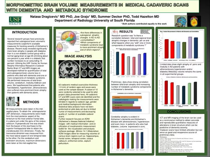
MORPHOMETRIC brain volume measurements in medical cadaveric scans WITH DEMENTIA and metabolic syndrome Natasa Dragicevic* MD PhD, Joe Grajo* MD, Summer Decker PhD, Todd Hazelton MD Department of Radiology University of South Florida * Both authors contributed equally to this work AD=Alzheimer's Disease AS=Atherosclerosis HTN=Hypertension DM=Type 2 Diabetes Pick's= Pick's dementia RESULTS INTRODUCTION • Are there differences in subregional atrophic changes in AD vs AD • +comorbidities? • Which components of metabolic syndrome lead to more prominent atrophy and of which regions?, Research question was “Is there a correlation between total and regional brain atrophic changes in dementia per se and what happens when you “add” one or more components of metabolic syndrome ?” Several research groups have previously reported that subregional brain volumetric measurements outperform available measures for tracking severity of Alzheimer’s disease. Recent study revealed significantly more brain atrophy in people with diabetes than in a non-diabetic control group with a normal aging brain at age 65 shrinking about one percent a year while in a diabetic that number increases to an astounding 15 percent .Utilizing the USF Center for Human Morpho-Informatics Research’s Cadaver Banks Brain CT and MRI images we developed method for quantification of total and subregional brain volume loss in patients who died with dementia and one or more components of metabolic syndrome. We performed measures of total brain volume, cerebellum, medulla,midbrainand pons. We hypothesized that patients who had diabetes, hypertension, atherosclerosis also suffered more prominent brain atrophy then patients with dementia alone. IMAGE ANALYSIS Limited data show slight atrophy of pons and medulla in AD patients with atherosclerosis,hypertension and/or diabetes.Medullary volume remains the same in all experimental groups. • 62 cadaveric medical scans(slice thickness 1.0 mm) of random ages and sexes were used as the sample dataset. A subset of 10 were randomly selected to be used for pilot project (data presented here). All cadaveric CT and MRI scans were de-identified and the radiologists making the measurements were blinded in regards to cadaver age, gender and medical background information • Statistical data analysis have not been preformed on pilot data set due to limited numer of measurements and decreasing quality of number of available cadaver scans. • Further research focuses on ADNI (Alzheimer’s disease National Initiative) images with medical information and on utilization of morphometric measurement toolkit for the visualization and analysis software package, Mimics 14.1 (Materialise). ADNI Images allow for measuring volumes of various brain regions such as :hippocampus, frontal cortex, temporal cortex, corpus callosum, enthorinalcortex, septum pellucidum. Preliminary data show strong correlation between total brain atrophy and increasing number of metabolic syndrome components in Alzheimer’s dementia. METHODS All measurements were taken in the mid-sagittal plane. To obtain the anteroposterior (AP) dimension, measurements were made from the most posterior aspect of the tentorium to the most anterior frontal lobe, in a plane just under the genu of the corpus callosum. A perpendicular line was drawn from the basion of the skull base to the superior frontoparietal lobe to obtain the crandiocaudal (CC) dimension. Finally, the transverse dimenion was measured from the most lateral aspect of one temporal lobe to the other. For the cerebellum and brainstem structures, maximum dimensions were taken at the mid-sagittal line. CONCLUSIONS Cerebellar atrophy is evident in Alzheimer’s dementia and Alzheimer’s disease with atherosclerosis, hypertension and even more in patients who also had diabetes. (volume expressed in ccm). • CT and MR imaging of the brain can be used as a noninvasive method to obtain accurate and reproducible quantitative measures of alterations in brain structure which can be predictive of dementia severity. • Cadaver scans have limitied utilization but can serve as good and inexpensive source of preliminary data. • References: • Launer LJ. Rates and risk factors for dementia and Alzheimer’s disease. • Neurology 1999; 52:78. • Launer LJ, White LR, Petrovitch H, Ross GW, Curb JD. Cholesterol and neuropathologic markers of AD. Neurology 2001; 57:1447–52. • Haag MDM, Hofman A, Koudstaal PJ, Stricker BHC, Breteler MMB. • Statins are associated with a reduced risk of Alzheimer disease • regardless of lipophilicity. The Rotterdam Study. J Neurol Neurosurg • Psychiatry 2009; 80:13–7. • Schnaider Beeri M, Goldbourt U, Silverman JM et al. Diabetes mellitus • in midlife and the risk of dementia three decades later. Neurology • 2004; 63:1902–7.