Download
1 / 45
460 likes | 616 Vues
Skeleton Development Patricia Ducy HHSC1616 x5-9299 pd2193@columbia.edu. Skeleton. ≥ 200 elements Two tissues: cartilage, bone Three cell types: chondrocytes, osteoblasts, osteoclasts Three “environments”: marrow, blood, SNS. Growth. Formation. Resorption.

E N D
Skeleton DevelopmentPatricia DucyHHSC1616x5-9299pd2193@columbia.edu
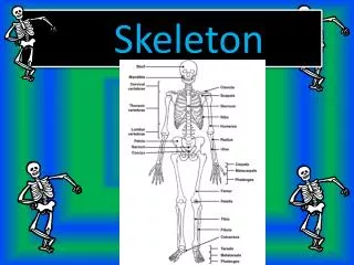
Skeleton • ≥ 200 elements • Two tissues: cartilage, bone • Three cell types: chondrocytes, osteoblasts, osteoclasts • Three “environments”: marrow, blood, SNS Growth Formation Resorption
Embryonic origin of the skeleton Cranial neural crest cells Somitic mesoderm Lateral plate mesoderm Monocyte lineage Craniofacial skeleton Axial skeleton Appendicular Skeleton Chondrocytes & Osteoblasts Osteoclasts
Skeleton Biology Patterning Location and shape of skeletal elements Development Skeletogenesis Differentiated cells Bone structure Growth/Modeling Birth Homeostasis Life Fracture repair Remodeling Balance between Formation/Resorption
Skeleton Pathologies Patterning Dysostoses Development Skeletogenesis Dysplasia Mineralization defects Degenerative diseases Life Homeostasis
Genetic defects associated with skeleton development (RUNX2)
Skeleton patterning • Condensation of mesenchymal cells to form the scaffold of each future skeletal element • Migration • Adhesion • Proliferation • Early steps use signaling molecules and pathways generally involved in patterning other tissues (FGFs, Wnts, BMPs) • Orchestrated by specific set of genes acting as territories organizers • When not embryonic lethal disorders often localized
Hox transcription factors • First described in Drosophila where they control body plan organization • Arranged in 4 genomic clusters in mammals • Expression patterns follow the cluster arrangement
Homeotic transformations in absence of Hox transcription factors Wellik, Dev. Dynamics 236 (2007)
Hox transcription factors control vertebrate limb patterning Genomic organization Site of expression
Mutations in HOXD13 cause synpolydactyly*in humans *OMIM 18600, 186300 Patient with increased number of Ala repeat in HOXD13 Muragaki et al., Science 272 (1996)
Skeletogenesis • Cell differentiation • Chondrocytes, osteoblasts, osteoclasts • Bone morphogenesis • Formation of growth plate cartilage, bone shaft and marrow cavity • Vascular invasion and innervation • Defects generalized
Two skeletogenetic mechanisms • Endochondral ossification • Differentiation of a cartilaginous scaffold (chondrocytes) later replaced by bone (osteoblasts) • Most of the skeletal elements • Intramembranous ossification • Direct differentiation of the condensed mesenchymal cells into osteoblasts • Many bones of the skull, clavicles
Sox9 • Transcription factor of the HMG family • Regulates the expression of chondrocyte-specific genes • Sox9 haploinsufficiency causes Campomelic dysplasia (OMIM 114290) • Earliest known regulator of chondrocyte differentiation
Sox9-deficient cells cannot differentiate into chondrocytes Bi et al., Nat. Genet 22 (1999)
Sox9 Sox5, 6 Hypertrophy
Accelerated chondrocyte hypertrophy in PTHrP-deficient mice +/+ PTHrP -/- Karp et al., Development 127 (2000)
PTHrP • Ubiquitously expressed growth factor • Shares the same receptor with PTH • Mice “knockout” only phenotype is a generalized growth plate cartilage defect • PTHrP protein signals to its receptor in the prehypertrophic chondrocytes and blocks their hypertrophic differentiation
Dwarfism in Ihh-deficient mice +/+ Ihh -/- St-Jacques et al. , Genes Dev. 13 (1999)
Indian hedgehog (Ihh) • One of 3 members of the Hedgehog family of growth factors • Widely expressed during development • Expression positively regulated by the transcription factor Runx2
Reduced chondrocyte proliferation and delayed chondrocyte hypertrophy in Ihh-deficient mice +/+ Ihh -/- +/+ Ihh -/- St-Jacques et al. , Genes Dev. 13 (1999)
No PTHrP No Ihh Chondrocyte maturation is regulated by a PTHrP/Ihh feedback loop Perichondrium Resting Proliferating Pre-hypertrophic PTHrP receptor Hypertrophic PTHrP Ihh
Mutations in the PTH/PTHrP receptor cause Jansen and Bloomstrand chondrodysplasia Activating mutations Loss-of-function mutations Jansen metaphyseal chondrodysplasia OMIM 156400 Blomstrand's lethal chondrodysplasia OMIM 215045 Schipani & Provost. Brith Defects Res. 69 (2003)
Sox9 Sox5, 6 Ihh/PTHrP Runx2 Hypertrophy VEGF
Arrest of osteoblast differentiation in Runx2-deficient mice +/+ Runx2 -/- Otto et al., Cell 89 (1997)
Runx2 • One of three members of the runt family of transcription factors • Identified as a regulator of the Osteocalcin promoter • Necessary and sufficient for osteoblast differentiation
Cleidocranial dysplasia (CCD, OMIM 119600)is caused by Runx2 haploinsufficiency +/+ +/- Mundlos et al., Cell 89 (1997) Lee et al. , Nat Genetics 16 (1997)
Intramembranous ossification Suture formation Growth Closure
Disorders of suture fusion Delay Msx2, Runx2 haploinsufficiency Acceleration = craniosynostosis FGFR1, 2, 3 activating mutations Msx2 activating mutations Twist haploinsufficiency
Twist (Saethre-Chotzen Syndrome OMIM 101400) Osx, ATF4 Osteoblast differentiation Runx2 Osteoprogenitor Pre-osteoblast Osteoblast
Delayedosteogenesisinabsence of Atf4 WT Atf4-/- E14 Yang et al., Cell 117 (2004)
E15 E16 P0 WT Atf4 -/- Delayed osteogenesis inAtf4-deficient mice Yang et al., Cell 117 (2004)
ATF4 • Divergent member of the ATF/CREB family of leucine-zipper transcription factors • Required for amino-acid import • Identified as a regulator of the Osteocalcin promoter • Activated by the Rsk2 kinase
ATF4 • Lack of ATF4 phosphorylation by inactivating mutations in Rsk2 causes the skeletal defects associated with Coffin-Lowry syndrome (OMIM 303600) • Increased ATF4 phosphorylation by Rsk2 causes the skeletal defects associated with Neurofibromatosis Type I (OMIM 162200)
ATF4 • Divergent member of the ATF/CREB family of leucine-zipper transcription factors • Required for amino-acid import • Identified as a regulator of the Osteocalcin promoter • Activated by the Rsk2 kinase
A high protein diet normalizes bone formation in Atf4-/- and Rsk2-/- mice High protein diet BV/TV BFR Ob.S/BS Elefteriou et al., CellMetab. 4 (2006)
A low protein diet normalizes bone formation in a mouse model of Neurofibromatosis type I Normal diet Low protein diet wt Nf1ob-/- wt Nf1ob-/- 15.3±1 19.6±0.4* 14.8±0.7 15.3±0.6 BV/TV 153.3±11 313.9±7.0* 157.0±11 186.2±22 BFR 19.1±0.8 31.8±1.6* 19.0±0.5 19.7±1.0 ObS/BS Elefteriou et al., CellMetab. 4 (2006)
Structure of a growing long bone articular cartilage (chondrocytes) secondary ossification centre (osteoblasts/osteoclasts) reserve cartilage proliferating cells Growth plate Cartilage (chondrocytes) hypertrophic cells calcified cartilage trabecular bone (osteoblasts/osteoclasts) cortical bone (osteoblasts)
Osteopenia in OPG-deficient mice Bucay et al., Genes Dev. 12 (1998)
Osteopetrosis in RANK-L deficient mice Lacey et al., Cell 93 (1998)
Osteoblast progenitor OPG RANK-L Inactive complex A ctive complex RANK TRAF 6 TRAF 2/5 Osteoclast progenitor NF NF B B k k c-src JNK JNK Osteoclast AP1 activation Maturation
Research directions Knowledge Diseases Patterning Development Skeletogenesis Life Homeostasis