Download
1 / 1
10 likes | 136 Vues
G q and G 13 Activation of NHE1. EGFR. a 13 GTP. g. g. g. LPA. b. SOS. ß. PE. Ras. a q GTP. GRB. b. a 13 GDP. a. b. P. P. P. a q GDP. PTK. PLCß1. Raf. RhoGEF. GDP. GTP. GDP. GTP. DAG + IP 3. PIP 2. ?. RhoA. MEK. P. ?. RhoA. PKC a. ROCK. PLD. PKC n.

E N D
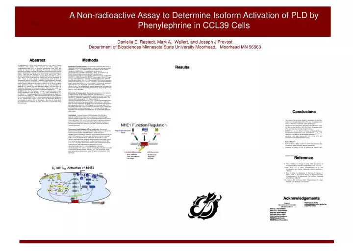
Gq and G13 Activation of NHE1 EGFR a13 GTP g g g LPA b SOS ß PE Ras aq GTP GRB b a13 GDP a b P P P aq GDP PTK PLCß1 Raf RhoGEF GDP GTP GDP GTP DAG + IP3 PIP2 ? RhoA MEK P ? RhoA PKCa ROCK PLD PKCn PA PC PLD DAG ERK ROCK PC PA RSK P ERM Na+ NHE-1 P H+ Introduction Conclusions • The solvent that provides superior separation of free fatty acids from phosphatidylcholine (PC) and phospatidic acid (PA) is chloroform: methanol: water (63:34:3;v/v) • The fluorescent lipids were detected by densitometry using UV light on the Gel Doc 1000 (Bio-Rad). Phospatidic acid (PA) was only seen visually at higher doses. • BODIPY C1, C12 4,4-difluoro-5-methyl-4-bora-3a,4a-diaza-s-inda-cene-3-dodecanoic acid not BODIPY C8, C5 4,4-difluoro-5-octyl-4-bora-3a,4a-diaza-s-indacene-3-pentanoic acid was incorporated into living cells and converted to phosphatidylcholine ((PC). • Future Studies: • Further studies will be needed to finish characterizing the fluorescent lipids (solvents, labeling, sensitivity). • Examine the ability of PLD to catalyze its reaction with BODIPY PC in cells. NHE1 Function/Regulation Migration/Proliferative Signal GPCR RTK Integrins H+ NHE1 Reference Na+ ERM F-Actin pHi Homeostasis Cytoskeletal Remodeling Focal Adhesion Actin Stress Fibers Cell Shape Proliferation Migration Invasion K. Meier, T.Gibbs, S. Knoepp, K. Ella. 1999. Expression of Phospholipase D Isoforms in Mammalian Cells 200-211 Foster, D.A., Xu, L. 2003. Phospholipase D in Cell Proliferation and Cancer. Molecular Cancer Research.1: 789-800 K. Ella, G. Meier, C. Bradshaw, K. Huffman, R. Spivey, K. Meier. 1994. A Fluorescent Assay for Agonist-Activated Phospholipase D in Mammalian Cell Extracts. Analytical Biochemistry. 136-142. GM. Jenkins, MA. Frohman. 2005. Phospholipase D: A Lipid Review. Cell Molecular Life Science Acknowledgements Thank you to all the undergraduates in the lab for the support provided Support NIH-1-R15-HL074924-01A1 NSF-MCB0080243 NSF-ILI – DUE 9750937 NSF-CCLI - DUE 0088654 NSF-RUI - MCB 0080243 NSF-MRI - DBI 0110537 Dille Fund for Excellence MSUM Faculty Grants MSUM Alumni Foundation Phospholipase D (PLD) is an enzyme found in the cells of high mammals and plants. PLD is activated in mammalian cells in response to treatment of mammalian cells with a variety of hormones and growth factors (K.E Meier et al., 1994). PLD acts on phosphatidylcholine (PC) it produces phospatidic acid (PA) and choline. PA acts as a bioactive lipid activating a number of protein kinases and other effectors and can be further metabolized to diacylglycerol, an activator of protein kinase C (PKC). PC is a common phospholipids. It and other phospholipids make up a major part of a cell’s membrane system, including the plasma membrane and cytoplasmic organelles. There are two isoforms of PLD, PLD1 and PLD2. PLD1 is activated by the small G proteins RhoA and ARF as well as PKC; PLD2 is constitutively active and can be further stimulated by ARF(Foster & Xu, 2003). There is great interest in understanding which isoform is activated by the different hormones and and neurotransmitters that impact cells. Because of its importance, numerous methods to determine its enzymatic activity have been developed. The methods differ in their means of isolating and quantifying the reaction products used to measure PLD activity. There are several in vitro methods but few in vivo methods. The currently accepted method used to determine enzymatic activity is an in vivo PLD assay using radioactive lipids. In the present study our plan is to use fluorescent labels to measure in vivo PLD activity in a non-radioactive assay. A Non-radioactive Assay to Determine Isoform Activation of PLD by Phenylephrine in CCL39 Cells Danielle E. Rastedt, Mark A. Wallert, and Joseph J Provost Department of Biosciences Minnesota State University Moorhead, Moorhead MN 56563 Abstract Methods Phospholipase D (PLD) is an enzyme found in the cells of higher mammals and plants. The process of PLD acting on phosphatidylcholine (PC) to produce phospatidic acid (PA) and choline is important in cell signaling. PA acts as a bioactive lipid activating a number of protein kinases and other effectors and can be further metabolized to diacylglycerol, an activator of protein kinase C (PKC). There are two isoforms of PLD, PLD1 and PLD2. PLD1 activity is activated by the small G proteins RhoA and ARF as well as PKC, while PLD2 is constitutively active and can be stimulated by ARF. There is great interest in understanding which isoforms is activated by various hormones. Therefore, several different methods have been developed to determine its enzymatic activity. The current method used to determine enzymatic activity is an in vivo PLD assay using radioactive lipids. Our plan is to use fluorescent labels to measure PLD activity in a non-radioactive assay. The three types of fluorescent lipids used in these experiments. Two free fatty acids 4,4-difluoro-5-octyl-4-bora-3a,4a-diaza-s-indacene-3-pentanoic acid (BODIPY C8, C5); 4,4-difluoro-5-methyl-4-bora-3a,4a-diaza-s-inda-cene-3-dodecanoic acid (BODIPY C1, C12); and 1-myristoyl-2-[, 12-[(7-nitro-2-1, 3-benzoxadiazol-4-yl)amino]dodecanoyl]-sn-glycero-3-phosphocholine (NBD-PC). We found that both NBD-PC and BODIPY C1, C12 but not BODIPY C8, C5 were incorporated into the membrane as PC. Furthermore, there is a dose and time dependent manner in the labeling of starved CCL39 fibroblasts. We plan to show which PLD isoforms(s) is activated by stress hormones in CCL39 cells using this technique. Results Separation: Solvent system- A separation of the free fatty acid 4,4-difluoro-5-methyl-4-bora-3a,4a-diaza-s-inda-cene-3-dodecanoic acid (BODIPY C1, C12), 2-(4,4-difluoro-5-methyl-4-bora-3a,4a-diaza-s-indacene-3-dodecanoy0-1-hexadecanoyl-sn-glycero-3-phosphocholine (-BODIPY 500/510 C12-HPC), 2-(4,4-difluoro-5-methyl-4-bora-3a,4a-diaza-s-indacene-3-dodecanoy0-1-hexadecanoyl-sn-glycero-3-phosphate, diammonium salt ((-BODIPY 500/510 C5 –HPA), and ptd BD BODIPY were tested. 15l of 0.1g/l or 1g/l was applied to a thin layer chromatography. Four different solvents were tested. Solvent 1- ethyl acetate:2,2,4 trimethyl pentane: acetic acid: water (65:10:15:50; v/v). Solvent 2- chloroform: methanol: water: acetic acid (45:45:10:1; v/v). Solvent 3- Chloroform: methanol: water (63:34:3; v/v). Solvent 4- chloroform: methanol: water (45:45:10; v/v). The solvents were used to develop the TLC plate The fluorescent lipids were detected by densitometry using UV light on the Gel Doc 1000 (Bio-Rad). Sensitivity of Visualization- Appropriate amounts of 2-(4,4-difluoro-5-methyl-4-bora-3a,4a-diaza-s-indacene-3-dodecanoy0-1-hexadecanoyl-sn-glycero-3-phosphate, diammonium salt ((-BODIPY 500/510 C5 –HPA)were applied to a thin layer chromatography. The solvent used to develop the TLC plate consisted of chloroform/methanol/water (63:34:3; v/v). The fluorescent lipids were detected by densitometry using UV light on the Gel Doc 1000 (Bio-Rad). A further analysis of the detection of fluorescents was achieved by scraping the silica from the TLC plate into microfuge tube. The silica was rinsed with 0.3mL of methanol and then vortex, centrifuged, and incubated at room temperature for 10 minutes. 200 l was extracted from the solution and measured for fluorescents using the plate reader. Cell Culture- Chinese hamster lung fibroblasts (CCL39) were cultured in attachment culture using Dulbecco’s modified Eagle medium (DMEM, Sigma) and 10% heat-activated fetal bovine serum (FBS, GibocBRL) 37oC in a 5% CO2 incubator. Cells were allowed to grow to 60% confluence in 35mm dishes. Appropriate amounts of resuspended lipids were added to each dish, and were allowed to incubate overnight. Fluorescent Lipid Analysis of Free Fatty Acids- Appropriate amounts of fluorescent lipids were dried under nitrogen in sterile test tube along with DMEM complete media. Lipids were then resuspended by sonication, added to 35mm dishes containing CCL39 cells at 60% percent confluence, and allowed to incubate overnight. At the end of incubation period, cells were rinsed with ice-cold calcium magnesium free phospho-buffered saline (CMF-PBS). Lipids were extracted and separated with methanol/chloroform/0.1M HCl (1:1:1;v/v). The lower phase of the solution was extracted and dried under nitrogen and lipids were resuspended in 30 l of chloroform/methanol (2:1; v/v) and applied to a thin layer chromatography. The solvent used to develop the TLC plate consisted of chloroform/methanol/water (63:34:3; v/v). The fluorescent lipids were detected by densitometry using UV light on the Gel Doc 1000 (Bio-Rad).