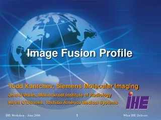
Image Fusion Profile
160 likes | 378 Vues
Image Fusion Profile. Todd Kantchev, Siemens Molecular Imaging Jerold Wallis, Mallinckrodt Institute of Radiology Kevin O’Donnell, Toshiba America Medical Systems. Image Registration and Fusion. Translate Rotate Rigid Registration Scale Warp- optional (Deformable Registration)

Image Fusion Profile
E N D
Presentation Transcript
Image Fusion Profile Todd Kantchev, Siemens Molecular Imaging Jerold Wallis, Mallinckrodt Institute of Radiology Kevin O’Donnell, Toshiba America Medical Systems
Image Registration and Fusion • Translate • Rotate Rigid Registration • Scale • Warp- optional(Deformable Registration) • Fuse
Image Fusion- General Use Case CT MR • Tech acquires two or more series of images, possibly separated in time. • Tech reconstructs images into volumes, performs registration, and stores results data. • Radiologist reads study or Oncologist completes RT planning on RT Planning System. • Referring Physician or Researcher reviews results.
Fusion- The Integration Problem • Results not readily transferable between systems • Proprietary or inconsistent implementations • Lack of guidance • Results presented on different systems may not appear the same. • Systems must correctly handle issues such as • Identifying and retrieving the right datasets • Matching data between single-slice and multi-slice datasets • Performing spatial translations • Rendering the fused display
Fusion- The Integration Solution • A collection of DICOM objects exists to address the problem. • Their use is constrained and clarified to avoid misinterpretation and promote compatibility.
Essential Clinical Features: MDs will be Ecstatic • MPR • Fusion • Side-by-side • Registered
Value Proposition • Interoperable DICOM encoding for registered/fused data • Consistent and reliable acquisition, storage and presentation of fused data across vendors • Allows separation of workflow steps: Acquisition on “A”; Fusion on “B”; Display on “C” • Wider distribution of results • Separable registration for re-doing if necessary • Can easily request basic interoperable fusion features. E.g. blended display, MPR, side by side display, etc. • Supports PET-CT (primary initial interest) viewing on general radiology PACS displays • Modality independent • Supports general needs in radiation oncology pre-planning
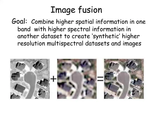
Image FusionTechnical Basis Spatial Registration Object • Encodes transformation to align images in a common registered space • Identifies the registered space • References the images to be aligned • Not required for already-registered PET-CT data from a hybrid scanner
Image FusionTechnical Basis Blending Presentation Object • References one set of underlying images (e.g. CT) • References one set of superimposed images (e.g. PET) • References (optionally) a Registration Object to align both image sets before blending. • Encodes the window level for each set • Encodes the blending factor (0 to 100%) of the overlay transparency
Blending PresentationOverlay Transparency 20% 50% 80% 50% 50% 100% 50%
Use Case- The IT Solution Modality CT Modality MR Image Archive Evidence Creator Image Display Store CT Store MR Retrieve CT, MR Register MR to CT Fuse CT and MR Store SRO Store BSPS Retrieve CT, MR, SRO, BSPS Apply T to MR Fuse CT and MR
Image Fusion Actors and Transactions Acquisition Modality Evidence Creator [RAD 8] Modality Images Stored ↓ [RAD 57] Blending Presentation States Stored ↓ [RAD 56] Spatial Registrations Stored ↓[RAD 10] Storage Commitment ↓ ↓ [RAD 18] Creator Images Stored ↓ [RAD 57] Blending Presentation States Stored ↓ [RAD 56] Spatial Registrations Stored ↓ [RAD 10] Storage Commitment Image Manager Image Archive ↓ [RAD 14] Query Images ↓ [RAD 16] Retrieve Images ↓ [RAD 15] Query Presentation States ↓ [RAD 17] Retrieve Presentation States ↓ [RAD 58] Retrieve Spatial Registrations Image Display
Load CT and PET images Align images (automatically or manually) and fuse with desired blending factor Store the alignment, including Registration object Blending State (defining the blending factor, window levels and referencing all other objects) Storage can be on PACS or disk. Images do not need to be stored if already stored. Fusion- The Creating Process CT PET Blending State Registration Object
Blending state can be searched and retrieved from PACS Can be used to retrieve the image data and the alignment automatically. User selects a Blending state. Automatically load the referenced CT and PET images and the Registration object. Alignment can be applied, PET re-sampled to CT and displayed fused automatically . Display is required to provide Scrolling through volume Adjustment of fusion blending and window levels MPR Side-by-side display Fusion- The Displaying Process Blending State Registration Object CT PET