Download
1 / 44
440 likes | 1.1k Vues
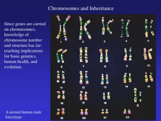
Chapter 14 Chromosomes and Human Inheritance. 14.1 Shades of Skin. Variations in skin color may have evolved as a balance between vitamin D production and UV protection More than 100 gene products are involved in the synthesis of melanin, and the formation and deposition of melanosomes

E N D
14.1 Shades of Skin • Variations in skin color may have evolved as a balance between vitamin D production and UV protection • More than 100 gene products are involved in the synthesis of melanin, and the formation and deposition of melanosomes • Mutations in some of these genes may have contributed to regional variations in human skin color
Genetic Abnormalities and Disorders • A genetic abnormality is an uncommon version of a trait that is not inherently life-threatening, • A genetic disorder causes medical problems that may be severe • A genetic disorder is often characterized by a specific set of symptoms (a syndrome)
Types of Genetic Variation • Single genes on autosomes or sex chromosomes govern more than 6,000 genetic abnormalities • Most human traits, including skin color, are polygenic (influenced by multiple genes) and some have epigenetic contributions or causes
Patterns of Inheritance • Based on variations in single genes (Mendelian patterns) • Autosomal dominant • Autosomal recessive • X-linked recessive • X-linked dominant • Based on variations in whole chromosomes • Changes in chromosome number • Changes in chromosome structure
14.3 Autosomal Inheritance Patterns • An allele is inherited in an autosomal dominant pattern if the trait it specifies appears in heterozygous people • An allele is inherited in an autosomal recessive pattern if the trait it specifies appears only in homozygous people
Autosomal Dominant Inheritance • A dominant autosomal allele is expressed in homozygotes and heterozygotes • Tends to appear in every generation • With one homozygous recessive and one heterozygous parent, children have a 50% chance of inheriting and displaying the trait • Examples: • Achondroplasia • Huntington’s disease • Hutchinson–Gilford progeria
normal mother affected father meiosis and gamete formation affected child normal child disorder-causing allele (dominant) Stepped Art Figure 14-3a p222
Autosomal Recessive Inheritance • Autosomal recessive alleles are expressed only in homozygotes • Heterozygotes are carriers and do not have the trait • A child of two carriers has a 25% chance of expressing the trait • Examples: • Albinism • Tay-Sachs didease
carrier father carrier mother meiosis and gamete formation affected child carrier child normal child disorder-causing allele (recessive) Stepped Art Figure 14-4a p223
Take-Home Message: How do we know a trait is associated with an allele on an autosome? • With an autosomal dominant inheritance pattern, persons heterozygous for an allele have the associated trait; the trait appears in every generation • With an autosomal recessive inheritance pattern, only persons who are homozygous for an allele have the associated trait, which can skip generations
X-Linked Recessive Pattern • More males than females have X-linked recessive genetic disorders • Males have only one X chromosome and can express a single recessive allele • A female heterozygote has two X chromosomes and may not show symptoms • Males transmit an X only to their daughters, not to their sons
carrier mother normal father meiosis and gamete formation normal daughter or son carrier daughter affected son recessive allele on X chromosome Stepped Art Figure 14-6a1 p224
Some X-Linked Recessive Disorders • Red-green color blindness • Inability to distinguish certain colors caused by altered photoreceptors in the eyes • Duchenne muscular dystrophy • Degeneration of muscles caused by lack of the structural protein dystrophin • Hemophilia A • Bleeding caused by lack of blood-clotting protein
Take-Home Message: Is a trait associated with an allele on an X chromosome? • Men who have an X-linked recessive allele have the trait associated with the allele; heterozygous women do not, they have a normal allele on their second X chromosome – the trait appears more often in men • Men transmit an X-linked allele to their daughters, but not to their sons
14.5 Heritable Changes in Chromosome Structure • On rare occasions, a chromosome’s structure changes; such changes are usually harmful or lethal, rarely neutral or beneficial • A segment of a chromosome may be duplicated, deleted, inverted, or translocated
Duplication • DNA sequences that are repeated two or more times • Duplication may be caused by unequal crossovers in prophase
Deletion • Loss of some portion of a chromosome • Usually causes serious or lethal disorders • Example: Cri-du-chat
Inversion • Part of the sequence of DNA becomes oriented in the reverse direction, with no molecular loss
Translocation • If a chromosome breaks, the broken part may get attached to a different chromosome, or to a different part of the same one • Most translocations are reciprocal, or balanced, which means that two chromosomes exchange broken parts • A reciprocal translocation between chromosomes 8 and 14 is the usual cause of Burkitt’s lymphoma
Translocation D With a translocation, a broken piece of a chromosome gets reattached in the wrong place. This example shows a reciprocal translocation, in which two chromosomes exchange chunks.
Chromosome Changes in Evolution • Changes in chromosome structure can reduce fertility in heterozygotes; but accumulation of multiple changes in homozygotes may result in new species • Certain duplications may allow one copy of a gene to mutate while the other carries out its original function • Example: X and Y chromosomes were once homologous autosomes in reptile-like ancestors of mammals
Differences Among Closely Related Organisms • Humans have 23 pairs of chromosomes; chimpanzees, gorillas, and orangutans have 24 • Two chromosomes fused end-to-end telomere sequence human chimpanzee
14.6 Heritable Changes in Chromosome Number • Occasionally, abnormal events occur before or during meiosis, and new individuals end up with the wrong chromosome number • Consequences range from minor to lethal changes in form and function
Polyploidy and Aneuploidy • Many flowering plant species, and some insects, fishes, and other animals, are polyploid – they have three or more complete sets of chromosomes • Trisomy and monosomy are examples of aneuploidy, in which an individual’s cells have too many or too few copies of a chromosome
Nondisjunction • Changes in chromosome number can be caused by nondisjunction, when a pair of chromosomes fails to separate properly during mitosis or meiosis • Affects the chromosome number at fertilization • Monosomy (n-1 gamete) • Trisomy (n+1 gamete)
Metaphase I Anaphase I Telophase I Metaphase II Anaphase II Telophase II Nondisjunction Stepped Art
Autosomal Change and Down Syndrome • Only trisomy 21 (Down syndrome) allows survival to adulthood • Characteristics include physical appearance, mental impairment, and heart defects • Incidence of nondisjunction increases with maternal age • Can be detected through prenatal diagnosis
Change in Sex Chromosome Number • Changes in sex chromosome number may impair learning or motor skills, or be undetected • Female sex chromosome abnormalities • Turner syndrome (XO) • XXX syndrome (three or more X chromosomes) • Male sex chromosome abnormalities • Klinefelter syndrome (XXY) • XYY syndrome
Take-Home Message: What are the effects of chromosome number changes? • Nondisjunction can change the number of autosomes or sex chromosomes in gametes; such changes usually cause genetic disorders in offspring • Sex chromosome abnormalities are usually associated with some degree of learning difficulty and motor skill impairment
Detecting Genetic Disorders • Surgery, prescription drugs, hormone replacement therapy, and dietary controls can minimize and in some cases eliminate the symptoms of a genetic disorder • Some disorders can be detected early enough to start countermeasures before symptoms develop • Example: Most hospitals in the United States now screen newborns for mutations that cause phenylketonuria (PKU)
Prenatal Diagnosis • In prenatal diagnosis, an embryo or fetus is tested before birth to screen for sex or genetic problems • Noninvasive techniques include obstetric sonography • Invasive procedures in which samples of tissue or blood are taken involve risks to mother and fetus • Fetoscopy • Amniocentesis • Chorionic villus sampling (CVS)
amniotic sac chorion Figure 14-14 p230