Download

1 / 75
770 likes | 1.47k Vues
Fundamentals of the NS and Nervous Tissue Chapter 12 Part II Basic Divisions of the Nervous System Figure 12.2 Figure 12.3 Nervous Tissue Cells are densely packed and tightly intertwined Two main cell types 1) neurons, the excitable nerve cells that transit electrical signals

E N D
Fundamentals of the NS and Nervous Tissue Chapter 12 Part II
Basic Divisions of the Nervous System Figure 12.2
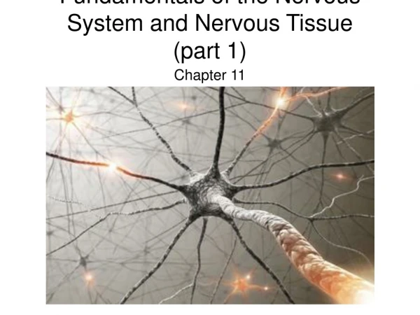
Nervous Tissue • Cells are densely packed and tightly intertwined • Two main cell types 1) neurons, the excitable nerve cells that transit electrical signals 2) neuroglia, nonexcitable supporting cells that surround and wrap the neurons • Both developed from the embryonic neural tube and neural crest
The Neuron • The human body contains billions of neurons, or nerve cells • Basic structural unit of the NS - specialized cells conduct signals from one part of the body to another - signals are transmitted along the plasma membrane as nerve impulses, or action potentials
Neuron - Special Characteristics 1. Longevity - can live and function for a lifetime 2. Do not divide – fetal neurons lose their ability to undergo mitosis; neural stem cells are an exception 3. High metabolic rate – require abundant oxygen and glucose. Neurons cannot survive for more than a few minutes without oxygen • Neurons typically are large, complex cells and all have a cell body with one or more processes
The Cell Body • Also called a soma, all have a single nucleus surrounded by cytoplasm (perikaryon) - size of cell body varies from 5 – 140 um - contain usual organelles - plus chromatophilic (Nissl) bodies, clusters of RER and free ribosomes that stain darkly and continually renew the membranes of the cell • Neurofibrils – bundles of intermediate filaments (neurofilaments) - form a network between chromatophilic bodies
The Cell Body • Most neuronal cell bodies are located within the CNS - protected by bones of the skull and vertebral columns • Ganglia (sing. ganglion) – clusters of cell bodies - lie along the nerves in the PNS
Structure of a Typical Large Neuron Figure 12.4
Neuron Processes • Armlike processes extend from the cell bodies of all neurons – dendrites and axons • Dendrites - extensively branch from the cell body - transmit electrical signals toward the cell body - chromatophilic bodies – only extend into the basal part of dendrites and to the base of the axon hillock - function as receptive sites for receiving signals from other neurons
Axons • Neuron has only one - initial segment called the axon hillock • Impulse generator and conductor • Transmits impulses away from the cell body • Chromatophilic bodies are absent - lack GA, ribosomes, all organelles involved in protein synthesis • No protein synthesis occurs in the axon
Axons • Neurofilaments, actin microfilaments, and microtubules - provide strength along length of axon - aid in the transport of substances to and from the cell body (axonal transport) • Branches along the length are infrequent - axon collaterals • Multiple branches at the end or terminus - terminal branches (telodendria) end in knots called axon terminals (aka end bulbs or boutons)
Nerve Impulse • Generated at the initial segment of the axon • Conducted along the axon • Releases neurotransmitters at axon terminals - neurotransmitters excite or inhibit the neuron or target organ • Axon diameter varies - larger diameter faster the impulse according to the basic law of physics: resistance to the passage of an electrical current decreases as the diameter of any ‘cable’ increases • Neuron receives and sends signals
Synapses • Synapse (‘union’) - site at which neurons communicate • Signals pass across synapse in one direction • Presynaptic (information-sending) neuron – conducts the signal toward a synapse • Postsynaptic (information-receiving) neuron – transmits electrical signals away from a synapse • Most CNS neurons function as both presynaptic and postsynaptic - getting signals from some neurons and dispatching it to others
Two Neurons Communicating at a Synapse Figure 12.6
Types of Synapses Two main types: • Axodendritic – between axon terminals of one neuron and dendrites of another - most common type of synapse • Axosomatic – between axons and neuronal cell bodies • Axoaxonic, dendrodendritic, and dendrosomatic are uncommon types of synapses
Synapse Structurally synapses are elaborate cell junctions • Axodendritic synapses – representative type • Synaptic vesicles on the presynaptic side - membrane-bound sacs containing neurotransmitters - abundant mitochondria in axon terminals • Synaptic cleft - separates the PM of the two neurons • Presynaptic and postsynaptic densities – regions of dense material of opposing cell membranes - presynaptic region a honeycombed grid - postsynaptic region a perforate plate
Structure of a Synapses Figure 12.8a, b
The Signals Carried by Neurons • PMs of neurons conduct electrical signals • Resting neuron – membrane is polarized - inner, cytoplasmic side is negatively charged - high [K+] intercellular and high [Na+] extracellular • Stimulation of the neuron depolarization - permeability of PM changes allowing Na+ ions to rush in
Signals Carried by Neurons Figure 12.9a, b
Action Potential • Strong stimulus applied to the axon triggers - nerve impulse/action potential • Membrane at the axon’s initial segment is depolarized - positive charge inside the axon - negative charge outside the axon • Impulse travels the length of the axon - impulse stimulates the synaptic vesicle to fuse with the presynaptic membrane, fused area ruptures - NT diffuses across the synaptic cleft binds to the postsynaptic membrane at the postsynaptic density - binding changes the postsynaptic membrane charge
Excitatory Synapses • Neurotransmitters alter the permeability of the postsynaptic membrane to certain ions - influx of positive ions depolarizes the postsynaptic membrane - drives the postsynaptic neuron toward impulse generation
Inhibitory Synapses • Increases membrane polarization - the external surface of the postsynaptic membrane becomes more positive - reduces the ability of the postsynaptic neuron to generate an action potential • 1000s of exitatory and inhibitory synapses act on every neuron
Structural Classification of Neurons According to the number of processes they contain • Multipolar – possess more than two processes - numerous dendrites and one axon - 99% of neurons including motor and most interneurons • Bipolar – possess two processes - rare neurons found in some special sensory organs (inner ear, olfactory epithelium, retina) • Unipolar (pseudounipolar) – possess one short single process - most start as bipolar neurons, 2 processes fuse together - make up the typical sensory neurons
Neurons Classified by Structure Figure 12.10a
Neurons Classified by Structure Figure 12.10b
Neurons Classified by Structure Figure 12.10c
Neurons Classifed by Function According to the direction the nerve impulse travels: sensory neurons, motor neurons, interneurons • Sensory or afferent (arriving) neurons make up the sensory division of the PNS - transmit impulses toward the CNS - short, single process divides into the central process that runs centrally into the CNS - the peripheral process extends peripherally to the receptors
Motor Neurons • Or efferent (exiting) neurons make up the motor division of the PNS - carry impulses away from the CNS to effector organs - most are multipolar - cell bodies are within the CNS (except for some in the ANS) - form junctions with effector cells, stimulating muscle contraction or glandular secretion
Interneurons • Or association neurons lie between motor and sensory neurons - confined entirely to the CNS - link into chains that form complex neuronal pathways - make up 99.98% of all neurons - almost all are multipolar but have a great diversity in size and branching patterns of their processes
Neurons Classified by Function Figure 12.11
Supporting Cells • Six types – 4 in the CNS and 2 in the PNS • Provide supportive functions for neurons • Cover nonsynaptic regions of the neurons
Neuroglial in the CNS • Or glial cells include the: astrocyte, ependymal cells, microglia, and oligodendrocytes - have branching processes and a central cell body - outnumber neurons 10 to 1 - make up half the mass of the brain - can divide throughout life
Astrocytes • The most abundant glial cell type - sense when neurons release glutamate - extract blood sugar from capillaries for energy - take up and release ions in order to control environment around neurons - involved in synapse formation in developing neural tissue - produce molecules necessary for neuronal growth (brain-derived trophic factor, BDTF) - propagate calcium signals involved with memory
Figure 12.12b • Microglia – smallest, least abundant glial cell • Macrophages of the CNS • Engulf invading microorganisms and dead neurons • Derive from blood cells called monocytes
Ependymal Cells and Oligodendrocytes • Ependymal cells - line the central cavity of the spinal cord and brain - bear cilia – help circulate the CSF • Oligodendrocytes – have few branches - wrap their cell processes around axons in the CNS - produce myelin sheaths
Neuroglia in the PNS • Satellite cells –surround neuron cell bodies within ganglia • Schwann cells (neurolemmocytes) – surround PNS axons - form myelin sheath around axons of the PNS Figure 12.13
Myelin Sheaths • Segmented structures composed of the lipoprotein myelin • Surround thicker axons • Form an insulating layer • Prevent leakage of electrical current • Increase the speed of impulse conduction along the axon - makes impulse propagation more energy-efficient
Myelin Sheaths in the PNS • Formed by Schwann cells (neurolemmacytes) • Develop during fetal period and in the first year of postnatal life • Schwann cells (neurolemmacytes) wrap in concentric layers around the axon • cover the axon in a tightly packed coil of membranes • Neurilemma – nucleus and most of the cytoplasm of the neurolemmacytes end up just external to the myelin layers
Myelin Sheaths in the PNS Figure 12.14a, b
Unmyelinated Axons in the PNS Figure 12.14c, d
Myelin Sheaths in the PNS Figure 12.15a
Myelin Sheaths in the PNS • Nodes of Ranvier – gaps along axon • Thick axons are myelinated - nerve impulses jump from the membrane of one node to the next • Thin axons are unmyelinated - conduct impulses more slowly
Myelin Sheaths in the CNS Figure 12.15b • Oligodendrocytes form myelin sheaths in the CNS - have multiple processes, coil around several different axons - Nodes of Ranier present but more widely spaced