Download
1 / 37
390 likes | 1.04k Vues
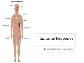
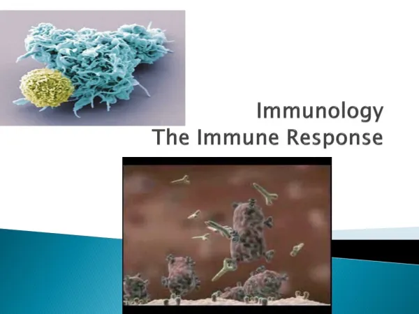
Immunology The Immune Response. Defense Mechanisms of the Host. Figure 14.1. The Immune System. Immunology: the study of all features of the body’s second and third lines of defense Healthy functioning immune system is responsible for: Surveillance of the body

E N D
Defense Mechanisms of the Host Figure 14.1
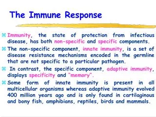
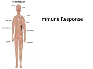
The Immune System • Immunology: the study of all features of the body’s second and third lines of defense • Healthy functioning immune system is responsible for: • Surveillance of the body • Recognition of foreign material • Destruction of entities deemed to be foreign
Self and Nonself White blood cells must distinguish self from nonself cells Evaluates cells by examining markers on their surfaces
Blood components White blood cells • B lymphocytes • T lymphocytes
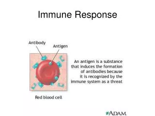
Antigen Anything that is foreign to the body (non self) and will elicit an immune response. ie. an infectious agent
Antibody A group of serum proteins whose function is to attack and inactivate non-self entities.
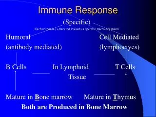
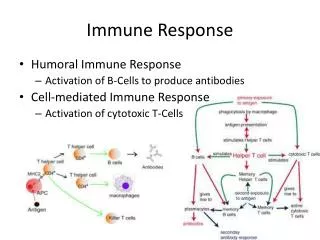
The Immune Repsonse Two types of Immune Responses Cell Mediated Humoral
Components of Blood • The cells and cell fragments in plasma are called formed elements • Three types of formed elements • Erythrocytes – carry oxygen and carbon dioxide in the blood • Platelets – involved in blood clotting • Leukocytes – involved in defending the body against invaders • Divided into granulocytes and agranulocytes
Cell Mediated Immune Response T lymphocytes There are a variety of different immune cells whose functions are to recognize, tag/label, and inactivate non-self: • T helper • T cytolytic • T suppressor
Role of the T-Helper The most important component of the immune system is the T helper cells. They wander around body as sentinels, looking for things that aren’t supposed to be there. When they find something they alert the cytolytic cells to destroy.
Synopsis of Roles of T-Lymphocytes T lymphocytes (cell mediated) • T-helper (recognition) • T-cytolytic (inactivation) • T-supressor (regulation and modification of cell mediated immune response)
Defense Components of BloodLeukocytes • Granulocytes • Contain large granules that stain different colors based on the dye used • Three types • Basophils – stain blue with the basic dye methylene blue • Eosinophils – stain red/orange with the acidic dye eosin • Neutrophils – stain lilac with a mixture of acidic and basic dyes
Defense Components of BloodLeukocytes • Granulocytes • Neutrophils and eosinophils can phagocytize pathogens • Neutrophils and eosinophils are capable of diapedesis
Defense Components of Blood • Defensive blood cells: leukocytes • Lab analysis of leukocytes • The differential white blood cell count test can signal signs of disease • Increased eosinophils can indicate allergies or parasitic worm infection • Bacterial diseases often show increase in leukocytes and in neutrophils • Viral infections show increase in lymphocytes
Defense of Blood Components Phagocytosis • Can be divided into five stages • Chemotaxis • Adherence • Ingestion • Killing • Elimination
Nonphagocytic Killing - Esinophilia • Killing by eosinophils • Mainly attack parasitic helminths (worms) by attaching to their surface • Secrete toxins that weaken or kill the helminth • Eosinophilia, or elevated eosinophil levels, is often indicative of a helminth infestation
Nonphagocytic Killing - NETs • Killing by neutrophils • Produce chemicals that kill nearby invaders • Generate extracellular fibers called neutrophil extracellular traps (NETs) that bind to and kill bacteria
Defense of Blood Components [INSERT FIGURE: 15.7]
Nonspecific Chemical Defenses Against Pathogens Complement • Activation of complement • Body’s own cells withstand complement cascade • Membrane-bound proteins on many cells bind with and break down activated complement proteins
The Body’s Second Line of Defense [INSERT FIGURE: 15.11]
Major Histocompatibility Complex • Set of genes that codes for human cell receptors • Gives rise to a series of glycoproteins (MHC molecules) found on all cells except red blood cells • Class I genes- code for markers that display unique characteristics of self
Cytokines Cytokines are critical to the development and functioning of both the innate and adaptive immune response, although not limited to just the immune system. They are often secreted by immune cells that have encountered a pathogen, thereby activating and recruiting further immune cells to increase the system's response to the pathogen.
Humoral Immune Response Role of Beta lymphocytes Formed in response to a foreign entity in the body. Antibodies formed against toxins, bacteria, viruses, and fungi.
Humoral Immune Response Beta lymphocytes produce/secrete plasma cells which make immunoglobulins aka – antibodies 5 different classes of antibodies • Ig μ – Mu • Ig G - Gamma • Ig E – Epsilon • Ig A – Alpha • Ig D – Delta
IgM Structure is normally pentameric. This is considered to be the largest of all the antibody molecules and accounts for about 5-10% of the immunoglobulin pool. Located in the blood, lymph, and on B cell surfaces, these are the first antibodies to be produced in the first few days of a primary immune response to an infecting organism. IgM antibodies are effective against microbes and agglutinating antigens. As a consequence of its structure, IgM is also a good agglutinator (combiner) and can clump microorganisms together for eventual elimination from the body. Microbiology and Immunology Online. http://pathmicro.med.sc.edu/mayer/IgStruct2000.htm (Accessed on July 22, 2007)
IgG IgG (structure is monomeric) IgG is the smallest and most predominant immunoglobulin of internal components such as blood, cerebrospinal fluid, and peritoneal fluid. IgG makes up 80% of the total immunoglobulins and is considered the most versatile due to its capacity for carrying out the same functions as other immunoglobulin molecules. IgG is the only class of immunoglobulin that crosses the placenta conferring the mother's immunity on the fetus. Capable of diffusing into the interstitial fluid due to its very small molecular weight, it enhances phagocytosis, neutralizes toxins and viruses, and protects the fetus and newborn
IgA IgA (structure can be mono- or dimeric) IgA represents 10 to 15% of the total circulatory immunoglobulin pool. IgA predominates in body secretions and is mainly concerned with defending the exposed external surfaces of the body. It is found in the secretions of saliva, tears, nasal fluids, colostrums breast milk, sweat, genito-urinary and gastro-intestinal tracts, secretions of the lungs, etc. IgA plays an important role in protection against respiratory, urinary tract and bowel infections and it is probably also important in preventing absorption of potential antigens in the food we eat. Its significant presence in colostrum and breast milk indicates that it can be transferred across the gut mucosa in the neonate and plays an important role in protecting the neonate from infection.
IgD IgD (structure is monomeric) IgD is primarily a cell membrane immunoglobulin found on the surface of B lymphocytes and accounts for less than 1% of the total immunoglobulin pool. While their serum function is not fully understood, they are known to initiate immune response on the B-cell surface (is expressed on B cells as an antigen receptor). IgD antibodies are found in small amounts in the tissues that line the belly or chest.
IgE IgE (structure is monomeric) Athough difficult to find (constituting about .002% of immunoglobulins), IgE antibodies are of major importance as mediators of allergic reactions and are also generally responsible for an individual's immunity to invading parasites. IgE antibodies are found in the lungs, skin, and mucous membranes. They cause the body to react against foreign substances such as pollen, fungus spores, and animal dander by triggering the mast cells to release histamine along with a variety of other mediators that result in allergic symptoms such as increased vascular permeability, skin rashes, respiratory tract constriction (wheezing), and increased secretions from epithelium (watery eyes, runny nose). This may occur in allergic reactions to milk, some medicines, and some poisons. Thus, IgE antibody levels are often high in people with allergies. IgE also plays a role in parasitic helminth diseases. Since serum IgE levels rise in parasitic diseases, measuring IgE levels is helpful in diagnosing parasitic infections.
Monitoring Antibody Production over Time: Primary and Secondary Response to Antigens Figure 15.15
Principal Activity of an Antibody Figure 15.13