Download

1 / 24
240 likes | 616 Vues
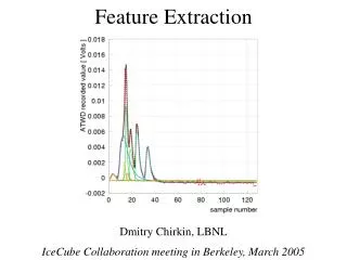
A Computer Aided Detection System For Mammograms Based on Asymmetry and Feature Extraction Techniques. By Mohammed Jirari Benidorm, Spain Sept 9th, 2005. Why This Project?. Breast Cancer is the most common cancer and is the second leading cause of cancer deaths

E N D
A Computer Aided Detection System For Mammograms Based on Asymmetry and Feature Extraction Techniques By Mohammed Jirari Benidorm, Spain Sept 9th, 2005
Why This Project? • Breast Cancer is the most common cancer and is the second leading cause of cancer deaths • Mammographic screening reduces the mortality of breast cancer • But, mammography has low positive predictive value PPV (only 35% have malignancies) • Goal of Computer Aided Detection (CAD) is to provide a second reading, hence reducing the false positive rate
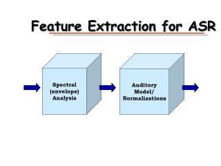
Basic Components of the System • Mammogram Normalization • Mammogram Registration • Mammogram Subtraction • Feature Extraction • Morphological Closing • Morphological Opening • Size Test • Border Test • ROC Analysis
What is a Mammogram? • A Mammogram is an x-ray image of the breast. Mammography is the procedure used to generate a mammogram • The equipment used to obtain a mammogram, however, is very different from that used to perform an x-ray of chest or bones
Mammograms (cont.) • In order to get a good image, the breast must be flattened or compressed • In a standard examination, two images of each breast are taken: one from the top (CC) and one from the side(MLO)
Mammogram Examples Mammogram of a left breast, cranio-caudal (from the top) view Mammogram of a left breast, medio-lateral oblique (from the side) view
Purpose of CAD • Mammography is the most reliable method in early detection of breast cancer • But, due to the high number of mammograms to be read, the accuracy rate tends to decrease • Double reading of mammograms has been proven to increase the accuracy, but at high cost • CAD can assist the medical staff to achieve high efficiency and effectiveness • The physician/radiologist makes the call not CAD
Proposed Method • The proposed method will assist the physician by providing a second opinion on reading the mammogram, by pointing out area(s) that are different between the right and left breasts • If the two readings are similar, no more work is to be done • If they are different, the radiologist will take a second look to make the final diagnosis
Data Used • The dataset used is the Mammographic Image Analysis Society (MIAS) MINIMIAS database containing Medio-Lateral Oblique (MLO) views for each breast for 161 patients for a total of 322 images Each image is: 1024 pixels X 1024 pixels
Normalization The images were corrected/normalized to avoid differences in brightness between the right and left mammograms
Mammogram Registration • Thermodynamic concepts are used • Match a model M with a scene S(M must be deformed to resemble Sas much as possible) • Use diffusion process technique as follows:
Mammogram Registration (cont.) 1. Select pixels to be demons 2. For each demon, store displacement then apply Gaussian filter 3. Use trilinear interpolation to estimate intermediate intensities 4. The demon force is given by optical flow
Registration Example Mammogram of left breast Mammogram of right breast
Registration Example (cont.) Grid of displacement Registered images
Mammogram Subtraction • Simple linear subtraction is used • Flipped right – left • Most common gray level is 0 • Masses in right breast are in lower gray level region of subtraction image histogram, while left breast masses are in the higher gray level region
Mammogram Subtraction Example Flipped right breast Left breast showing mass
Mammogram Subtraction Example (cont.) Subtraction image Superimposed subtraction image
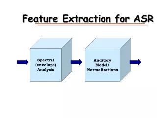
Feature Extraction • Many features are not masses • Morphological filtering using a 3X3 kernel • Size test (100 pixels) • Border test for border misalignment
Results • 102 registered pairs of mammograms used • Verified by expert radiologists
Future work • Use more features like brightness and directionality • Try and reduce False Negatives on the basis of region characteristics size, difference in homogeneity and entropy • Use larger database that contains both MLO and CC to train/learn, since most commercial CADs use hundreds of thousands of mammograms to try and recognize foreign samples