Download

1 / 6
60 likes | 76 Vues
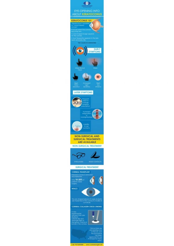
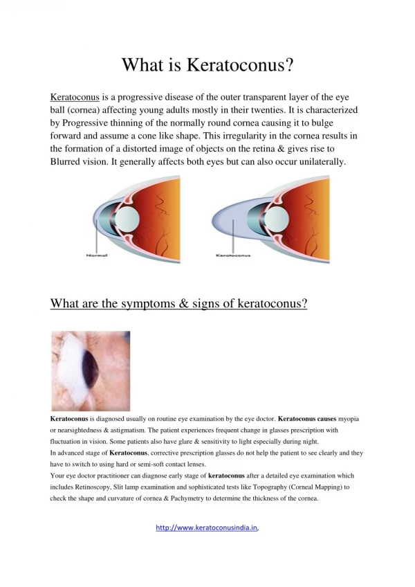
Keratoconus is a vision disorder that occurs when the normally round cornea (the front part of the eye) becomes thin and irregular (cone) shaped. This abnormal shape prevents the light entering the eye from being focused correctly on the retina and causes distortion of vision. It is characterized by para-central corneal thinning and ectasia so that the cornea takes the shape of a cone. Visual loss occurs primarily from myopia and irregular astigmatism and secondarily from corneal scarring. Keratoconus often begins at puberty and most often is seen in teenagers or young adults.

E N D
Keratoconus Treatment In Ghatkopar from Cornea Specialist in Mumbai - Dr. JatinAshar Keratoconus Treatment In Ghatkopar Keratoconus is a vision disorder that occurs when the normally round cornea (the front part of the eye) becomes thin and irregular (cone) shaped. This abnormal shape prevents the light entering the eye from being focused correctly on the retina and causes distortion of vision. It is characterized by para-central corneal thinning and ectasia so that the cornea takes the shape of a cone. Visual loss occurs primarily from myopia and irregular astigmatism and secondarily from corneal scarring. Keratoconus often begins at puberty and most often is seen in teenagers or youngadults. Keratoconus causes distorted vision that cannot be corrected with eyeglasses. Tiny fibers of protein in your eye called collagen help hold your cornea in place. When these fibers get weak, they can’t hold their shape. Your cornea gets more and morecone-like. It happens when you don’t have enough protective antioxidants in your cornea. Its cells produce harmful byproducts, the same way a car puts outexhaust. Normally, antioxidants get rid of them and protect the collagen fibers. But if levels are low, the collagen weakens and the corneabulges. Symptoms Signs and symptoms of keratoconus may change as the disease progresses. Theyinclude: Blurred or distortedvision A need for frequent changes in eyeglass prescriptions Sudden worsening or clouding ofvision
The swelling occurs when the strain of the cornea's protruding cone-like shape causes a tiny crack to develop. The swelling may last for weeks or months as the crack heals and is gradually replaced by scartissue. Monocular polyopia (perception of multiple ‘ghost’ images in the eye). Streaking and flaring distortion around lightsources. Marked anisometropia (difference in vision of two eyes). Photophobia (increased sensitivity to light). Eyestrain, in order to readclearly. Risk factors Heredity. One in 10 keratoconus sufferers has a close family relative with the disorder. Frequent eye rubbing, especially aggressive “knuckling” eyerubbing. Having a history of asthma, allergies, Ehlos Danlers syndrome, Down’ssyndrome Keratoconus is categorised clinically as: Latent stage: Latent stage was recognisable by placido disconly. Early stage: Early stages were subdivided into two categoriesas: Keratoconus fruste, which entailed 1- to 4-degree deviation of horizontal axis of the placidodisc. Early or mild keratoconus, which entailed 5- to 8-degree deviation of horizontal axis. Causes A family history of keratoconus has been established in some cases. Most researchers believe that multiple, complex factors are required for the development of keratoconus including both genetic and environmental factors. With the advent of videokeratography to assess family members, however, pedigrees have been analysed. These studies show cornealchanges
consistent with keratoconus in some family members, which suggest an autosomal dominant pattern ofinheritance. Keratoconus may be associated with wide variety of systemic and ocularconditions. Systemicassociations: Atopy (a genetic predisposition to develop an allergic reaction): Eye rubbing seen in systemic atopy may play a role in the development ofkeratoconus. Down syndrome (Trisomy 21): In Down syndrome (Trisomy 21), frequency of acute hydrops is higher, perhaps because of eye rubbing and/or these patients are treated infrequently with keratoplasty and their disease is allowed to progress further. Ehlers-Danlossyndrome. Ocularassociations: Retinitispigmentosa. Retinopathy ofprematurity. Fuchs’ corneal endothelial dystrophy. Posterior polymorphousdystrophy. Contributory factors such as: Enzyme abnormalities in corneal epithelium: Enzyme abnormalities such as increased expression of lysosomal enzymes (catalase and cathepsin) and decreased levels of inhibitors of proteolytic enzymes (tissue inhibitor matrix metalloproteinases), may play a role in corneal stromaldegradation. Differentially expressed corneal epithelium: Differentially expressed corneal epithelium between keratoconus and myopes (as controls) in both genetic expression and proteinexpression. Molecular defect: Molecular defect producing unusual absence of water channel protein aquaporin 5 in keratoconus as compared to normal cornealepithelium.
Gelatinolytic activity: Gelatinolytic activity in stroma has been described, which may be due to decreased function of enzymeinhibitors. Abnormalities in corneal collagen and its cross-linking: Abnormalities in corneal collagen and its cross-linking may be the cause ofkeratoconus. Hard contact lenswear. Pathophysiology: First is thinning of the corneal stroma then fragmentation of the Bowman layer and the deposition of iron in the basal epithelial cells, forming the Fleischer ring. Folds and breaks in the Descemet’s membrane result in acute hydrops and striae, which produces variable amount of diffusescarring. How diagnosis is made? Certain tests like refraction, keratometry, corneal topography/Computerised videokeratography, ultrasound pachymetry and slit lamp microscopy help in reaching finalconclusion. Computerized videokeratography, which takes pictures of your cornea so a map can be made of the surface while also measuring the thickness of your cornea Severity of keratoconus depends on shape of cone: Nipplecones Oval cones Globuscones Treatment If your keratoconus is progressing, corneal collagen cross-linking might be indicated to slow or stop the progression. Contact lenses can be used to correct astigmatism and mild near-sightedness. Improving your vision depends on the severity of keratoconus. Mild to moderate keratoconus can be treated with eyeglasses or contactlenses.
Hard contact lenses. Hard lenses may feel uncomfortable at first, but many people adjust to wearing them and they can provide excellent vision. This type of lens can be made to fit yourcorneas. Piggyback lenses. If rigid lenses are uncomfortable, your doctor may recommend "piggybacking" a hard contact lens on top of a softone. Eyeglasses or soft contact lenses. Glasses or soft contact lenses can correct blurry or distorted vision in early keratoconus. But people frequently need to change their prescription for eyeglasses or contacts as the shape of their corneas change. Hybrid lenses. These contact lenses have a rigid center with a softer ring around the outside for increased comfort. People who can't tolerate hard contact lenses may prefer hybridlenses. Scleral lenses. These lenses are useful for very irregular shape changes in your cornea in advanced keratoconus. Instead of resting on the cornea like traditional contact lenses do, scleral lenses sit on the white part of the eye (sclera) and vault over the cornea without touchingit. SurgicalInterventions Some form of surgery may become necessary if the cornea progresses in its shape-changing until it is so steep that contacts cannot be tolerated atall. NTACS are described as arc-like and plastic. These pieces are inserted into the center of the cornea to flatten it, thereby making the eye more contact lens- tolerant. Collagen crosslinking (CXL) with UVA is a complex surgery that involves removing the topmost layer of your cornea, adding vitamin drops and then exposing the eye to a special UV lamp that helps the cornea fibers multiply, strengthening thecornea. Corneal transplant surgery is the last resort for most doctors. In this procedure cornea would be removed and replaced with a healthy, normal-shaped cornea. This surgery has a long recovery time, a year or more in some cases, for clear vision. Lenses
Penetrating keratoplasty. If you have corneal scarring or extreme thinning, you'll likely need a cornea transplant (keratoplasty). Penetrating keratoplasty is a full-cornea transplant. In this procedure, doctors remove a full-thickness portion of your central cornea and replace it with donortissue Deep anterior lamellar keratoplasty (DALK). The DALK procedure preserves the inside lining of the cornea (endothelium). This helps avoid the rejection of this critical inside lining that can occur with a full-thicknesstransplant. Important Reminder: This information is only intended to provide guidance, not a definitive medical advice. Please consult eye doctor about your specific condition. Only a trained, experienced board certified eye doctor can determine an accurate diagnosis and propertreatment. To schedule an appointment with our experts for Keratoconus Treatment in Ghatkopar, please call us at +91 8451045935, +91- 8451045934 or visit our clinic atAddress. Tag = cornea surgery in mumbai, cornea specialist in mumbai, eye clinic in ghatkopar, Keratoconus Treatment In Ghatkopar For more information =https://www.mumbaieyecare.com/