Download
1 / 74
740 likes | 1.54k Vues
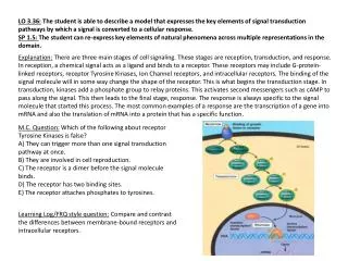
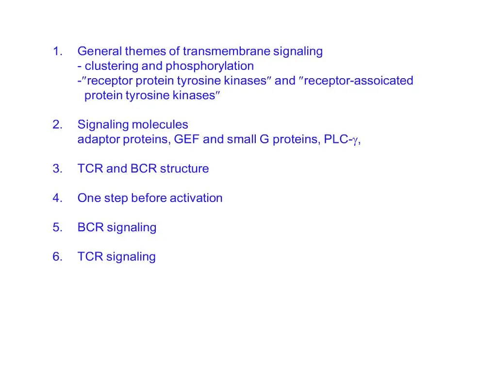
General Principles of Transmembrane Signaling. Binding of antigen leads to clustering of antigen receptors on lymphocytesClustering of antigen receptors leads to activation of intracellular signal moleculesPhosphorylation of receptor cytoplasmic tails by tyrosine kinases concentratesintracellular signaling molecules around the receptorsintracellular signaling components recruited to activated receptors transmitthe signal onward from the membrane and amplify it.

E N D
2. Many receptors undergo a change in protein conformation on binding their ligand (Ag binding results in receptor conformation change ?????? ?????).
In some types of receptor, this CONFORMATIONAL CHANGE OPENS AN ION CHANNEL into the cell and the resulting change in the concentration of important ions within the cell acts as the intracellular signal, which is then converted into an intracellular response.
In other receptors, the CONFORMATIONAL CHANGE AFFECTS THE CYTOPLASMIC PORTION OF THE RECEPTOR, enabling it to associate with and activate intracellular signaling proteins and enzymes.Many receptors undergo a change in protein conformation on binding their ligand (Ag binding results in receptor conformation change ?????? ?????).
In some types of receptor, this CONFORMATIONAL CHANGE OPENS AN ION CHANNEL into the cell and the resulting change in the concentration of important ions within the cell acts as the intracellular signal, which is then converted into an intracellular response.
In other receptors, the CONFORMATIONAL CHANGE AFFECTS THE CYTOPLASMIC PORTION OF THE RECEPTOR, enabling it to associate with and activate intracellular signaling proteins and enzymes.
3. Figure legend:
The requirement for receptor cross-linking is illustrated by (???????????, ??? illustrate) the use of anti-immunoglobulin antibodies to activate the B-cell antigen receptor (BCR). As shown in the left panel, Fab fragments of an anti-immunoglobulin can bind to the receptors but cannot cross-link them; they also fail to activate B cells. F(ab�)2 fragments of the same anti-immunoglobulin, which have two binding sites, can bridge two receptors (center panel), and thus signal, albeit weakly, to the B cell. The most effective activation occurs when receptors are extensively cross-linked by first adding the F(ab�)2 fragments and then rabbit antibody molecules that bind and cross-link the bound F(ab�)2 fragments (right panel). The use of antibodies generally to stimulate receptors is described in Appendix I, Section A-19.
?how antigen receptors are clustered (?????clustering, in stead of cross-linking) in vivo when B and T cells encounter their specific antigens is not yet completely understood.Figure legend:
The requirement for receptor cross-linking is illustrated by (???????????, ??? illustrate) the use of anti-immunoglobulin antibodies to activate the B-cell antigen receptor (BCR). As shown in the left panel, Fab fragments of an anti-immunoglobulin can bind to the receptors but cannot cross-link them; they also fail to activate B cells. F(ab�)2 fragments of the same anti-immunoglobulin, which have two binding sites, can bridge two receptors (center panel), and thus signal, albeit weakly, to the B cell. The most effective activation occurs when receptors are extensively cross-linked by first adding the F(ab�)2 fragments and then rabbit antibody molecules that bind and cross-link the bound F(ab�)2 fragments (right panel). The use of antibodies generally to stimulate receptors is described in Appendix I, Section A-19.
?how antigen receptors are clustered (?????clustering, in stead of cross-linking) in vivo when B and T cells encounter their specific antigens is not yet completely understood.
4. figure legend:
Kit (CD117) is a transmembrane protein with an external ligand-binding domain specific for stem-cell factor (SCF) and a cytoplasmic domain with intrinsic tyrosine kinase activity. In the unbound state, the kinase part of the receptor is inactive (??????active?)(top panel). When SCF binds to Kit, it causes the receptor proteins to dimerize; this allows the two tyrosine kinase domains to phosphorylate one another and so become activated (???phosphorylate???????????????phosporylated, ???????phosphorylate??????????). Transactivation of protein kinases by transphosphorylation is an important step in signaling from many cell-surface kinases (cluster).
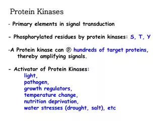
Most of the receptors discussed in this chapter initiate intracellular signaling by the activation of protein tyrosine kinases, enzymes that affect the activity of other proteins by adding a phosphate group to certain tyrosine residues. They have cytoplasmic domains that contain an intrinsic tyrosine kinase activity. These enzyme domains are normally inactive, but when brought together by receptor clustering they are able to activate each other by transphosphorylation (??????, why?) (Fig. 6.2). Once activated, these tyrosine kinases can phosphorylate and activate other cytoplasmic signaling molecules (??transphosphorylation?????,????! ??????phosphorylate ???phosphorylate ??).
figure legend:
Kit (CD117) is a transmembrane protein with an external ligand-binding domain specific for stem-cell factor (SCF) and a cytoplasmic domain with intrinsic tyrosine kinase activity. In the unbound state, the kinase part of the receptor is inactive (??????active?)(top panel). When SCF binds to Kit, it causes the receptor proteins to dimerize; this allows the two tyrosine kinase domains to phosphorylate one another and so become activated (???phosphorylate???????????????phosporylated, ???????phosphorylate??????????). Transactivation of protein kinases by transphosphorylation is an important step in signaling from many cell-surface kinases (cluster).
Most of the receptors discussed in this chapter initiate intracellular signaling by the activation of protein tyrosine kinases, enzymes that affect the activity of other proteins by adding a phosphate group to certain tyrosine residues. They have cytoplasmic domains that contain an intrinsic tyrosine kinase activity. These enzyme domains are normally inactive, but when brought together by receptor clustering they are able to activate each other by transphosphorylation (??????, why?) (Fig. 6.2). Once activated, these tyrosine kinases can phosphorylate and activate other cytoplasmic signaling molecules (??transphosphorylation?????,????! ??????phosphorylate ???phosphorylate ??).
5. All these receptor protein-tyrosine kinases share a common structural organization: an N-terminal extracellular ligand binding domain, a single transmembrane a-helix, and a cytosolic C-terminal domain with protein tyrosine kinase activity.
The EGF receptor and insulin receptor both have cysteine rich extracellular domains.
The insulin receptor is unusual in being a dimer of two pairs of polypeptide chains.
All these receptor protein-tyrosine kinases share a common structural organization: an N-terminal extracellular ligand binding domain, a single transmembrane a-helix, and a cytosolic C-terminal domain with protein tyrosine kinase activity.
The EGF receptor and insulin receptor both have cysteine rich extracellular domains.
The insulin receptor is unusual in being a dimer of two pairs of polypeptide chains.
6. Dimerization and Autophosphorylation of Receptor Protein-Tyrosine Kinases The first step in signaling from most receptor protein tyrosine kinase is ligand-induced receptor dimerization.
Such autophosphorylation plays two roles in signaling from these receptors: ?phosphorylation of tyrosine residue within catalytic domain increases protein kinase activity ?phosphorylation of tyrosine residue outside the catalytic domain creates binding sites for �
The first step in signaling from most receptor protein tyrosine kinase is ligand-induced receptor dimerization.
Such autophosphorylation plays two roles in signaling from these receptors: ?phosphorylation of tyrosine residue within catalytic domain increases protein kinase activity ?phosphorylation of tyrosine residue outside the catalytic domain creates binding sites for �
7. Association of Downstream Signaling Molecules with Receptor Protein-Tyrosine
Figures_Hi-res\ch13\cell3e13150.jpg
Figures_Hi-res\ch13\cell3e13150.jpg
8. Receptor activation and the phosphorylation of membrane-associated proteins can, however, create binding sites for these target proteins. Cytosolic proteins that bind to phosphorylated sites at the membrane are thus concentrated near to the kinase and can in their turn be phosphorylated and activated (??many kinases involved in signaling are associated with the inner surface of the cell membrane and can act only inefficiently upon their target proteins when these are free in the cytosol.)(Fig. 6.3). They can also, in some cases, be activated simply by binding to phosphotyrosine. This is an example of allosteric activation, as binding the phosphotyrosine leads to an alteration in their molecular conformation.
???Lck????phosphorylated, ????phosphorylate adaptor protein, ???adaptor protein ?? phosphate ??? target protein,???????Receptor activation and the phosphorylation of membrane-associated proteins can, however, create binding sites for these target proteins. Cytosolic proteins that bind to phosphorylated sites at the membrane are thus concentrated near to the kinase and can in their turn be phosphorylated and activated (??many kinases involved in signaling are associated with the inner surface of the cell membrane and can act only inefficiently upon their target proteins when these are free in the cytosol.)(Fig. 6.3). They can also, in some cases, be activated simply by binding to phosphotyrosine. This is an example of allosteric activation, as binding the phosphotyrosine leads to an alteration in their molecular conformation.
???Lck????phosphorylated, ????phosphorylate adaptor protein, ???adaptor protein ?? phosphate ??? target protein,???????
9. figure legend:
Adaptor proteins are specialized signaling molecules that usually have no enzymatic activity themselves. Instead, they allow other molecules to become associated with the activated receptors. Adaptors, as shown in the first panel, often contain SH2 domains flanked by SH3 domains. Once a receptor, in this case Kit, has been activated and trans-phosphorylated (second panel), adaptors can bind to the phosphotyrosines through their SH2 domains (third panel). Other molecules that contain proline-rich regions can now bind to the adaptors through the SH3 domains and be activated by the receptor-associated kinases (fourth panel) ?????adaptor protein ?phosphorylation? attract target protein.
Many of the cellular processes that are activated in lymphocytes when antigen binds to its receptor are common to many cell types. For example, resting lymphocytes proliferate when exposed to antigen, whereas other cell types proliferate in response to particular growth factors; what differs in each case is the receptor that initiates the common response pathway in the different cell types.
? To link these different receptors to common intracellular signaling components, specialized adaptor proteins are needed (are adaptor protein common to all these signal pathway?).
figure legend:
Adaptor proteins are specialized signaling molecules that usually have no enzymatic activity themselves. Instead, they allow other molecules to become associated with the activated receptors. Adaptors, as shown in the first panel, often contain SH2 domains flanked by SH3 domains. Once a receptor, in this case Kit, has been activated and trans-phosphorylated (second panel), adaptors can bind to the phosphotyrosines through their SH2 domains (third panel). Other molecules that contain proline-rich regions can now bind to the adaptors through the SH3 domains and be activated by the receptor-associated kinases (fourth panel) ?????adaptor protein ?phosphorylation? attract target protein.
Many of the cellular processes that are activated in lymphocytes when antigen binds to its receptor are common to many cell types. For example, resting lymphocytes proliferate when exposed to antigen, whereas other cell types proliferate in response to particular growth factors; what differs in each case is the receptor that initiates the common response pathway in the different cell types.
? To link these different receptors to common intracellular signaling components, specialized adaptor proteins are needed (are adaptor protein common to all these signal pathway?).
11. figure legend:
Ras is a small GTP-binding protein with intrinsic GTPase activity. In its GTP-bound state, it is active (first panel), whereas in the GDP-bound state it is inactive. Most of the time, it is in the inactive state owing to its intrinsic GTPase activity (second panel). Receptor signaling activates guanine-nucleotide exchange factors (GEFs, e.g. SOS ??Vav), which can bind to small G proteins such as Ras and displace GDP, allowing GTP to bind in its place (right panel). In the time before the intrinsic GTPase activity converts GTP to GDP, the Ras protein is active and transmits the signal onward.
Small GTP-binding proteins (= small G proteins ) are another class of protein that serves to propagate signals from tyrosine kinase-associated receptors. The family of small single-chain GTP-binding proteins is distinct from the heterotrimeric G proteins that associate with seven-span transmembrane receptors such as the anaphylotoxin or chemokine receptors (see Section 6-16). The best-known small G protein is Ras.
small G proteins do not normally stay permanently activated; instead they eventually turn themselves off. Thus the small G proteins are usually found in the inactive GDP-containing state, and they are activated only transiently in response to activating ligands. However, mutation at a single residue can lock them in the active state, causing them to become oncogenic (???Ras ?protooncogene).figure legend:
Ras is a small GTP-binding protein with intrinsic GTPase activity. In its GTP-bound state, it is active (first panel), whereas in the GDP-bound state it is inactive. Most of the time, it is in the inactive state owing to its intrinsic GTPase activity (second panel). Receptor signaling activates guanine-nucleotide exchange factors (GEFs, e.g. SOS ??Vav), which can bind to small G proteins such as Ras and displace GDP, allowing GTP to bind in its place (right panel). In the time before the intrinsic GTPase activity converts GTP to GDP, the Ras protein is active and transmits the signal onward.
Small GTP-binding proteins (= small G proteins ) are another class of protein that serves to propagate signals from tyrosine kinase-associated receptors. The family of small single-chain GTP-binding proteins is distinct from the heterotrimeric G proteins that associate with seven-span transmembrane receptors such as the anaphylotoxin or chemokine receptors (see Section 6-16). The best-known small G protein is Ras.
small G proteins do not normally stay permanently activated; instead they eventually turn themselves off. Thus the small G proteins are usually found in the inactive GDP-containing state, and they are activated only transiently in response to activating ligands. However, mutation at a single residue can lock them in the active state, causing them to become oncogenic (???Ras ?protooncogene).
13. figure legend:
The enzyme phospholipase C-? cleaves inositol phospholipids to generate two important signaling molecules. Phosphatidylinositol bisphosphate (PIP2) is a component of the inner leaflet of the plasma membrane. When phospholipase C-? (PLC-?) is activated, it cleaves PIP2 into two parts, inositol trisphosphate (IP3), which diffuses away from the membrane, and diacylglycerol (DAG), which stays in the membrane. Both these molecules are important in signaling.
?IP3 binds to calcium channels in the endoplasmic reticulum (ER) membrane, opening the channels and allowing Ca2+ to enter the cytosol from stores in the ER (IP3 ??bind?cell membrane ??Ca channel ! ).
?The depletion of Ca2+ from the ER in turn triggers the opening of channels in the plasma membrane that allows Ca2+ in from the external fluid. Raised Ca2+ levels also activate a protein known as calmodulin, a ubiquitous Ca2+-binding protein that is responsible for activating other Ca2+-dependent enzymes within the cell.
?DAG attracts a protein kinase C (PKC) to the cell membrane where it is activated, often with the help of the increased level of Ca2+. The active forms of protein kinase C are serine/threonine kinases with several roles in cell activation. figure legend:
The enzyme phospholipase C-? cleaves inositol phospholipids to generate two important signaling molecules. Phosphatidylinositol bisphosphate (PIP2) is a component of the inner leaflet of the plasma membrane. When phospholipase C-? (PLC-?) is activated, it cleaves PIP2 into two parts, inositol trisphosphate (IP3), which diffuses away from the membrane, and diacylglycerol (DAG), which stays in the membrane. Both these molecules are important in signaling.
?IP3 binds to calcium channels in the endoplasmic reticulum (ER) membrane, opening the channels and allowing Ca2+ to enter the cytosol from stores in the ER (IP3 ??bind?cell membrane ??Ca channel ! ).
?The depletion of Ca2+ from the ER in turn triggers the opening of channels in the plasma membrane that allows Ca2+ in from the external fluid. Raised Ca2+ levels also activate a protein known as calmodulin, a ubiquitous Ca2+-binding protein that is responsible for activating other Ca2+-dependent enzymes within the cell.
?DAG attracts a protein kinase C (PKC) to the cell membrane where it is activated, often with the help of the increased level of Ca2+. The active forms of protein kinase C are serine/threonine kinases with several roles in cell activation.
14. Several classes of protein are typically recruited to the activated receptors and participate in signal propagation. The enzyme phospholipase C-? (PLC-?) contains TWO SH2 DOMAINS through which it can bind to phosphotyrosine (???); it is thus recruited to the site of receptor-associated tyrosine kinase activity at the plasma membrane. PLC-? has a crucial role in propagating the signal onward from the membrane and in amplifying it. PHOSPHORYLATION OF A TYROSINE RESIDUE IN PLC-? ACTIVATES THE ENZYME.
Increased intracellular free Ca2+ leads to the activation of the Ca2+- binding protein calmodulin, which in turn binds to and regulates the activity of several other proteins and enzymes in the cell, transmitting the signal onward along pathways that eventually converge on the nucleus. One protein that is regulated by the calcium pathway is the nuclear factor of activated T cells (NFAT), a transcription factor we will discuss further in Section 6-11.Several classes of protein are typically recruited to the activated receptors and participate in signal propagation. The enzyme phospholipase C-? (PLC-?) contains TWO SH2 DOMAINS through which it can bind to phosphotyrosine (???); it is thus recruited to the site of receptor-associated tyrosine kinase activity at the plasma membrane. PLC-? has a crucial role in propagating the signal onward from the membrane and in amplifying it. PHOSPHORYLATION OF A TYROSINE RESIDUE IN PLC-? ACTIVATES THE ENZYME.
Increased intracellular free Ca2+ leads to the activation of the Ca2+- binding protein calmodulin, which in turn binds to and regulates the activity of several other proteins and enzymes in the cell, transmitting the signal onward along pathways that eventually converge on the nucleus. One protein that is regulated by the calcium pathway is the nuclear factor of activated T cells (NFAT), a transcription factor we will discuss further in Section 6-11.
15. One Ig? chain and one Ig? chain associates with each surface immunoglobulin molecule. Thus the complete B-cell receptor is thought to be a complex of six chains-two identical light chains, two identical heavy chains, one Ig?, and one Ig? (Fig. 6.7).
ITAMs were originally identified in the cytoplasmic tails of Ig? and Ig?, but are now known to be present in the accessory chains involved in signaling from the T-cell receptor, and in the Fc receptors on mast cells, macrophages, monocytes, and natural killer (NK) cells that bind antibody constant regions.
ITAMs are composed of two tyrosine residues separated by around 9-12 a.a.; the canonical ITAM sequence is �YXX[L/V]X6-9YXX[L/V]�. Ig? and Ig? each have a single ITAM in their cytosolic tails, giving the B-cell receptor a total of two ITAMs.
??????:
When antigen binds, the tyrosines in these ITAMs become phosphorylated by receptor-associated Src-family tyrosine kinases Blk, Fyn, or Lyn. The ITAMs, by virtue of their two precisely spaced tyrosines, are then able to bind with high affinity to the tandem SH2 domains of members of a second family of protein tyrosine kinases; ? Syk in B cells and ZAP70 in T cells.
One Ig? chain and one Ig? chain associates with each surface immunoglobulin molecule. Thus the complete B-cell receptor is thought to be a complex of six chains-two identical light chains, two identical heavy chains, one Ig?, and one Ig? (Fig. 6.7).
ITAMs were originally identified in the cytoplasmic tails of Ig? and Ig?, but are now known to be present in the accessory chains involved in signaling from the T-cell receptor, and in the Fc receptors on mast cells, macrophages, monocytes, and natural killer (NK) cells that bind antibody constant regions.
ITAMs are composed of two tyrosine residues separated by around 9-12 a.a.; the canonical ITAM sequence is �YXX[L/V]X6-9YXX[L/V]�. Ig? and Ig? each have a single ITAM in their cytosolic tails, giving the B-cell receptor a total of two ITAMs.
??????:
When antigen binds, the tyrosines in these ITAMs become phosphorylated by receptor-associated Src-family tyrosine kinases Blk, Fyn, or Lyn. The ITAMs, by virtue of their two precisely spaced tyrosines, are then able to bind with high affinity to the tandem SH2 domains of members of a second family of protein tyrosine kinases; ? Syk in B cells and ZAP70 in T cells.
16. The receptor associated with cytokine receptors belong to the Janus kinase, or JAK family. Members of the JAK family appear to be universally required for signaling from cytokine receptors. Additional nonreceptor protein tyrosine kinases belong to Src family, which consists of Src and eight closely related proteins.
The receptor associated with cytokine receptors belong to the Janus kinase, or JAK family. Members of the JAK family appear to be universally required for signaling from cytokine receptors. Additional nonreceptor protein tyrosine kinases belong to Src family, which consists of Src and eight closely related proteins.
17. the MHC class I molecule is a heterodimer of a membrane-spanning ? chain (molecular weight 43 kDa) bound noncovalently to ?2-microglobulin (12 kDa), which does not span the membrane. The ? chain folds into three domains: ?1, ?2, and ?3. The ?3 domain and ?2-microglobulin show similarities in amino acid sequence to immunoglobulin C domains and have similar folded structures, whereas the ?1 and ?2 domains fold together into a single structure consisting of two segmented ? helices lying on a sheet of eight antiparallel ? strands. The folding of the ?1 and ?2 domains creates a long cleft or groove, which is the site at which peptide antigens bind to the MHC molecules. The transmembrane region and the short stretch of peptide that connects the external domains to the cell surface are not seen in panels a and b as they have been removed by the papain digestion. As can be seen in panel c, looking down on the molecule from above, the sides of the cleft are formed from the inner faces of the two helices; the ?-pleated sheet formed by the pairing of the ?1 and ?2 domains creates the floor of the cleft.
The MHC class II molecule is composed of two trans-membrane glycoprotein chains, ? (34 kDa) and ? (29 kDa), as shown schematically in panel d. Each chain has two domains, and the two chains together form a compact four-domain structure similar to that of the MHC class I molecule (compare with panel d of Fig. 3.20). The ?2 and ?2 domains, like the ?3 and ? 2-microglobulin domains of the MHC class I molecule, have amino acid sequence and structural similarities to immunoglobulin C domains; in the MHC class II molecule, the two domains forming the peptide-binding cleft are contributed by different chains and are therefore not joined by a covalent bond (see panels c and d). Another important difference, not apparent in this diagram, is that the peptide-binding groove of the MHC class II molecule is open at both ends.
the MHC class I molecule is a heterodimer of a membrane-spanning ? chain (molecular weight 43 kDa) bound noncovalently to ?2-microglobulin (12 kDa), which does not span the membrane. The ? chain folds into three domains: ?1, ?2, and ?3. The ?3 domain and ?2-microglobulin show similarities in amino acid sequence to immunoglobulin C domains and have similar folded structures, whereas the ?1 and ?2 domains fold together into a single structure consisting of two segmented ? helices lying on a sheet of eight antiparallel ? strands. The folding of the ?1 and ?2 domains creates a long cleft or groove, which is the site at which peptide antigens bind to the MHC molecules. The transmembrane region and the short stretch of peptide that connects the external domains to the cell surface are not seen in panels a and b as they have been removed by the papain digestion. As can be seen in panel c, looking down on the molecule from above, the sides of the cleft are formed from the inner faces of the two helices; the ?-pleated sheet formed by the pairing of the ?1 and ?2 domains creates the floor of the cleft.
The MHC class II molecule is composed of two trans-membrane glycoprotein chains, ? (34 kDa) and ? (29 kDa), as shown schematically in panel d. Each chain has two domains, and the two chains together form a compact four-domain structure similar to that of the MHC class I molecule (compare with panel d of Fig. 3.20). The ?2 and ?2 domains, like the ?3 and ? 2-microglobulin domains of the MHC class I molecule, have amino acid sequence and structural similarities to immunoglobulin C domains; in the MHC class II molecule, the two domains forming the peptide-binding cleft are contributed by different chains and are therefore not joined by a covalent bond (see panels c and d). Another important difference, not apparent in this diagram, is that the peptide-binding groove of the MHC class II molecule is open at both ends.
20. CD4 binds MHC class II molecules through a region that is located mainly on a lateral face of the first domain, D1. Because CD4 binds to a site on the ?2 domain of the MHC class II molecule that is well away from the site where the T-cell receptor binds (Fig. 3.16a), the CD4 molecule and the T-cell receptor can bind the same peptide:MHC class II complex.
CD4 interacts strongly with a cytoplasmic tyrosine kinase called Lck, and can deliver this tyrosine kinase into close proximity with the signaling components of the T-cell receptor complex. This results in enhancement of the signal that is generated when the T-cell receptor binds its peptide:MHC class II ligand, as we will discuss further in Chapter 6. W
When CD4 and the T-cell receptor can simultaneously bind to the same MHC class II:peptide complex, the sensitivity of a T cell to antigen presented by MHC class II molecules is markedly increased; [the T-cell in this case requires 100-fold less antigen for activation.]
CD8 binds weakly to an invariant site in the ?3 domain of an MHC class I molecule (Fig. 3.16b), which is equivalent to the site in MHC class II molecules to which CD4 binds. Although only the interaction of the CD8? homodimer with MHC class I is so far known in detail, it is clear from this that the MHC class I binding site of the CD8 ?:? heterodimer will be formed by the interaction of the CD8? and ? chains. In addition, CD8 (most probably through the ? chain) interacts with residues in the base of the ?2 domain of the MHC class I molecule. Binding in this way, CD8 leaves the upper surface of the MHC class I molecule exposed and free to interact simultaneously with a T-cell receptor, as shown in Fig. 3.18. Like CD4, CD8 also binds Lck through the cytoplasmic tail of the ? chain and brings it into close proximity with the T-cell receptor. And as with CD4, the presence of CD8 increases the sensitivity of T cells to antigen presented by MHC class I molecules by about 100-fold. Thus, CD4 and CD8 have similar functions and bind to the same approximate location in MHC class I and MHC class II molecules even though the structures of the two co-receptor proteins are only distantly relatedCD4 binds MHC class II molecules through a region that is located mainly on a lateral face of the first domain, D1. Because CD4 binds to a site on the ?2 domain of the MHC class II molecule that is well away from the site where the T-cell receptor binds (Fig. 3.16a), the CD4 molecule and the T-cell receptor can bind the same peptide:MHC class II complex.
CD4 interacts strongly with a cytoplasmic tyrosine kinase called Lck, and can deliver this tyrosine kinase into close proximity with the signaling components of the T-cell receptor complex. This results in enhancement of the signal that is generated when the T-cell receptor binds its peptide:MHC class II ligand, as we will discuss further in Chapter 6. W
When CD4 and the T-cell receptor can simultaneously bind to the same MHC class II:peptide complex, the sensitivity of a T cell to antigen presented by MHC class II molecules is markedly increased; [the T-cell in this case requires 100-fold less antigen for activation.]
CD8 binds weakly to an invariant site in the ?3 domain of an MHC class I molecule (Fig. 3.16b), which is equivalent to the site in MHC class II molecules to which CD4 binds. Although only the interaction of the CD8? homodimer with MHC class I is so far known in detail, it is clear from this that the MHC class I binding site of the CD8 ?:? heterodimer will be formed by the interaction of the CD8? and ? chains. In addition, CD8 (most probably through the ? chain) interacts with residues in the base of the ?2 domain of the MHC class I molecule. Binding in this way, CD8 leaves the upper surface of the MHC class I molecule exposed and free to interact simultaneously with a T-cell receptor, as shown in Fig. 3.18. Like CD4, CD8 also binds Lck through the cytoplasmic tail of the ? chain and brings it into close proximity with the T-cell receptor. And as with CD4, the presence of CD8 increases the sensitivity of T cells to antigen presented by MHC class I molecules by about 100-fold. Thus, CD4 and CD8 have similar functions and bind to the same approximate location in MHC class I and MHC class II molecules even though the structures of the two co-receptor proteins are only distantly related
23. The situation in the antigen receptors is somewhat more complex. As we will see later, they do not themselves have intrinsic tyrosine kinase activity. Instead, the cytoplasmic portions of some of the receptor (?????? receptor? ) components bind to intracellular protein tyrosine kinases, which are therefore known as receptor-associated tyrosine kinases ( ?????? ). When the receptors cluster, these enzymes are brought together and act on each other and on the receptor cytoplasmic tails to initiate the signaling process as in the example above.The situation in the antigen receptors is somewhat more complex. As we will see later, they do not themselves have intrinsic tyrosine kinase activity. Instead, the cytoplasmic portions of some of the receptor (?????? receptor? ) components bind to intracellular protein tyrosine kinases, which are therefore known as receptor-associated tyrosine kinases ( ?????? ). When the receptors cluster, these enzymes are brought together and act on each other and on the receptor cytoplasmic tails to initiate the signaling process as in the example above.
24. In the case of the antigen receptors, the first tyrosine kinase associated with the receptor are members of the Src family of tryrosine kinases.
( ???signal transduction ?????phosphorylation? )phosphorylation of enzymes and other proteins by protein kinases has many advantages as a control mechanism. It is rapid, not requiring new protein synthesis or protein degradation to change the biochemical activity of a cell. It can also be easily reversed by the action of protein phosphatases, which remove the phosphate group.In the case of the antigen receptors, the first tyrosine kinase associated with the receptor are members of the Src family of tryrosine kinases.
( ???signal transduction ?????phosphorylation? )phosphorylation of enzymes and other proteins by protein kinases has many advantages as a control mechanism. It is rapid, not requiring new protein synthesis or protein degradation to change the biochemical activity of a cell. It can also be easily reversed by the action of protein phosphatases, which remove the phosphate group.
25. Src-family kinases are usually anchored to the cell membrane by a lipid moiety attached to their amino-terminal region (??????B cell ??Blk, Fyn, Lyn ???????anchor to cell membrance, ???, ???cyteine palmit). They are distributed over the inner surface of the cell membrane; during cell activation they become localized to sites of receptor signaling by binding to phosphotyrosine (??phosphotyrosine?) via their SH2 domains.Src-family kinases are usually anchored to the cell membrane by a lipid moiety attached to their amino-terminal region (??????B cell ??Blk, Fyn, Lyn ???????anchor to cell membrance, ???, ???cyteine palmit). They are distributed over the inner surface of the cell membrane; during cell activation they become localized to sites of receptor signaling by binding to phosphotyrosine (??phosphotyrosine?) via their SH2 domains.
26. figure legend:
Src-family kinases contain two tyrosine residues (red bars) that are targets for phosphorylation. Phosphorylation of the tyrosine in the kinase domain (bottom left panel) stimulates kinase activity, and this tyrosine is a target for phosphorylation by receptor-associated tyrosine kinases (which one?). The second tyrosine lies near the carboxy terminus and has a regulatory function. When it has been phosphorylated, the kinase is inactive AS A RESULT OF AN INTERACTION BETWEEN THE INHIBITORY PHOSPHOTYROSINE AND THE SH2 DOMAIN, as shown in the lower right panel.
The enzyme activity of the Src-family kinases is itself regulated by the phosphorylation status of the kinase domain and the carboxy-terminal region, each of which has regulatory tyrosine residues.
Even after being phosphorylated at the activating tyrosine, Src-family kinases can be kept inactive by a protein tyrosine kinase called Csk (C-terminal Src kinase), which phosphorylates the inhibitory tyrosine (Fig. 6.10). As CSK ACTIVITY IS CONSTITUTIVE IN RESTING CELLS, the Src proteins are generally inactive. Csk ??phosphorylate Src kinases?
An agent that counteracts the effects of Csk is the transmembrane protein tyrosine phosphatase CD45, also known as LEUKOCYTE COMMON ANTIGEN, which is required for receptor signaling in lymphocytes and other cells. CD45 can remove the phosphate from phosphotyrosines, especially from the inhibitory tyrosine residue of Src-family kinases ( ??????? ). Thus, the balance between Csk, which prevents the activation of Src-family kinases, and CD45, which restores their potential for being activated, determines the signaling activity of Src-family kinases in response to receptor aggregation. This is one way in which the threshold for initiating receptor signaling is regulated in lymphocytes. For example, as we will see in Chapter 10, activated effector T lymphocytes and memory T lymphocytes express an isoform of CD45 that associates with the T-cell receptor complex, thus facilitating signaling through the receptor. By contrast, naive T cells express a CD45 isoform that does not have this property.
figure legend:
Src-family kinases contain two tyrosine residues (red bars) that are targets for phosphorylation. Phosphorylation of the tyrosine in the kinase domain (bottom left panel) stimulates kinase activity, and this tyrosine is a target for phosphorylation by receptor-associated tyrosine kinases (which one?). The second tyrosine lies near the carboxy terminus and has a regulatory function. When it has been phosphorylated, the kinase is inactive AS A RESULT OF AN INTERACTION BETWEEN THE INHIBITORY PHOSPHOTYROSINE AND THE SH2 DOMAIN, as shown in the lower right panel.
The enzyme activity of the Src-family kinases is itself regulated by the phosphorylation status of the kinase domain and the carboxy-terminal region, each of which has regulatory tyrosine residues.
Even after being phosphorylated at the activating tyrosine, Src-family kinases can be kept inactive by a protein tyrosine kinase called Csk (C-terminal Src kinase), which phosphorylates the inhibitory tyrosine (Fig. 6.10). As CSK ACTIVITY IS CONSTITUTIVE IN RESTING CELLS, the Src proteins are generally inactive. Csk ??phosphorylate Src kinases?
An agent that counteracts the effects of Csk is the transmembrane protein tyrosine phosphatase CD45, also known as LEUKOCYTE COMMON ANTIGEN, which is required for receptor signaling in lymphocytes and other cells. CD45 can remove the phosphate from phosphotyrosines, especially from the inhibitory tyrosine residue of Src-family kinases ( ??????? ). Thus, the balance between Csk, which prevents the activation of Src-family kinases, and CD45, which restores their potential for being activated, determines the signaling activity of Src-family kinases in response to receptor aggregation. This is one way in which the threshold for initiating receptor signaling is regulated in lymphocytes. For example, as we will see in Chapter 10, activated effector T lymphocytes and memory T lymphocytes express an isoform of CD45 that associates with the T-cell receptor complex, thus facilitating signaling through the receptor. By contrast, naive T cells express a CD45 isoform that does not have this property.
27. figure legend:
The membrane-bound Src-family kinases Fyn, Blk, and Lyn (membrane-bound? But not integrated into membrane) associate with the B-cell antigen receptor by binding to ITAM motifs, either through their amino-terminal domains (binding to ITAM ????through SH2-phosphotyrosine) or, as shown in the figure, by binding a single phosphorylated tyrosine through their SH2 domains (??????? widely distributed ??? ! ???bind ?receptor??). After ligand binding and receptor clustering, they phosphorylate tyrosines ( ??phosphorylate each other? ??????? phosphorylate each other )in the ITAMs on the cytoplasmic tails of Ig? and Ig.?
In B cells, three protein tyrosine kinases of the Src family-Fyn, Blk, and Lyn-are thought to be responsible for this . These Src-family kinases can associate with resting receptors through a low-affinity interaction with an unphosphorylated ITAM (Src ? B cell ??? associate with ITAM, even in resting B cells ), to which they bind through a site in their amino-terminal domain.
When the receptors cluster after antigen binding, the receptor-associated kinases phosphorylate and ACTIVATE EACH OTHER and are then thought to phosphorylate the tyrosine residues in the ITAMs of the cytoplasmic tails of Ig? and Ig?. Phosphorylation of a single tyrosine in an ITAM allows the binding of a Src-family kinase through its SH2 domains, and kinases bound in this way are in turn activated to phosphorylate further ITAM tyrosine residues (???Src????bind ? ITAM ??phosphorylate ITAM ??? tyrosine? ??????phosphorylated).
The initial events in T-cell receptor signaling are also implemented by two Src-family kinases-Lck, which is constitutively associated with the cytoplasmic domain of the co-receptor molecules CD4 and CD8 (see Chapter 3), and Fyn, which associates with the cytoplasmic domains of the ? and CD3? chains upon receptor clustering.
figure legend:
The membrane-bound Src-family kinases Fyn, Blk, and Lyn (membrane-bound? But not integrated into membrane) associate with the B-cell antigen receptor by binding to ITAM motifs, either through their amino-terminal domains (binding to ITAM ????through SH2-phosphotyrosine) or, as shown in the figure, by binding a single phosphorylated tyrosine through their SH2 domains (??????? widely distributed ??? ! ???bind ?receptor??). After ligand binding and receptor clustering, they phosphorylate tyrosines ( ??phosphorylate each other? ??????? phosphorylate each other )in the ITAMs on the cytoplasmic tails of Ig? and Ig.?
In B cells, three protein tyrosine kinases of the Src family-Fyn, Blk, and Lyn-are thought to be responsible for this . These Src-family kinases can associate with resting receptors through a low-affinity interaction with an unphosphorylated ITAM (Src ? B cell ??? associate with ITAM, even in resting B cells ), to which they bind through a site in their amino-terminal domain.
When the receptors cluster after antigen binding, the receptor-associated kinases phosphorylate and ACTIVATE EACH OTHER and are then thought to phosphorylate the tyrosine residues in the ITAMs of the cytoplasmic tails of Ig? and Ig?. Phosphorylation of a single tyrosine in an ITAM allows the binding of a Src-family kinase through its SH2 domains, and kinases bound in this way are in turn activated to phosphorylate further ITAM tyrosine residues (???Src????bind ? ITAM ??phosphorylate ITAM ??? tyrosine? ??????phosphorylated).
The initial events in T-cell receptor signaling are also implemented by two Src-family kinases-Lck, which is constitutively associated with the cytoplasmic domain of the co-receptor molecules CD4 and CD8 (see Chapter 3), and Fyn, which associates with the cytoplasmic domains of the ? and CD3? chains upon receptor clustering.
28. figure legend:
On clustering of the receptors, the receptor-associated tyrosine kinases Blk, Fyn, and Lyn phosphorylate the ITAMs on the cytoplasmic tails of Ig? and Ig? (shown in blue and orange, respectively). Subsequently, Syk binds to the phosphorylated ITAMs of the Ig? chain. Because there are at least two receptor complexes in each cluster, Syk molecules become bound in close proximity and can activate each other by transphosphorylation, thus initiating further signaling.
These two proteins define a second family of protein tyrosine kinases expressed mainly in hematopoietic cells (Syk) or mainly in T lymphocytes (ZAP-70); they have two SH2 domains in their amino-terminal halves and a carboxy-terminal kinase domain. As each SH2 domain binds to one phosphotyrosine, these proteins preferentially bind to motifs with two phosphotyrosines spaced a precise distance apart; tyrosines spaced correctly are found in the ITAM motif. Thus, ZAP-70 or Syk molecules are recruited to the receptor complex upon full phosphorylation of the ITAMs.
To become active it must itself be phosphorylated, and this is thought to occur by transphosphorylation mediated by Syk itself or by Src kinases. Each B-cell receptor complex contains two molecules of Syk, bound to the Ig? and Ig? chains. Once the receptors are clustered, these receptor-associated kinases are brought into contact with each other and are thus able to phosphorylate, and hence activate, each other (Fig. 6.13).
figure legend:
On clustering of the receptors, the receptor-associated tyrosine kinases Blk, Fyn, and Lyn phosphorylate the ITAMs on the cytoplasmic tails of Ig? and Ig? (shown in blue and orange, respectively). Subsequently, Syk binds to the phosphorylated ITAMs of the Ig? chain. Because there are at least two receptor complexes in each cluster, Syk molecules become bound in close proximity and can activate each other by transphosphorylation, thus initiating further signaling.
These two proteins define a second family of protein tyrosine kinases expressed mainly in hematopoietic cells (Syk) or mainly in T lymphocytes (ZAP-70); they have two SH2 domains in their amino-terminal halves and a carboxy-terminal kinase domain. As each SH2 domain binds to one phosphotyrosine, these proteins preferentially bind to motifs with two phosphotyrosines spaced a precise distance apart; tyrosines spaced correctly are found in the ITAM motif. Thus, ZAP-70 or Syk molecules are recruited to the receptor complex upon full phosphorylation of the ITAMs.
To become active it must itself be phosphorylated, and this is thought to occur by transphosphorylation mediated by Syk itself or by Src kinases. Each B-cell receptor complex contains two molecules of Syk, bound to the Ig? and Ig? chains. Once the receptors are clustered, these receptor-associated kinases are brought into contact with each other and are thus able to phosphorylate, and hence activate, each other (Fig. 6.13).
29. figure legend:
Binding of the cleaved complement fragment C3d to antigen allows the tagged antigen to bind to both the B-cell receptor and the complement cell-surface protein CD21 (complement receptor 2, CR2), a component of the B-cell co-receptor complex. Cross-linking and clustering of the co-receptor with the antigen receptor results in phosphorylation of tyrosine residues in the cytoplasmic domain of CD19 by protein kinases associated with the B-cell receptor ( ????phosphorylate CD19 ? ); other Src-family kinases can bind to phosphorylated CD19 and so augment signaling through the B-cell receptor. Phosphorylated CD19 can also bind the enzyme phosphatidylinositol 3-OH kinase (PI 3-kinase), which is instrumental in recruiting the guanine-nucleotide exchange factor Vav to the receptor complex.
Co-ligation of the B-cell receptor with its CD19/CD21/CD81 co-receptor increases signaling 1000- to 10,000-fold. The role of the third component of the B-cell receptor complex, CD81 (TAPA-1), is as yet unknown.
Another small G protein is activated via the B-cell co-receptor complex (see Section 6-8). Phosphorylated CD19 binds a multifunctional intracellular signaling molecule called Vav; this is an adaptor protein that also contains guanine-nucleotide exchange factor activity.
When Vav is activated, it can activate the small G protein Rac. Small G proteins such as Ras and Rac activate a cascade of protein kinases that leads directly to the phosphorylation and activation of transcription factors; we will discuss this in the next section. Vav and Rac can also influence changes in the actin cytoskeleton.figure legend:
Binding of the cleaved complement fragment C3d to antigen allows the tagged antigen to bind to both the B-cell receptor and the complement cell-surface protein CD21 (complement receptor 2, CR2), a component of the B-cell co-receptor complex. Cross-linking and clustering of the co-receptor with the antigen receptor results in phosphorylation of tyrosine residues in the cytoplasmic domain of CD19 by protein kinases associated with the B-cell receptor ( ????phosphorylate CD19 ? ); other Src-family kinases can bind to phosphorylated CD19 and so augment signaling through the B-cell receptor. Phosphorylated CD19 can also bind the enzyme phosphatidylinositol 3-OH kinase (PI 3-kinase), which is instrumental in recruiting the guanine-nucleotide exchange factor Vav to the receptor complex.
Co-ligation of the B-cell receptor with its CD19/CD21/CD81 co-receptor increases signaling 1000- to 10,000-fold. The role of the third component of the B-cell receptor complex, CD81 (TAPA-1), is as yet unknown.
Another small G protein is activated via the B-cell co-receptor complex (see Section 6-8). Phosphorylated CD19 binds a multifunctional intracellular signaling molecule called Vav; this is an adaptor protein that also contains guanine-nucleotide exchange factor activity.
When Vav is activated, it can activate the small G protein Rac. Small G proteins such as Ras and Rac activate a cascade of protein kinases that leads directly to the phosphorylation and activation of transcription factors; we will discuss this in the next section. Vav and Rac can also influence changes in the actin cytoskeleton.
32. In one experimental in vitro system, for example, 104 molecules of mIgM had to be engaged by antigen for B-cell activation to occur when the co-receptor was not involved. But when CD19/CR/2/TAPA-1 co-receptor was cross-linked to the BCR, only 102 molecules of mIgM had to be engaged for B-cell activation.In one experimental in vitro system, for example, 104 molecules of mIgM had to be engaged by antigen for B-cell activation to occur when the co-receptor was not involved. But when CD19/CR/2/TAPA-1 co-receptor was cross-linked to the BCR, only 102 molecules of mIgM had to be engaged for B-cell activation.
34. Figure legned:
Cross-linking of surface immunoglobulin molecules activates the receptor-associated Src-family protein tyrosine kinases Blk, Fyn, and Lyn. The CD45 phosphatase can remove an inhibitory phosphate from these kinases, thus allowing their activation. The receptor-associated kinases phosphorylate the ITAMs in the receptor complex, which bind and activate the cytosolic protein kinase Syk, whose activation has been described in Fig. 6.13. Syk then phosphorylates other targets, including the adaptor protein BLNK, which help to recruit Tec kinases that in turn phosphorylate and activate the enzyme phospholipase C-?. PLC-? cleaves the membrane phospholipid PIP2 into IP3 and DAG, thus initiating two of the three main signaling pathways to the nucleus. IP3 releases Ca2+ from intracellular and extracellular sources, and Ca2+-dependent enzymes are activated, whereas DAG activates protein kinase C with the help of Ca2+. The third main signaling pathway is initiated by guanine-nucleotide exchange factors (GEFs) that become associated with the receptor and activate small GTP-binding proteins such as Ras. These in turn trigger protein kinase cascades (MAP kinase cascades) that lead to the activation of MAP kinases that move into the nucleus and phosphorylate proteins that regulate gene transcription. This scheme is a simplification of the events that actually occur during signaling, showing only the main events and pathways.
In B lymphocytes, the adaptor protein Shc binds to tyrosine residues that have been phosphorylated by the receptor-associated tyrosine kinases. Another adaptor protein, Grb2, which has an SH2 domain flanked on both sides by SH3 domains, forms a complex with Shc and this complex binds the guanine-nucleotide exchange factor SOS. SOS, in turn, is involved in activating Ras by the mechanism shown in Fig. 6.6.
In T lymphocytes, the adaptor protein GADS, a homologue of Grb2, is recruited by phosphorylated LAT, which again uses SOS to recruit Ras to the pathway. Adaptor proteins thus form the scaffolding of a signaling complex, associated with lipid rafts, that links ligand binding by the antigen receptor at the cell surface to the activation of Ras, which then triggers further signaling events downstream.
Another small G protein is activated via the B-cell co-receptor complex (see Section 6-8). Phosphorylated CD19 binds a multifunctional intracellular signaling molecule called Vav; this is an adaptor protein that also contains guanine-nucleotide exchange factor activity. When Vav is activated, it can activate the small G protein Rac. Small G proteins such as Ras and Rac activate a cascade of protein kinases that leads directly to the phosphorylation and activation of transcription factors; we will discuss this in the next section. Vav and Rac can also influence changes in the actin cytoskeleton.
Figure legned:
Cross-linking of surface immunoglobulin molecules activates the receptor-associated Src-family protein tyrosine kinases Blk, Fyn, and Lyn. The CD45 phosphatase can remove an inhibitory phosphate from these kinases, thus allowing their activation. The receptor-associated kinases phosphorylate the ITAMs in the receptor complex, which bind and activate the cytosolic protein kinase Syk, whose activation has been described in Fig. 6.13. Syk then phosphorylates other targets, including the adaptor protein BLNK, which help to recruit Tec kinases that in turn phosphorylate and activate the enzyme phospholipase C-?. PLC-? cleaves the membrane phospholipid PIP2 into IP3 and DAG, thus initiating two of the three main signaling pathways to the nucleus. IP3 releases Ca2+ from intracellular and extracellular sources, and Ca2+-dependent enzymes are activated, whereas DAG activates protein kinase C with the help of Ca2+. The third main signaling pathway is initiated by guanine-nucleotide exchange factors (GEFs) that become associated with the receptor and activate small GTP-binding proteins such as Ras. These in turn trigger protein kinase cascades (MAP kinase cascades) that lead to the activation of MAP kinases that move into the nucleus and phosphorylate proteins that regulate gene transcription. This scheme is a simplification of the events that actually occur during signaling, showing only the main events and pathways.
In B lymphocytes, the adaptor protein Shc binds to tyrosine residues that have been phosphorylated by the receptor-associated tyrosine kinases. Another adaptor protein, Grb2, which has an SH2 domain flanked on both sides by SH3 domains, forms a complex with Shc and this complex binds the guanine-nucleotide exchange factor SOS. SOS, in turn, is involved in activating Ras by the mechanism shown in Fig. 6.6.
In T lymphocytes, the adaptor protein GADS, a homologue of Grb2, is recruited by phosphorylated LAT, which again uses SOS to recruit Ras to the pathway. Adaptor proteins thus form the scaffolding of a signaling complex, associated with lipid rafts, that links ligand binding by the antigen receptor at the cell surface to the activation of Ras, which then triggers further signaling events downstream.
Another small G protein is activated via the B-cell co-receptor complex (see Section 6-8). Phosphorylated CD19 binds a multifunctional intracellular signaling molecule called Vav; this is an adaptor protein that also contains guanine-nucleotide exchange factor activity. When Vav is activated, it can activate the small G protein Rac. Small G proteins such as Ras and Rac activate a cascade of protein kinases that leads directly to the phosphorylation and activation of transcription factors; we will discuss this in the next section. Vav and Rac can also influence changes in the actin cytoskeleton.
35. figure legend:
When T-cell receptors become clustered on binding MHC:peptide complexes on the surface of an antigen-presenting cell, activation of receptor-associated kinases such as Fyn leads to phosphorylation of the CD3?, ?, ?,and ? ITAMs as well as those on the ? chain. The tyrosine kinase ZAP-70 binds to the phosphorylated ITAMs of the ? chain, but is not activated until binding of the co-receptor to the MHC molecule on the antigen-presenting cell (here shown as CD4 binding to an MHC class II molecule) brings the kinase Lck into the complex. Lck then phosphorylates and activates ZAP-70.
?About 100 identical specific peptide:MHC complexes are required on a target cell to trigger a T cell expressing the appropriate co-receptor. In the absence of the co-receptor, 10,000 identical complexes (about 10% of all the MHC molecules on a cell) are required for optimal T-cell activation. This density is rarely, if ever, achieved in vivo (?????). ?
figure legend:
When T-cell receptors become clustered on binding MHC:peptide complexes on the surface of an antigen-presenting cell, activation of receptor-associated kinases such as Fyn leads to phosphorylation of the CD3?, ?, ?,and ? ITAMs as well as those on the ? chain. The tyrosine kinase ZAP-70 binds to the phosphorylated ITAMs of the ? chain, but is not activated until binding of the co-receptor to the MHC molecule on the antigen-presenting cell (here shown as CD4 binding to an MHC class II molecule) brings the kinase Lck into the complex. Lck then phosphorylates and activates ZAP-70.
?About 100 identical specific peptide:MHC complexes are required on a target cell to trigger a T cell expressing the appropriate co-receptor. In the absence of the co-receptor, 10,000 identical complexes (about 10% of all the MHC molecules on a cell) are required for optimal T-cell activation. This density is rarely, if ever, achieved in vivo (?????). ?
36. In antigen receptor signaling, the phosphotyrosines generated by tyrosine kinase action form binding sites for a protein domain known as an SH2 domain (Src homology 2 domain). This is found in many intracellular signaling proteins including the Src-family kinases, in which SH2 domains were first discovered.
Binding of SH2 domains to phosphotyrosines is a crucial mechanism for recruiting intracellular signaling molecules to an activated receptor. As well as the SH2 domain, Src-family kinases possess another binding domain known as SH3 or Src homology 3. This domain, which is also found in other proteins, binds to proline-rich regions in diverse proteins and can thus recruit these proteins into the signaling pathway, as we will see later.In antigen receptor signaling, the phosphotyrosines generated by tyrosine kinase action form binding sites for a protein domain known as an SH2 domain (Src homology 2 domain). This is found in many intracellular signaling proteins including the Src-family kinases, in which SH2 domains were first discovered.
Binding of SH2 domains to phosphotyrosines is a crucial mechanism for recruiting intracellular signaling molecules to an activated receptor. As well as the SH2 domain, Src-family kinases possess another binding domain known as SH3 or Src homology 3. This domain, which is also found in other proteins, binds to proline-rich regions in diverse proteins and can thus recruit these proteins into the signaling pathway, as we will see later.
37. A second method by which the activity of Src-family kinases is regulated is by controlling the level at which they are present in the cell.
Src-family kinases can be covalently modified with ubiquitin, a signal that targets proteins for degradation by the proteasome, and this degradation pathway is controlled through association with a regulatory protein, Cbl.
Cbl itself does not appear to add the ubiquitin to the Src-family kinases; rather, it acts as an adaptor between the kinases and ubiquitin ligase enzymes. This process may be used to set a maximum level of response by limiting the concentration of kinases within the cell. However, Cbl is itself a target of tyrosine phosphorylation after receptor aggregation, and it seems more likely that its role is to switch off cascades of activated Src-family kinases after the cell has become activated.A second method by which the activity of Src-family kinases is regulated is by controlling the level at which they are present in the cell.
Src-family kinases can be covalently modified with ubiquitin, a signal that targets proteins for degradation by the proteasome, and this degradation pathway is controlled through association with a regulatory protein, Cbl.
Cbl itself does not appear to add the ubiquitin to the Src-family kinases; rather, it acts as an adaptor between the kinases and ubiquitin ligase enzymes. This process may be used to set a maximum level of response by limiting the concentration of kinases within the cell. However, Cbl is itself a target of tyrosine phosphorylation after receptor aggregation, and it seems more likely that its role is to switch off cascades of activated Src-family kinases after the cell has become activated.
38. Following TCR ligation, ZAP-70 is activated and phosphorylates a number of substrates, including LAT and SLP-76. Phosphorylated LAT then recruits a number of molecules to the cell surface, including Grb2 and phospholipase-C1 (PLC1). Recruitment of Grb2 to LAT may localize Grb2-associated Sos to Ras, resulting in Ras activation and the initiation of distal signaling events. Phosphorylated SLP-76 recruits Vav, although the functional consequence of this association remains unknown. (Stripes) SH2 domains; (stipples) SH3 domainsFollowing TCR ligation, ZAP-70 is activated and phosphorylates a number of substrates, including LAT and SLP-76. Phosphorylated LAT then recruits a number of molecules to the cell surface, including Grb2 and phospholipase-C1 (PLC1). Recruitment of Grb2 to LAT may localize Grb2-associated Sos to Ras, resulting in Ras activation and the initiation of distal signaling events. Phosphorylated SLP-76 recruits Vav, although the functional consequence of this association remains unknown. (Stripes) SH2 domains; (stipples) SH3 domains
40. figure legend:
In many signaling systems, Ras activation has been shown to involve the binding of the SH2 domain of the Grb2 adapter protein with tyrosine-phosphorylated proteins at the cell surface. Ras is constitutively associated with the plasma membrane via a carboxy-terminal modification. Thus, recruitment of Grb2 along with Sos, ( associated with the SH3 domains is Grb2), results in activation of Ras. In T cells, the interaction of Shc with the tyrosine phosphorylated ITAMs of the TCR has been suggested to mediate the translocation of Grb2 to the plasma membrane. Shc contains a phosphotyrosine binding domain (????PTB) and SH2 domain which may function to interact with tyrosine phosphorylated residues of the TCR. In addition, Shc itself is tyrosine phosphorylated and may interact with the SH2 domain of Grb2 to allow its translocation to the cell surface.
?In B lymphocytes, the adaptor protein Shc binds to tyrosine residues that have been phosphorylated by the receptor-associated tyrosine kinases (TCR??phosphotyrosine???Src family kinase ? Zap-70 bind ?, shc ?bind).
Another adaptor protein, Grb2, which has an SH2 domain flanked on both sides by SH3 domains, forms a complex with Shc and this complex binds the guanine-nucleotide exchange factor SOS. SOS, in turn, is involved in activating Ras by the mechanism shown in Fig. 6.6.
?In T lymphocytes, the adaptor protein GADS, a homologue of Grb2, is recruited by phosphorylated LAT, which again uses SOS to recruit Ras to the pathway. figure legend:
In many signaling systems, Ras activation has been shown to involve the binding of the SH2 domain of the Grb2 adapter protein with tyrosine-phosphorylated proteins at the cell surface. Ras is constitutively associated with the plasma membrane via a carboxy-terminal modification. Thus, recruitment of Grb2 along with Sos, ( associated with the SH3 domains is Grb2), results in activation of Ras. In T cells, the interaction of Shc with the tyrosine phosphorylated ITAMs of the TCR has been suggested to mediate the translocation of Grb2 to the plasma membrane. Shc contains a phosphotyrosine binding domain (????PTB) and SH2 domain which may function to interact with tyrosine phosphorylated residues of the TCR. In addition, Shc itself is tyrosine phosphorylated and may interact with the SH2 domain of Grb2 to allow its translocation to the cell surface.
?In B lymphocytes, the adaptor protein Shc binds to tyrosine residues that have been phosphorylated by the receptor-associated tyrosine kinases (TCR??phosphotyrosine???Src family kinase ? Zap-70 bind ?, shc ?bind).
Another adaptor protein, Grb2, which has an SH2 domain flanked on both sides by SH3 domains, forms a complex with Shc and this complex binds the guanine-nucleotide exchange factor SOS. SOS, in turn, is involved in activating Ras by the mechanism shown in Fig. 6.6.
?In T lymphocytes, the adaptor protein GADS, a homologue of Grb2, is recruited by phosphorylated LAT, which again uses SOS to recruit Ras to the pathway.
41. At least three major species become tyrosine phosphorylated and associate with Grb2 following TCR ligation.
Phosphorylated pp36 (??LAT) is found exclusively at the plasma membrane and, in addition to binding Grb2, has been shown to associate with an SH2 domain of PLC?1. These observations suggest that LAT may link TCR-stimulated PTKs with the phosphatidylinositol second messenger (ZAP70????phosphorylate PLC-?????Shc-Grb2-??) and/or Ras pathways.
A second major Grb2-associated protein which becomes tyrosine phosphorylated following TCR engagement is SLP-76, a 76-kDa hematopoietic cell-specific phosphoprotein which contains a highly acidic amino-terminal domain with tyrosine phosphorylation sites, a central proline-rich region responsible for the interaction with Grb2, and a carboxy-terminal SH2 domain which binds to two other, as yet unidentified, substrates of the TCR-stimulated PTK. Overexpression of SLP-76 augment ERK activation and AP-1 promoter activity, suggesting that SLP-76 impacts the Ras/ERK signaling pathway.
The third Grb2-binding protein which is phosphorylated following TCR engagement is Cbl, At least three major species become tyrosine phosphorylated and associate with Grb2 following TCR ligation.
Phosphorylated pp36 (??LAT) is found exclusively at the plasma membrane and, in addition to binding Grb2, has been shown to associate with an SH2 domain of PLC?1. These observations suggest that LAT may link TCR-stimulated PTKs with the phosphatidylinositol second messenger (ZAP70????phosphorylate PLC-?????Shc-Grb2-??) and/or Ras pathways.
A second major Grb2-associated protein which becomes tyrosine phosphorylated following TCR engagement is SLP-76, a 76-kDa hematopoietic cell-specific phosphoprotein which contains a highly acidic amino-terminal domain with tyrosine phosphorylation sites, a central proline-rich region responsible for the interaction with Grb2, and a carboxy-terminal SH2 domain which binds to two other, as yet unidentified, substrates of the TCR-stimulated PTK. Overexpression of SLP-76 augment ERK activation and AP-1 promoter activity, suggesting that SLP-76 impacts the Ras/ERK signaling pathway.
The third Grb2-binding protein which is phosphorylated following TCR engagement is Cbl,
42.
Once activated, ZAP-70 phosphorylates the substrate LAT (linker of activation in T cells) and the protein called SLP-76, a second linker or adaptor protein in T cells. LAT is a cytoplasmic protein containing multiple tyrosines and associates with the inside face of the plasma membrane through cysteine residues that are palmitoylated and thus become associated with membrane lipid rafts.
The association of LAT with lipid rafts, small, cholesterol-rich areas in the membrane, is essential for recruitment of a signaling complex and the propagation of signals away from the membrane and on into the interior of the cell. By virtue of its MANY TYROSINE RESIDUES, phosphorylated LAT is able to recruit proteins that bind through their SH2 domains and transmit the signal from the T-cell membrane to downstream targets.
A protein serving the same function in B cells has recently been identified as SLP-65 or BLNK for B-cell linker protein. This, again, has multiple sites for tyrosine phosphorylation, and interacts with many of the same proteins as LAT and SLP-76, to which it has strong homology in sequence.
43. The activation of small G proteins is mediated by guanine-nucleotide exchange factors (GEFs), which exchange GDP for GTP (Fig. 6.6). In lymphocytes, Ras and other small G proteins are recruited to the receptor site by ADAPTOR PROTEINS and are activated by GEFs bound to these adaptor proteins. Thus, G proteins can act as molecular switches, becoming switched on only when the cell-surface receptor is activated.The activation of small G proteins is mediated by guanine-nucleotide exchange factors (GEFs), which exchange GDP for GTP (Fig. 6.6). In lymphocytes, Ras and other small G proteins are recruited to the receptor site by ADAPTOR PROTEINS and are activated by GEFs bound to these adaptor proteins. Thus, G proteins can act as molecular switches, becoming switched on only when the cell-surface receptor is activated.
45. ?ZAP-70?PLC-?
ZAP-70 phosphorylates LAT and SLP-76. Tec kinase contains SH2 and SH3, which enable Tec kinase to interact with LAT and SLP-76, therefore clustered around the activated TCR and phosphorylated and activated by Src. Once activated, Tec kinases phosphorylate and activate PLC-?.
A family of Src-like tyrosine kinases called Tec kinases, after a member of the family which is expressed in T and B cells. Other Tec kinases with an important role in lymphocytes are Btk, which is expressed in B lymphocytes and Itk, which is expressed in T lymphocytes.
These kinases contain a domain, called a pleckstrin-homology (PH) domain, that allows them to interact with phosphorylated lipids on the inner face of the cell membrane. They also carry SH2 and SH3 domains that allow them to interact with the adaptor proteins LAT, SLP-76, and BLNK. These interactions allow Tec kinases to cluster around the activated antigen receptor where they are phosphorylated and activated by other Src-family tyrosine kinases. The importance of the Tec kinases in lymphocytes can be seen from inherited deficiencies in these enzymes. The human immunodeficiency disease X-linked agammaglobulinemia (XLA), in which B cells fail to mature (see Section 11-8), results from a mutation of Btk, while the same gene mutated in mice leads to a similar immunodeficiency called xid.?ZAP-70?PLC-?
ZAP-70 phosphorylates LAT and SLP-76. Tec kinase contains SH2 and SH3, which enable Tec kinase to interact with LAT and SLP-76, therefore clustered around the activated TCR and phosphorylated and activated by Src. Once activated, Tec kinases phosphorylate and activate PLC-?.
A family of Src-like tyrosine kinases called Tec kinases, after a member of the family which is expressed in T and B cells. Other Tec kinases with an important role in lymphocytes are Btk, which is expressed in B lymphocytes and Itk, which is expressed in T lymphocytes.
These kinases contain a domain, called a pleckstrin-homology (PH) domain, that allows them to interact with phosphorylated lipids on the inner face of the cell membrane. They also carry SH2 and SH3 domains that allow them to interact with the adaptor proteins LAT, SLP-76, and BLNK. These interactions allow Tec kinases to cluster around the activated antigen receptor where they are phosphorylated and activated by other Src-family tyrosine kinases. The importance of the Tec kinases in lymphocytes can be seen from inherited deficiencies in these enzymes. The human immunodeficiency disease X-linked agammaglobulinemia (XLA), in which B cells fail to mature (see Section 11-8), results from a mutation of Btk, while the same gene mutated in mice leads to a similar immunodeficiency called xid.
47.
48. figure legend:
All MAP kinase cascades share the same general features. They are initiated by a small G protein, which is switched from an inactive state to an active state by a guanine-nucleotide exchange factor (GEF). The small G protein activates the first enzyme of the cascade, a protein kinase called a MAP kinase kinase kinase (MAPKKK). As expected from its name, the MAP kinase kinase kinase phosphorylates a second enzyme, MAP kinase kinase (MAPKK), which in its turn phosphorylates and activates the mitogen-activated protein kinase (MAP kinase, MAPK) on two sites, a tyrosine and a threonine separated by a single amino acid. This MAP kinase, when doubly phosphorylated, is both activated as a kinase and enabled to translocate from the cytosol into the nucleus, where it can phosphorylate and activate nuclear transcription factors.figure legend:
All MAP kinase cascades share the same general features. They are initiated by a small G protein, which is switched from an inactive state to an active state by a guanine-nucleotide exchange factor (GEF). The small G protein activates the first enzyme of the cascade, a protein kinase called a MAP kinase kinase kinase (MAPKKK). As expected from its name, the MAP kinase kinase kinase phosphorylates a second enzyme, MAP kinase kinase (MAPKK), which in its turn phosphorylates and activates the mitogen-activated protein kinase (MAP kinase, MAPK) on two sites, a tyrosine and a threonine separated by a single amino acid. This MAP kinase, when doubly phosphorylated, is both activated as a kinase and enabled to translocate from the cytosol into the nucleus, where it can phosphorylate and activate nuclear transcription factors.
49. Figure legend:
Signaling through the antigen receptors of both B and T cells (left panel) leads to the activation of the guanine-nucleotide exchange factor SOS, which activates the small G protein Ras. Ras activates the MAP kinase cascade, where Raf, Mek, and Erk activate each other in turn. Erk finally phosphorylates and activates the transcription factor Elk (Elk ??phosphorlated ???? nucleus), which enters the nucleus to initiate new gene transcription, particularly of the Fos gene. Signaling through the B-cell co-receptor or through the co-stimulatory receptor CD28 on T cells leads to the recruitment and activation of a different guanine-nucleotide exchange factor, Vav (right panel). Activation of Vav initiates a second MAP kinase cascae as Vav activates the small G protein Rac, which initiates the sequential activation of Mekk, Jnkk, and Jnk. Finally, Jnk (which stands for Jun N-terminal kinase) phosphorylates and activates Jun, which, together with Fos, forms the AP-1 transcription factor.
the MAP kinase cascade triggered by signals from the B-cell co-receptor through Vav and Rac uses different kinases from those in the antigen receptor pathway, and activates different transcription factors (Fig. 6.17, right panel). The MAP kinase cascade activated by the antigen receptor activates the transcription factor Elk, which in turn up-regulates the synthesis of the transcription factor Fos. By contrast, the MAP kinase pathway activated by co-ligation of the B-cell co-receptor activates the transcription factor Jun. These pathways can combine in their effects because heterodimers of Fos and Jun form AP-1 transcription factors, which regulate the expression of many genes involved in cell growth. It appears that both pathways are required to drive the clonal expansion of naive lymphocytes. Figure legend:
Signaling through the antigen receptors of both B and T cells (left panel) leads to the activation of the guanine-nucleotide exchange factor SOS, which activates the small G protein Ras. Ras activates the MAP kinase cascade, where Raf, Mek, and Erk activate each other in turn. Erk finally phosphorylates and activates the transcription factor Elk (Elk ??phosphorlated ???? nucleus), which enters the nucleus to initiate new gene transcription, particularly of the Fos gene. Signaling through the B-cell co-receptor or through the co-stimulatory receptor CD28 on T cells leads to the recruitment and activation of a different guanine-nucleotide exchange factor, Vav (right panel). Activation of Vav initiates a second MAP kinase cascae as Vav activates the small G protein Rac, which initiates the sequential activation of Mekk, Jnkk, and Jnk. Finally, Jnk (which stands for Jun N-terminal kinase) phosphorylates and activates Jun, which, together with Fos, forms the AP-1 transcription factor.
the MAP kinase cascade triggered by signals from the B-cell co-receptor through Vav and Rac uses different kinases from those in the antigen receptor pathway, and activates different transcription factors (Fig. 6.17, right panel). The MAP kinase cascade activated by the antigen receptor activates the transcription factor Elk, which in turn up-regulates the synthesis of the transcription factor Fos. By contrast, the MAP kinase pathway activated by co-ligation of the B-cell co-receptor activates the transcription factor Jun. These pathways can combine in their effects because heterodimers of Fos and Jun form AP-1 transcription factors, which regulate the expression of many genes involved in cell growth. It appears that both pathways are required to drive the clonal expansion of naive lymphocytes.
50. In T cells, the MAP kinase pathway that activates Jun is activated through a cell-surface molecule known as CD28, which interacts with co-stimulatory molecules that are induced on the surface of antigen-presenting cells during the innate immune response to infection (see Section 2-18).
In T cells, the MAP kinase pathway that activates Jun is activated through a cell-surface molecule known as CD28, which interacts with co-stimulatory molecules that are induced on the surface of antigen-presenting cells during the innate immune response to infection (see Section 2-18).
51. A transcription factor that is activated through antigen-receptor signaling, but by a different pathway, is NFAT (nuclear factor of activated T cells). This is something of a misnomer, as NFAT transcription factors are found not only in T cells but also in B cells, NK cells, mast cells, and monocytes, as well as in some nonhematopoietic cells (NFAT ??? T cells ??).
The function of NFAT is regulated by its intracellular location. NFAT contains a.a. sequence motifs specifying a nuclear localization signal, which enables it to be translocated into the nucleus, and a nuclear export signal, which directs the movement of NFAT back into the cytosol. In un-stimulated cells, the nuclear localization signal is rendered inoperative by phosphorylation at serine/threonine residues; hence NFAT is retained in the cytosol after its synthesis. In addition, should any NFAT make its way to the nucleus, the nuclear export sequence also becomes phosphorylated by a kinase, glycogen synthase kinase 3 (GSK3), which resides primarily within the nucleus. The phosphorylated NFAT is then rapidly exported from the nucleus. The sum of these two effects is that all the NFAT in a resting cell is found in the cytosol (?????).
NFAT is released from the cytosol by the action of the enzyme calcineurin, a serine/threonine protein phosphatase. Calcineurin is itself activated by the increase in intracellular free Ca2+ that accompanies lymphocyte activation. Once NFAT has been dephosphorylated by calcineurin, it enters the nucleus, where it can act as a transcriptional regulatory protein. However, activation of calcineurin alone is insufficient to allow NFAT to function within the nucleus. In the absence of other signals, the NFAT is phosphorylated by GSK3 and exported back out of the nucleus again. Other signals mediated through the T-cell receptor are required to downregulate the activity of GSK3 and permit NFAT to remain in the nucleus. These other signals, and signals generated through co-stimulation, may contribute to NFAT function in another way, as NFAT acts as a transcriptional regulator in combination with AP-1 transcription factors, which, as we have seen above, are dimers of Fos and Jun. In lymphocytes, NFAT interacts with Fos:Jun heterodimers that have been activated by the MAP kinase know as Jnk. This illustrates how complex the process of signal transduction can be, and how proteins that regulate transcription can integrate signals that come from different pathways.
The importance of NFAT in T-cell activation is illustrated by the effects of the selective inhibitors of NFAT called cyclosporin A and FK506 (tacrolimus). These drugs inhibit calcineurin and hence prevent the formation of active NFAT. T cells express low levels of calcineurin, so they are more sensitive to inhibition of this pathway than are many other cell types. Both cyclosporin A and FK506 thus act as effective inhibitors of T-cell activation with only limited side-effects (see Chapter 14). These drugs are used widely to prevent graft rejection, which they do by inhibiting the activation of alloreactive T cells. A transcription factor that is activated through antigen-receptor signaling, but by a different pathway, is NFAT (nuclear factor of activated T cells). This is something of a misnomer, as NFAT transcription factors are found not only in T cells but also in B cells, NK cells, mast cells, and monocytes, as well as in some nonhematopoietic cells (NFAT ??? T cells ??).
The function of NFAT is regulated by its intracellular location. NFAT contains a.a. sequence motifs specifying a nuclear localization signal, which enables it to be translocated into the nucleus, and a nuclear export signal, which directs the movement of NFAT back into the cytosol. In un-stimulated cells, the nuclear localization signal is rendered inoperative by phosphorylation at serine/threonine residues; hence NFAT is retained in the cytosol after its synthesis. In addition, should any NFAT make its way to the nucleus, the nuclear export sequence also becomes phosphorylated by a kinase, glycogen synthase kinase 3 (GSK3), which resides primarily within the nucleus. The phosphorylated NFAT is then rapidly exported from the nucleus. The sum of these two effects is that all the NFAT in a resting cell is found in the cytosol (?????).
NFAT is released from the cytosol by the action of the enzyme calcineurin, a serine/threonine protein phosphatase. Calcineurin is itself activated by the increase in intracellular free Ca2+ that accompanies lymphocyte activation. Once NFAT has been dephosphorylated by calcineurin, it enters the nucleus, where it can act as a transcriptional regulatory protein. However, activation of calcineurin alone is insufficient to allow NFAT to function within the nucleus. In the absence of other signals, the NFAT is phosphorylated by GSK3 and exported back out of the nucleus again. Other signals mediated through the T-cell receptor are required to downregulate the activity of GSK3 and permit NFAT to remain in the nucleus. These other signals, and signals generated through co-stimulation, may contribute to NFAT function in another way, as NFAT acts as a transcriptional regulator in combination with AP-1 transcription factors, which, as we have seen above, are dimers of Fos and Jun. In lymphocytes, NFAT interacts with Fos:Jun heterodimers that have been activated by the MAP kinase know as Jnk. This illustrates how complex the process of signal transduction can be, and how proteins that regulate transcription can integrate signals that come from different pathways.
The importance of NFAT in T-cell activation is illustrated by the effects of the selective inhibitors of NFAT called cyclosporin A and FK506 (tacrolimus). These drugs inhibit calcineurin and hence prevent the formation of active NFAT. T cells express low levels of calcineurin, so they are more sensitive to inhibition of this pathway than are many other cell types. Both cyclosporin A and FK506 thus act as effective inhibitors of T-cell activation with only limited side-effects (see Chapter 14). These drugs are used widely to prevent graft rejection, which they do by inhibiting the activation of alloreactive T cells.
52. Figure legend:
Cells other than B and T cells have receptors that pair with accessory chains containing ITAMs, which are phosphorylated when the receptor is cross-linked. These receptors deliver activating signals. The Fc? receptor III (CD16) is found on NK cells, macrophages, and neutrophils. Binding of IgG to this receptor activates the killing function of the NK cell, leading to the process known as antibody-dependent cell-mediated cytotoxicity (ADCC). Killer activatory receptors (KARs) are also found on NK cells. The Fc? receptor (Fc?RI) is found on mast cells and basophils. It binds to IgE antibodies with very high affinity. When antigen subsequently binds to the IgE, the mast cell is triggered to release granules containing inflammatory mediators. The ? chain associated with the Fc receptors, and the DAP12, or similar DAP10, chain that associates with the NK killer activatory receptors contain one ITAM per chain and are present as homodimers.
Other receptors on immune system cells also use the ITAM motif to transduce activating signals (Fig. 6.18). One example is Fc?RIII (CD16); this is a receptor for IgG that triggers antibody-dependent cell-mediated cytotoxicity (ADCC) by NK cells, which we will learn about in Chapter 9; it is also found on macrophages and neutrophils, where it facilitates the uptake and destruction of antibody-bound pathogens. Fc?RIII is associated with either an ITAM-containing ? chain like those found in the T-cell receptor complex or with a second member of the same protein family known as the ? chain. The ? chain is also associated with another type of receptor- the Fc? receptor I (Fc?RI)on mast cells.
Other members of the ITAM-containing ?-chain family have recently been discovered in NK cells. These proteins, called DAP10 and DAP12, each have only one ITAM, but in other respects they are similar to ? chains and can form ?-like homodimers in the membranes of NK cells. Both DAP10 and DAP12 associate with receptors called killer activatory receptors (KARs). As we discussed in Section 2-27, these receptors can activate NK cells to kill infected or abnormal target cells. The killer activatory receptors signal through their associated ITAM-containing homodimer for the release of the cytotoxic granules by which NK cells kill their targets.
Figure legend:
Cells other than B and T cells have receptors that pair with accessory chains containing ITAMs, which are phosphorylated when the receptor is cross-linked. These receptors deliver activating signals. The Fc? receptor III (CD16) is found on NK cells, macrophages, and neutrophils. Binding of IgG to this receptor activates the killing function of the NK cell, leading to the process known as antibody-dependent cell-mediated cytotoxicity (ADCC). Killer activatory receptors (KARs) are also found on NK cells. The Fc? receptor (Fc?RI) is found on mast cells and basophils. It binds to IgE antibodies with very high affinity. When antigen subsequently binds to the IgE, the mast cell is triggered to release granules containing inflammatory mediators. The ? chain associated with the Fc receptors, and the DAP12, or similar DAP10, chain that associates with the NK killer activatory receptors contain one ITAM per chain and are present as homodimers.
Other receptors on immune system cells also use the ITAM motif to transduce activating signals (Fig. 6.18). One example is Fc?RIII (CD16); this is a receptor for IgG that triggers antibody-dependent cell-mediated cytotoxicity (ADCC) by NK cells, which we will learn about in Chapter 9; it is also found on macrophages and neutrophils, where it facilitates the uptake and destruction of antibody-bound pathogens. Fc?RIII is associated with either an ITAM-containing ? chain like those found in the T-cell receptor complex or with a second member of the same protein family known as the ? chain. The ? chain is also associated with another type of receptor- the Fc? receptor I (Fc?RI)on mast cells.
Other members of the ITAM-containing ?-chain family have recently been discovered in NK cells. These proteins, called DAP10 and DAP12, each have only one ITAM, but in other respects they are similar to ? chains and can form ?-like homodimers in the membranes of NK cells. Both DAP10 and DAP12 associate with receptors called killer activatory receptors (KARs). As we discussed in Section 2-27, these receptors can activate NK cells to kill infected or abnormal target cells. The killer activatory receptors signal through their associated ITAM-containing homodimer for the release of the cytotoxic granules by which NK cells kill their targets.
53. Figure legend:
Several receptors that transduce signals that inhibit lymphocyte or NK cell activation contain ITIMs (immunoreceptor tyrosine-based inhibitory motifs) in their cytoplasmic tails. These bind to various phosphatases that, when activated, inhibit signals derived from ITAM-containing receptors. Analysis of the mode of action of these receptors has implicated lipid and protein phosphatases that contain SH2 domains.
ITIM motif contains a large hydrophobic residue such as isoleucine or valine occurs two residues upstream of a tyrosine that is followed by two amino acids and a leucine to give the amino acid sequence ...[I/V]XYXXL... (Fig. 6.19). The ITIM motif is found in several receptors that modulate activation signals in lymphocytes. It functions by recruiting one or other of the inhibitory phosphatases SHP-1, SHP-2 and SHIP. These phosphatases carry one or more SH2 domains that preferentially bind the phosphorylated tyrosines in the ITIM. SHP-1 and SHP-2 are protein tyrosine phosphatases and remove the phosphate groups added by tyrosine kinases. SHIP is an inositol phosphatase and removes the 5� phsophate from PI-3,4,5-P. it is thought to inhibit the activation of PLC-? by inhibiting the recruitment of the Tec family of kinases, including Btk and Itk, and thus the production of DAG and IP3 and the associated mobilization of calcium.
One example of an ITIM-containing receptor that inhibits lymphocyte activation is the B-cell Fc receptor for IgG known as Fc?RIIB-1. It has long been known that the activation of naive B cells in response to antigen can be inhibited by soluble IgG antibodies that recognize the same antigen and therefore co-ligate the B-cell receptor with this Fc receptor. It was only recently, however, that the Fc?RIIB-1 ITIM motif was defined and shown to function by drawing SHIP into a complex with the B-cell receptor.
Several other receptors on B and T cells contain the ITIM motif and inhibit cell activation when they are ligated along with the antigen receptors. One example is CD22, a B-cell transmembrane protein that inhibits B-cell signaling. PIR-B, the paired immunoglobulin-like receptor, also expressed on B cells, contains an ITIM that allows it to interact with the tyrosine phosphatase SHP-1.
In T cells, the transmembrane protein CTLA-4 is induced by activation and then has a critical role in regulating T-cell signaling; it binds to the same co-stimulatory molecules as CD28 and inhibits signaling through the T-cell receptor by recruiting the tyrosine phosphatase SHP-2. Antibodies that block recognition by CTLA-4 of its ligands lead to massive increases in T-cell effector function.
The killer inhibitory receptors (KIR) on NK cells and T cells also have ITIM motifs; they recognize MHC class I molecules and transmit signals that inhibit the release of cytotoxic granules when NK cells recognize healthy uninfected cells (see Section 2-27). These examples illustrate how the ITIM motif is able to modify signals originating from the B-cell receptor, the T-cell receptor, Fc receptors, and NK cell receptors, even to the extent of completely negating them.
Figure legend:
Several receptors that transduce signals that inhibit lymphocyte or NK cell activation contain ITIMs (immunoreceptor tyrosine-based inhibitory motifs) in their cytoplasmic tails. These bind to various phosphatases that, when activated, inhibit signals derived from ITAM-containing receptors. Analysis of the mode of action of these receptors has implicated lipid and protein phosphatases that contain SH2 domains.
ITIM motif contains a large hydrophobic residue such as isoleucine or valine occurs two residues upstream of a tyrosine that is followed by two amino acids and a leucine to give the amino acid sequence ...[I/V]XYXXL... (Fig. 6.19). The ITIM motif is found in several receptors that modulate activation signals in lymphocytes. It functions by recruiting one or other of the inhibitory phosphatases SHP-1, SHP-2 and SHIP. These phosphatases carry one or more SH2 domains that preferentially bind the phosphorylated tyrosines in the ITIM. SHP-1 and SHP-2 are protein tyrosine phosphatases and remove the phosphate groups added by tyrosine kinases. SHIP is an inositol phosphatase and removes the 5� phsophate from PI-3,4,5-P. it is thought to inhibit the activation of PLC-? by inhibiting the recruitment of the Tec family of kinases, including Btk and Itk, and thus the production of DAG and IP3 and the associated mobilization of calcium.
One example of an ITIM-containing receptor that inhibits lymphocyte activation is the B-cell Fc receptor for IgG known as Fc?RIIB-1. It has long been known that the activation of naive B cells in response to antigen can be inhibited by soluble IgG antibodies that recognize the same antigen and therefore co-ligate the B-cell receptor with this Fc receptor. It was only recently, however, that the Fc?RIIB-1 ITIM motif was defined and shown to function by drawing SHIP into a complex with the B-cell receptor.
Several other receptors on B and T cells contain the ITIM motif and inhibit cell activation when they are ligated along with the antigen receptors. One example is CD22, a B-cell transmembrane protein that inhibits B-cell signaling. PIR-B, the paired immunoglobulin-like receptor, also expressed on B cells, contains an ITIM that allows it to interact with the tyrosine phosphatase SHP-1.
In T cells, the transmembrane protein CTLA-4 is induced by activation and then has a critical role in regulating T-cell signaling; it binds to the same co-stimulatory molecules as CD28 and inhibits signaling through the T-cell receptor by recruiting the tyrosine phosphatase SHP-2. Antibodies that block recognition by CTLA-4 of its ligands lead to massive increases in T-cell effector function.
The killer inhibitory receptors (KIR) on NK cells and T cells also have ITIM motifs; they recognize MHC class I molecules and transmit signals that inhibit the release of cytotoxic granules when NK cells recognize healthy uninfected cells (see Section 2-27). These examples illustrate how the ITIM motif is able to modify signals originating from the B-cell receptor, the T-cell receptor, Fc receptors, and NK cell receptors, even to the extent of completely negating them.
54. The Toll receptor was originally identified in the fruit fly Drosophila melanogaster on account of its role in determining dorsoventral body pattern during fly embryogenesis. Later, it was found to participate in signaling in response to infection in adult flies. A close homologue of Toll has been identified in mammals, and similar proteins are also used by plants in their defense against viruses, indicating that the Toll pathway is an ancient signaling pathway that is used in innate defenses in most multicellular organisms.
The cytoplasmic domain of the Toll receptor is known as a TIR domain because it is also found in the cytoplasmic tail of the receptor for the cytokine interleukin-1 (IL-1R)
At one end, the MyD88 protein also has a TIR domain through which it interacts with the TIR domain of Toll or IL-1R. At the other end of the MyD88 protein is another domain that mediates protein-protein interactions. This is known as a �death domain� because it was first found in proteins involved in programmed cell death. Through this death domain, the bound adaptor protein interacts with another death domain on a serine/threonine innate immunity kinase (SIIK) known as IRAK, or the IL-1R-associated kinase. This initiates a kinase activation cascade through which two kinases known as I?k? and I?k? are activated to form a dimer (I?k) that phosphorylates an inhibitory protein known as I?B��..
Also activated is the gene for I?B itself, which is rapidly synthesized and inactivates the NF?B signal.
The Toll receptor was originally identified in the fruit fly Drosophila melanogaster on account of its role in determining dorsoventral body pattern during fly embryogenesis. Later, it was found to participate in signaling in response to infection in adult flies. A close homologue of Toll has been identified in mammals, and similar proteins are also used by plants in their defense against viruses, indicating that the Toll pathway is an ancient signaling pathway that is used in innate defenses in most multicellular organisms.
The cytoplasmic domain of the Toll receptor is known as a TIR domain because it is also found in the cytoplasmic tail of the receptor for the cytokine interleukin-1 (IL-1R)
At one end, the MyD88 protein also has a TIR domain through which it interacts with the TIR domain of Toll or IL-1R. At the other end of the MyD88 protein is another domain that mediates protein-protein interactions. This is known as a �death domain� because it was first found in proteins involved in programmed cell death. Through this death domain, the bound adaptor protein interacts with another death domain on a serine/threonine innate immunity kinase (SIIK) known as IRAK, or the IL-1R-associated kinase. This initiates a kinase activation cascade through which two kinases known as I?k? and I?k? are activated to form a dimer (I?k) that phosphorylates an inhibitory protein known as I?B��..
Also activated is the gene for I?B itself, which is rapidly synthesized and inactivates the NF?B signal.
55. All receptors of this family use the same mechanism of signaling; ligand binding activates a member of a class of GTP-binding proteins called G proteins. These are sometimes called large G proteins, to distinguish them from the smaller Ras-like family of GTP-binding proteins, or heterotrimeric G proteins, as receptors and transmitting signals to different intracellular pathways.
In the resting state, the trimeric G protein is inactive, not associated with the receptor and has a molecule of GDP bound to the ? subunit. When the receptor binds its ligand, a conformational change in the receptor allows it now to bind the G protein, displacing the GDP molecule from the G protein and replacing it with a molecule of GTP. The G protein now dissociates into two components ( ???? ??? dissociate? ?GTP replace GDP? ), the ? subunit and the combined ?? subunits; each of these components is capable of interacting with other cellular components to transmit and amplify the signal.
Binding of the ? subunit to its ligand activates the GTPase activity of this subunit, cleaving the molecule of GTP to GDP, and thus allowing the ? and ?? subunits to reassociate (Fig. 6.21). The activated response ceases when ??? reassociate.
Important targets for the activated G protein subunits are adenylate cyclase and phospholipase C, whose activation gives rise to the second messengers cyclic AMP, IP3 and Ca2+.
All receptors of this family use the same mechanism of signaling; ligand binding activates a member of a class of GTP-binding proteins called G proteins. These are sometimes called large G proteins, to distinguish them from the smaller Ras-like family of GTP-binding proteins, or heterotrimeric G proteins, as receptors and transmitting signals to different intracellular pathways.
In the resting state, the trimeric G protein is inactive, not associated with the receptor and has a molecule of GDP bound to the ? subunit. When the receptor binds its ligand, a conformational change in the receptor allows it now to bind the G protein, displacing the GDP molecule from the G protein and replacing it with a molecule of GTP. The G protein now dissociates into two components ( ???? ??? dissociate? ?GTP replace GDP? ), the ? subunit and the combined ?? subunits; each of these components is capable of interacting with other cellular components to transmit and amplify the signal.
Binding of the ? subunit to its ligand activates the GTPase activity of this subunit, cleaving the molecule of GTP to GDP, and thus allowing the ? and ?? subunits to reassociate (Fig. 6.21). The activated response ceases when ??? reassociate.
Important targets for the activated G protein subunits are adenylate cyclase and phospholipase C, whose activation gives rise to the second messengers cyclic AMP, IP3 and Ca2+.
66. Cytokine binding to such receptors activates receptor-associated tyrosine kinases of the Janus kinase family (JAKs), so-called because they have two symmetrical kinase-like domains, and thus resemble the two-headed mythical Roman god Janus. These kinases then phosphorylate cytosolic proteins called signal transducers and activators of transcription (STATs). Phosphorylation of STAT proteins leads to their homo- and hetero-dimerization through interactions involving their SH2 domains; STAT dimers can then translocate to the nucleus, where they activate various genes.
In this pathway, gene transcription is activated very soon after the cytokine binds to its receptor, and specificity of signaling in response to different cytokines is achieved by using different combinations of JAKs and STATs. This signaling pathway is used by most of the cytokines that are released by T cells in response to antigen (IL-15?).
Cytokine binding to such receptors activates receptor-associated tyrosine kinases of the Janus kinase family (JAKs), so-called because they have two symmetrical kinase-like domains, and thus resemble the two-headed mythical Roman god Janus. These kinases then phosphorylate cytosolic proteins called signal transducers and activators of transcription (STATs). Phosphorylation of STAT proteins leads to their homo- and hetero-dimerization through interactions involving their SH2 domains; STAT dimers can then translocate to the nucleus, where they activate various genes.
In this pathway, gene transcription is activated very soon after the cytokine binds to its receptor, and specificity of signaling in response to different cytokines is achieved by using different combinations of JAKs and STATs. This signaling pathway is used by most of the cytokines that are released by T cells in response to antigen (IL-15?).
68. Figure 2�|�Signalling by the JAK/STAT pathway for a typical type I cytokine. � In this schematic, the receptor is a three-subunit receptor ( , and ); for example, interleukin (IL)-2 or IL-15. Many other cytokines have receptors that are homodimers or heterodimers. Binding of the cytokine induces the activation of JAKs (Janus-activated kinases), which then can phosphorylate cellular substrates, typically including at least one of the receptor chains. This allows the recruitment of STAT (signal transducer and activator of transcription) proteins to the phosphorylated receptor by their Src-homology 2 (SH2) domains, which, in turn, are also phosphorylated. The STAT proteins can dimerize, translocate to the nucleus and bind DNA. The TATA box allows the binding of basal transcription machinery by the transcription factor TF-IID. The 'accessory' transcription factor is shown to indicate that STAT proteins do not necessarily act alone, but that STAT-dependent responses can be differentially influenced by other transcription factors. Shown are SOCS (suppressor of cytokine signalling) family proteins, denoted as SOCS and CIS1 (cytokine-inducible SH2-containing protein 1), which can negatively regulate JAK/STAT signalling. CIS1 is shown as bound to the receptor, inhibiting STAT activation, and SOCS is shown as a JAK inhibitor36, 37. GAS motif, -interferon activated sequences (see Box 2); P, phosphorylated tyrosine (Y) (Animated online). Figure 2�|�Signalling by the JAK/STAT pathway for a typical type I cytokine. � In this schematic, the receptor is a three-subunit receptor ( , and ); for example, interleukin (IL)-2 or IL-15. Many other cytokines have receptors that are homodimers or heterodimers. Binding of the cytokine induces the activation of JAKs (Janus-activated kinases), which then can phosphorylate cellular substrates, typically including at least one of the receptor chains. This allows the recruitment of STAT (signal transducer and activator of transcription) proteins to the phosphorylated receptor by their Src-homology 2 (SH2) domains, which, in turn, are also phosphorylated. The STAT proteins can dimerize, translocate to the nucleus and bind DNA. The TATA box allows the binding of basal transcription machinery by the transcription factor TF-IID. The 'accessory' transcription factor is shown to indicate that STAT proteins do not necessarily act alone, but that STAT-dependent responses can be differentially influenced by other transcription factors. Shown are SOCS (suppressor of cytokine signalling) family proteins, denoted as SOCS and CIS1 (cytokine-inducible SH2-containing protein 1), which can negatively regulate JAK/STAT signalling. CIS1 is shown as bound to the receptor, inhibiting STAT activation, and SOCS is shown as a JAK inhibitor36, 37. GAS motif, -interferon activated sequences (see Box 2); P, phosphorylated tyrosine (Y) (Animated online).
69. Figure 3�|�Pathways for the induction of expression of IFN- . � Naive helper T (TH) cells activated under TH1-inducing conditions are exposed to interferon- (IFN- ) signalling during T-cell receptor (TCR) engagement, leading to the activation of signal transducer and activator of transcription 1 (STAT1). Downstream of STAT1, the expression of T-bet is induced, and T-bet acts in this model to induce remodelling of the repressed IFN- locus (shown) and induce expression of the interleukin-12 receptor 2-subunit (IL-12R 2) (not shown). Subsequently, the committed TH1 cell, which expresses receptors for IL-12 and IL-18, has at least two pathways available to induce the acute transcription of IFN- � the TCR signalling pathway or a cytokine signalling pathway that can include combined IL-12 and IL-18 signalling. TCR-induced transcription of the gene encoding IFN- is distinguishable pharmacologically from cytokine-induced transcription � the former is sensitive to cyclosporin A (CsA), whereas the latter is CsA resistant. Different nuclear factors might mediate each pathway. NFAT, nuclear factor of activated T cells; NF- B, nuclear factor- B. Figure 3�|�Pathways for the induction of expression of IFN- . � Naive helper T (TH) cells activated under TH1-inducing conditions are exposed to interferon- (IFN- ) signalling during T-cell receptor (TCR) engagement, leading to the activation of signal transducer and activator of transcription 1 (STAT1). Downstream of STAT1, the expression of T-bet is induced, and T-bet acts in this model to induce remodelling of the repressed IFN- locus (shown) and induce expression of the interleukin-12 receptor 2-subunit (IL-12R 2) (not shown). Subsequently, the committed TH1 cell, which expresses receptors for IL-12 and IL-18, has at least two pathways available to induce the acute transcription of IFN- � the TCR signalling pathway or a cytokine signalling pathway that can include combined IL-12 and IL-18 signalling. TCR-induced transcription of the gene encoding IFN- is distinguishable pharmacologically from cytokine-induced transcription � the former is sensitive to cyclosporin A (CsA), whereas the latter is CsA resistant. Different nuclear factors might mediate each pathway. NFAT, nuclear factor of activated T cells; NF- B, nuclear factor- B.
70. Figure 1�|�SOCS proteins are negative-feedback inhibitors of cytokine signal transduction. � a | In unstimulated cells, signalling molecules, such as the JAKs and STATs, are inactive and, typically, SOCS genes are not expressed. b | After binding of cytokines, receptor aggregation occurs; receptor-associated JAKs are brought together, which allows cross-phosphorylation (P) and activation. The active JAKs tyrosine (Y)-phosphorylate and activate several signalling proteins, including the cytokine receptors. Proteins such as the STATs are recruited through specific interactions with phosphorylated tyrosine residues on the receptors. The STATs become phosphorylated, which allows their dimerization and translocation to the nucleus, where they stimulate the transcription of cytokine-responsive genes, including those that encode SOCS proteins. The integration of inputs from the JAK�STAT pathway, as well as the other signalling cascades that are activated in this process, results in the appropriate biological response to the cytokine. c | The SOCS proteins act in a negative-feedback loop to extinguish signal transduction. SOCS1 interacts directly with JAKs and inhibits their catalytic activity; CIS is thought to bind receptor sites, which blocks the recruitment and activation of STATs; and SOCS3 seems to inhibit JAKs after gaining access by receptor binding. In addition, SOCS proteins also interact with the cellular ubiquitination machinery through the SOCS box and might direct associated proteins, such as JAKs or receptors, for ubiquitin-mediated proteasomal degradation. It is unclear whether the SOCS proteins are also targeted for degradation by this mechanism, although the blockade of signalling prevents further SOCS transcription. The disappearance of SOCS proteins once they have acted to shut down signalling is an appealing idea, as this would allow the cycle of cytokine stimulation to be completed and the cells to return to a cytokine-responsive state (a). CIS, cytokine-induced SRC-homology-2 protein; JAK, Janus kinase; SOCS, suppressor of cytokine signalling; STAT, signal transducer and activator of transcription Figure 1�|�SOCS proteins are negative-feedback inhibitors of cytokine signal transduction. � a | In unstimulated cells, signalling molecules, such as the JAKs and STATs, are inactive and, typically, SOCS genes are not expressed. b | After binding of cytokines, receptor aggregation occurs; receptor-associated JAKs are brought together, which allows cross-phosphorylation (P) and activation. The active JAKs tyrosine (Y)-phosphorylate and activate several signalling proteins, including the cytokine receptors. Proteins such as the STATs are recruited through specific interactions with phosphorylated tyrosine residues on the receptors. The STATs become phosphorylated, which allows their dimerization and translocation to the nucleus, where they stimulate the transcription of cytokine-responsive genes, including those that encode SOCS proteins. The integration of inputs from the JAK�STAT pathway, as well as the other signalling cascades that are activated in this process, results in the appropriate biological response to the cytokine. c | The SOCS proteins act in a negative-feedback loop to extinguish signal transduction. SOCS1 interacts directly with JAKs and inhibits their catalytic activity; CIS is thought to bind receptor sites, which blocks the recruitment and activation of STATs; and SOCS3 seems to inhibit JAKs after gaining access by receptor binding. In addition, SOCS proteins also interact with the cellular ubiquitination machinery through the SOCS box and might direct associated proteins, such as JAKs or receptors, for ubiquitin-mediated proteasomal degradation. It is unclear whether the SOCS proteins are also targeted for degradation by this mechanism, although the blockade of signalling prevents further SOCS transcription. The disappearance of SOCS proteins once they have acted to shut down signalling is an appealing idea, as this would allow the cycle of cytokine stimulation to be completed and the cells to return to a cytokine-responsive state (a). CIS, cytokine-induced SRC-homology-2 protein; JAK, Janus kinase; SOCS, suppressor of cytokine signalling; STAT, signal transducer and activator of transcription
71. Figure 5�|�The cross-talk between cytokine-signalling pathways. � a | Cross-talk between two Janus kinase (JAK)�signal transducer and activator of transcription (STAT) pathways. The interferon- (IFN- )-activated JAK�STAT pathway can induce the transcription of STAT1, which then positively regulates the JAK�STAT pathways triggered by subsequent stimulation with IFN- or IFN- . Similarly, IFN- treatment can induce the production of ISG15, which can sensitize the subsequent IFN- -activated JAK�STAT pathway. b | Cross-talk between IFN and lipopolysaccharide (LPS) pathways. Both IFN and LPS can induce the expression of suppressor of cytokine signalling 1 (SOCS1) through the JAK�STAT and nuclear factor- B (NF- B) pathways, respectively. SOCS1 is a negative regulator of both IFN and LPS pathways. c | Cross-talk between IFN- and integrin pathways. Activation of the integrin pathway leads to the phosphorylation of protein kinase C (PKC ), which can facilitate the phosphorylation of STAT1. The exact mechanism involved remains to be elucidated. IKK, inhibitor of NF- B kinase; TLR4, Toll-like receptor 4. Figure 5�|�The cross-talk between cytokine-signalling pathways. � a | Cross-talk between two Janus kinase (JAK)�signal transducer and activator of transcription (STAT) pathways. The interferon- (IFN- )-activated JAK�STAT pathway can induce the transcription of STAT1, which then positively regulates the JAK�STAT pathways triggered by subsequent stimulation with IFN- or IFN- . Similarly, IFN- treatment can induce the production of ISG15, which can sensitize the subsequent IFN- -activated JAK�STAT pathway. b | Cross-talk between IFN and lipopolysaccharide (LPS) pathways. Both IFN and LPS can induce the expression of suppressor of cytokine signalling 1 (SOCS1) through the JAK�STAT and nuclear factor- B (NF- B) pathways, respectively. SOCS1 is a negative regulator of both IFN and LPS pathways. c | Cross-talk between IFN- and integrin pathways. Activation of the integrin pathway leads to the phosphorylation of protein kinase C (PKC ), which can facilitate the phosphorylation of STAT1. The exact mechanism involved remains to be elucidated. IKK, inhibitor of NF- B kinase; TLR4, Toll-like receptor 4.
72. Schematic representation of signalling pathways activated by class II cytokine receptors. � High-affinity binding requires the association of two class II cytokine receptor chains. After ligand binding, the receptor-associated Janus-family tyrosine kinases (JAKs) become activated by auto- or cross-phosphorylation and phosphorylate the cytoplasmic domain of the receptor. Depending on the receptors, these phosphorylated tyrosine residues function as docking sites for cytoplasmic adaptors that trigger the different mitogen-activated protein kinase (MAPK) pathways, ERK (extracellular signal-related kinase), JNK (JUN N-terminal kinase) and p38 � for example, activation through type I interferon receptor (IFNR) and by cytokines that bind to interleukin-22 receptor (IL-22R) � or for signal transducer and activator of transcription (STAT) factors. After recruitment to the receptor, STATs become phosphorylated, form homo- or heterodimers and migrate to the nucleus to bind to specific sequences in the promoter of target genes. STAT1 and STAT3 are activated, to various extents, by all class II cytokine receptors and bind to consensus sequences known as GAS (IFN- activated sites). Type I IFNR chains and IL-28R also activate STAT2, which then associates with STAT1 and IFN regulatory factor 9 (IRF9), migrates to the nucleus and binds to IFN-stimulated response elements (ISREs), which are responsible for the induction of a series of antiviral genes. Schematic representation of signalling pathways activated by class II cytokine receptors. � High-affinity binding requires the association of two class II cytokine receptor chains. After ligand binding, the receptor-associated Janus-family tyrosine kinases (JAKs) become activated by auto- or cross-phosphorylation and phosphorylate the cytoplasmic domain of the receptor. Depending on the receptors, these phosphorylated tyrosine residues function as docking sites for cytoplasmic adaptors that trigger the different mitogen-activated protein kinase (MAPK) pathways, ERK (extracellular signal-related kinase), JNK (JUN N-terminal kinase) and p38 � for example, activation through type I interferon receptor (IFNR) and by cytokines that bind to interleukin-22 receptor (IL-22R) � or for signal transducer and activator of transcription (STAT) factors. After recruitment to the receptor, STATs become phosphorylated, form homo- or heterodimers and migrate to the nucleus to bind to specific sequences in the promoter of target genes. STAT1 and STAT3 are activated, to various extents, by all class II cytokine receptors and bind to consensus sequences known as GAS (IFN- activated sites). Type I IFNR chains and IL-28R also activate STAT2, which then associates with STAT1 and IFN regulatory factor 9 (IRF9), migrates to the nucleus and binds to IFN-stimulated response elements (ISREs), which are responsible for the induction of a series of antiviral genes.
73. Figure 5�|�Early death-inducing events after trophic-factor withdrawal. � In the first hours after loss of cytokine binding, signal transduction from the receptor ceases and changes in intracellular metabolism are detected. Glucose transport stops and glycolytic enzyme activity is decreased, which stalls glucose metabolism46. Stress, a consequence of lymphotrophin deprivation, induces the activity of p38 mitogen-activated protein kinase (MAPK), which phosphorylates the pH regulatory protein, sodium/hydrogen exchanger (NHE). As a result of NHE over-activity, a transient intracellular alkalinization occurs, which inhibits the import of ADP into mitochondria, increases the mitochondrial membrane potential ( m) and disrupts ATP synthesis. Both the loss of glucose and/or the rise in pH can promote the mitochondrial translocation of the death protein BAX (BCL-2-associated X protein)46, 58. Cytokine withdrawal also activates the apoptotic protein BAK (BCL-2 antagonist/killer). Although it is similar in structure to BAX, BAK is pre-associated with mitochondria before an apoptotic signal126. Induction of the BH3-only proteins BIM (BCL-2-interacting mediator of cell death) and BAD (BCL-2-antagonist of cell death) also contributes to death during cytokine deprivation. BIM contains a hydrophobic carboxyl terminus, and alternative splicing gives rise to three isoforms, BIMS, BIML and BIMEL127. In the absence of apoptosis, BIM assoicates with LC8, a component of the microtubule-associated dynein motor complex. After cytokine withdrawal, BIM is released and translocates to mitochondria, where it targets anti-apoptotic proteins such as BCL-2 (Ref. 128). Also, BIM is synthesized at a higher rate due to the activity of the forkhead transcription factor FKHRL1 (Ref. 129). In the absence of cytokine signalling, the unphosphorylated form of the death protein BAD targets mitochondria, also binding to anti-apoptotic proteins. The resulting mitochondrial breakdown, as detected by depolarization of the mitochondrial membrane and release of cytochrome c, induces the caspase cascade, which leads to cell death. GLUT1, glucose transporter protein 1; PI3K, phosphatidylinositol 3-kinase. Figure 5�|�Early death-inducing events after trophic-factor withdrawal. � In the first hours after loss of cytokine binding, signal transduction from the receptor ceases and changes in intracellular metabolism are detected. Glucose transport stops and glycolytic enzyme activity is decreased, which stalls glucose metabolism46. Stress, a consequence of lymphotrophin deprivation, induces the activity of p38 mitogen-activated protein kinase (MAPK), which phosphorylates the pH regulatory protein, sodium/hydrogen exchanger (NHE). As a result of NHE over-activity, a transient intracellular alkalinization occurs, which inhibits the import of ADP into mitochondria, increases the mitochondrial membrane potential ( m) and disrupts ATP synthesis. Both the loss of glucose and/or the rise in pH can promote the mitochondrial translocation of the death protein BAX (BCL-2-associated X protein)46, 58. Cytokine withdrawal also activates the apoptotic protein BAK (BCL-2 antagonist/killer). Although it is similar in structure to BAX, BAK is pre-associated with mitochondria before an apoptotic signal126. Induction of the BH3-only proteins BIM (BCL-2-interacting mediator of cell death) and BAD (BCL-2-antagonist of cell death) also contributes to death during cytokine deprivation. BIM contains a hydrophobic carboxyl terminus, and alternative splicing gives rise to three isoforms, BIMS, BIML and BIMEL127. In the absence of apoptosis, BIM assoicates with LC8, a component of the microtubule-associated dynein motor complex. After cytokine withdrawal, BIM is released and translocates to mitochondria, where it targets anti-apoptotic proteins such as BCL-2 (Ref. 128). Also, BIM is synthesized at a higher rate due to the activity of the forkhead transcription factor FKHRL1 (Ref. 129). In the absence of cytokine signalling, the unphosphorylated form of the death protein BAD targets mitochondria, also binding to anti-apoptotic proteins. The resulting mitochondrial breakdown, as detected by depolarization of the mitochondrial membrane and release of cytochrome c, induces the caspase cascade, which leads to cell death. GLUT1, glucose transporter protein 1; PI3K, phosphatidylinositol 3-kinase.
74. Figure 4�|�Signal-transduction pathways promoting lymphocyte survival. � Cytokine binding to its receptor triggers a cascade of signalling events that induce cell survival. The homeostatic survival effects of interleukin-7 (IL-7) and IL-15 are probably mediated through Janus kinase 3 (JAK3), because mice that express Jak3 in the thymus, but not in peripheral T cells, still have a peripheral T-cell deficiency121. Activated JAKs then activate signal transducers and activators of transcription (STATs). IL-3 drives synthesis of the anti-apoptotic protein BCL-XL through STAT5 (Ref. 122). Expression of the BCL-2 gene is induced by several transcription factors, including cyclic-AMP-responsive binding protein (CREB) and nuclear factor- B (NF- B)123. Cytokine binding further triggers the phosphatidylinositol 3-kinase (PI3K) pathway. PI3Ks consist of a regulatory subunit (p85 , p85 or p55 ) and a p110 catalytic subunit. Binding of the regulatory subunit to a phosphorylated site on a cytokine receptor engages the activity of the p110 subunit. PI3Ks phosphorylate phosphatidylinositol and its derivatives, producing lipid products that activate kinases. In turn, these kinases phosphorylate AKT, inducing its movement to membranes, where it is activated. Phosphatase and tensin homologue (PTEN) inhibits this process. There are three isoforms of AKT � AKT1, AKT2 and AKT3. Downstream targets of AKT phosphorylation include glycogen synthase kinase 3 (GSK3), which is inactivated by phosphorylation. GSK3 was identified initially as a regulator of glycogen metabolism, but it is now known to regulate gene transcription, for example through nuclear factor of activated T cells (NFAT)124, and cell cycling. AKT also phosphorylates and inactivates a forkhead transcription factor, FKHRL1, causing its cytosolic retention124. AKT can promote cell survival by phosphorylating the death inducer, BAD (BCL-2-antagonist of cell death), causing its sequestration by 14-3-3 proteins in the cytosol125, and by phosphorylating caspase-9, preventing its activation99. AKT is also thought to promote glucose transport and metabolism by enabling the uptake of glucose and its metabolic breakdown44. MAPK, mitogen-activated protein kinase; PtdIns3,4,5P3, phosphatidylinositol-3,4,5-triphosphate. Figure 4�|�Signal-transduction pathways promoting lymphocyte survival. � Cytokine binding to its receptor triggers a cascade of signalling events that induce cell survival. The homeostatic survival effects of interleukin-7 (IL-7) and IL-15 are probably mediated through Janus kinase 3 (JAK3), because mice that express Jak3 in the thymus, but not in peripheral T cells, still have a peripheral T-cell deficiency121. Activated JAKs then activate signal transducers and activators of transcription (STATs). IL-3 drives synthesis of the anti-apoptotic protein BCL-XL through STAT5 (Ref. 122). Expression of the BCL-2 gene is induced by several transcription factors, including cyclic-AMP-responsive binding protein (CREB) and nuclear factor- B (NF- B)123. Cytokine binding further triggers the phosphatidylinositol 3-kinase (PI3K) pathway. PI3Ks consist of a regulatory subunit (p85 , p85 or p55 ) and a p110 catalytic subunit. Binding of the regulatory subunit to a phosphorylated site on a cytokine receptor engages the activity of the p110 subunit. PI3Ks phosphorylate phosphatidylinositol and its derivatives, producing lipid products that activate kinases. In turn, these kinases phosphorylate AKT, inducing its movement to membranes, where it is activated. Phosphatase and tensin homologue (PTEN) inhibits this process. There are three isoforms of AKT � AKT1, AKT2 and AKT3. Downstream targets of AKT phosphorylation include glycogen synthase kinase 3 (GSK3), which is inactivated by phosphorylation. GSK3 was identified initially as a regulator of glycogen metabolism, but it is now known to regulate gene transcription, for example through nuclear factor of activated T cells (NFAT)124, and cell cycling. AKT also phosphorylates and inactivates a forkhead transcription factor, FKHRL1, causing its cytosolic retention124. AKT can promote cell survival by phosphorylating the death inducer, BAD (BCL-2-antagonist of cell death), causing its sequestration by 14-3-3 proteins in the cytosol125, and by phosphorylating caspase-9, preventing its activation99. AKT is also thought to promote glucose transport and metabolism by enabling the uptake of glucose and its metabolic breakdown44. MAPK, mitogen-activated protein kinase; PtdIns3,4,5P3, phosphatidylinositol-3,4,5-triphosphate.