Download
1 / 24
240 likes | 766 Vues
CNS tumors: pediatric vs. adult . Adults: 70% of tumors are supratentorialmeningiomapituitary adenomaHigh grade astrocytomaAnaplastic astrocytoma (grade III)Glioblastoma multiforme (grade IV astrocytoma)Pediatric: 70% in posterior fossapilocytic astrocytoma (cerebellar astrocytoma)medulloblastoma .

E N D
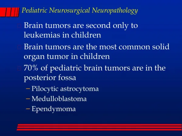
1. Pediatric Neurosurgical Neuropathology Brain tumors are second only to leukemias in children
Brain tumors are the most common solid organ tumor in children
70% of pediatric brain tumors are in the posterior fossa
Pilocytic astrocytoma
Medulloblastoma
Ependymoma
2. CNS tumors: pediatric vs. adult Adults: 70% of tumors are supratentorial
meningioma
pituitary adenoma
High grade astrocytoma
Anaplastic astrocytoma (grade III)
Glioblastoma multiforme (grade IV astrocytoma)
Pediatric: 70% in posterior fossa
pilocytic astrocytoma (cerebellar astrocytoma)
medulloblastoma
3. Brain tumors: intro Intracranial neoplasms
Primary
Secondary
Metastatic
Local invasion
Tumors of the spinal cord
4. Primary brain tumors: intro Primary brain tumors are rare
2.5% of all cancer deaths
Second most common type of tumor in children
There are over 100 different brain tumors
Most common types
Astrocytomas
Grades I-IV
Medulloblastomas
primitive neuroectodermal tumor-PNET
Meningiomas
Pituitary adenomas
5. Clinical presentation Clinical symptoms depend upon:
Age, location, and type of tumor and grade
Symptoms may include:
Increased intracranial pressure
secondary to obstruction of CSF at aqueduct
hydrocephalus (infants), headache, papilledema, vomiting
seizures
focal neurological deficits
hormonal changes (pituitary adenoma)
visual changes (diplopia, field defects)
Pituitary adenoma - pressure on optic chiasm
6. CNS tumors: diagnosis Symptoms prompt neuroimaging
CT and MRI
intra-axial vs. extra-axial
Location of tumor
contrast enhancement
typical of high grade
also in some low grade, i.e., pilocytic astrocytomas
7. CNS tumors: location Extra-axial
meningiomas
Cerebral hemispheres
grade II-III astrocytomas, GBM
Crossing corpus callosum - GBM
optic nerve - pilocytic astrocytoma (NF-1)
Sella - Pituitary adenoma
Peri-III ventricle - Pilocytic astrocytoma, GBM
8. CNS tumors: location posterior fossa (in children)
pilocytic astrocytoma
medulloblastoma
brainstem (pons)
pontine glioma (astrocytoma)
spinal cord
low-grade astrocytomas (grade I and II)
9. Pilocytic astrocytomas Most common in children
Grade I astrocytoma
Cerebellum (posterior fossa), optic nerve
Thalamic, spinal cord, cerebral
Discrete, well circumscribed mass
Often with associated cystic area
Contrast enhancing
Histologic appearance:
Biphasic: piloid cells and microcystic areas
Rosenthal fibers
no mitoses
10. Pilocytic astrocytomas Tumor of cerebellum, often with cyst, biphasic, Rosenthal fibers, piloid cells
11. Astrocytoma - high grade Astrocytoma grade II and III are very, very rare in the pediatric population
Grade IV - glioblastoma multiforme
Diffusely infiltrating glial tumor of cerebral hemispheres
Contrast enhancing tumor
Histological appearance:
Densely cellular, with marked nuclear pleomorphism
Numerous mitoses
Endothelial proliferation
Necrosis with pseudopallisading
12. Glioblastoma (grade IV) Less common in children than adults, typical pathology (necrosis with psuedopallisading)
13. Pontine glioma Diffuse expansion of pons, usually high grade astrocytoma (III-IV)
14. Medulloblastomas PNET of posterior fossa in children
Histologic appearance:
Densely cellular �small blue cell tumor�
Numerous mitoses
Apoptotic (karyorrhectic) cells
Endothelial proliferation
Necrosis
neuronal or glial differentiation
Homer Wright rosettes
GFAP positive cells
15. Medulloblastoma Mass arising in roof of fourth ventricle
Homer Wright rosettes
16. Ependymoma Mass arising in floor of fourth ventricle
Perivascular pseudorosettes
17. Meningiomas Discrete non-invasive tumor
Extra-axial, pushes into brain
Attached to dura
Hyperostosis or invasion of skull common
Histologic appearance:
Fibroblastic or menigothelial cells
Meningothelial whorls
Psammoma bodies
Rare in children, may be intraventricular (lateral ventricles)
18. Meningiomas Extra-axial tumor, meningothelial cells, whorls and psammoma bodies
19. Ganglioglioma Cerebrum, cervicomedullary, often with cystic component
Increased numbers of neurons (some binucleate) and increased glial cells (usually astrocytic)
20. Craniopharyngioma Heterogeneous, cystic mass in suprasellar region
Basiloid layer, stellate reticulum, �wet� keratin, often calcified
21. Choroid plexus papilloma Lateral ventricle in children (fourth ventricle in adults)
22. Germ cell tumors Pineal - 99% males, most are germinomas
Suprasellar - often mixed germ cell tumor, 50% female
Tertomas are rare
23. Metastatic tumors The most common �brain� tumor in adults is metastatic
Metastatic tumors are rare in children
The most common metastatic tumor in children is osteosarcoma
Local extension of malignant tumors of vertebral bodies (Ewing�s sarcoma) or paravertebral soft tissues (neuroblastoma) are not uncommon
24. Other tumors Subependymal giant cell astrocytoma (SEGA)
Intraventricular tumor in Tuberous sclerosis
Desmoplastic infantile ganglioglioma (DIG)
Superficial cerebral tumor in infants
Dysembryoplastic neuroepithelial tumor (DNET)
Hamartomatous lesion associated with seizures
Atypical teratoid rhabdoid tumor (ATR, AT/RT)
Infants, posterior fossa, very malignant
Eosinophilic granuloma
A type of Langerhans cell histiocytosis
Single discrete osteolytic lesion in skull
Meningioangiomatosis
Hamartomatous superficial cerebral lesion associated with seizures
25. Hereditary syndromes Neurofibromatosis type I
Caf�-au-lait spots
Dermatofibromas, multiple
optic nerve gliomas, bilateral
plexiform neurofibroma
Malignant peripheral nerve sheath tumor
Neurofibromatosis type II
bilateral acoustic neuroma
multiple meningiomas
ependymomas