Download

1 / 18
240 likes | 884 Vues
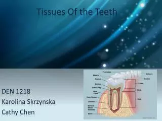
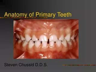
Anatomy of the Teeth. Tissues of the Tooth. Enamel – the hard tissue that covers the crown portion of the tooth (hardest substance in the body). Cementum – the substance covering the root surface of the tooth. Dentin – the material forming the main inner portion of the tooth structure.

E N D
Tissues of the Tooth • Enamel – the hard tissue that covers the crown portion of the tooth (hardest substance in the body). • Cementum – the substance covering the root surface of the tooth. • Dentin – the material forming the main inner portion of the tooth structure.
Pulp – the vital tissues of the tooth consisting of nerves, blood vessels, and connective tissue. • Pulp chamber – open area in center of tooth, found in the crown area; place for the pulpal tissues. • Pulp canal – small canal or trench area in the center of the root, containing the pulpal vessels.
Parts of a Tooth • Crown – top portion of the tooth covered by enamel. • Clinical crown - portion of the tooth that is visible in the mouth. • Anatomical crown – the total portion of the crown that is covered by the emanel.
Root – bottom part of the tooth; may be single-, double-, or triple-root. • Clinical root – portion of the root covered by the gingival (gum) tissue. • Anatomical root – total portion of the root covered by the cementum.
Cervix – neck – the neck of the tooth at the cementoenamel junction. • Apex – the anatomic area at the end of the tooth root.
Tissues surrounding the Teeth • Alveolar process – the extension of the maxilla and mandible that surrounds and supports the teeth to form the dental arches. • Alveolar bone – the part of the alveolar process that lines the bony sockets into which the roots of the teeth are embedded.
Periodontal ligament – dense connective tissue organized into fiber groups that connects the cementum covering of the root of the tooth with the alveolar bone of the socket wall.
Gingiva – the mucous membrane tissue that immediately surrounds the tooth. • Attached gingiva – the portion of the gingiva extending from the gingival margin to the alveolar mucosa. • Free gingiva – the part of the gingiva that surrounds the tooth and is not directly attached to the tooth surface.
Important Oral Cavity Structures • Labia – lips • Superior oris – upper lip • Inferior oris – lower lip • Commissure – area at the corners of the mouth where the lips meet. • Vermillion – area where the pink-red lip tissue meets the facial skin. • Philtrum – the median groove on the external surface of the upper lip.
Frenum – a tissue fold attachment that connects two parts. • Labial frenum – tissue that attaches the inside of lip to the mucous membrane in the anterior of the oral cavity. • Lingual fremun – attaches the lower side of the tongue to the floor membrane. • Buccal frenum – attaches the side of the cheeck to the oral cavity in the maxillary first molar area.
Palate – roof of the mouth • Hard palate – composed of palatine processes of the maxillae bones. • Rugae – irregular folds or bumps on the anterior surface of the hard palate. • Soft palate – flexible portion located in the posterior region of the oral cavity. • Gag reflex – protective mechanism located in the posterior region of the soft palate.
Uvula – tissue structure hanging from from the soft palate in the posterior of the oral cavity. • Retromolar pad – located at the rear of the mouth distal to the molars on the mandibular arch. • Tuberosities – rounded area on the outer surface of the maxillary bones in the area of the posterior teeth.
Surfaces of the Teeth • Facial – pertaining to the surface of the cheek and lips (face). • Labial – pertaining to the lips: anterior surface of the anterior teeth. • Buccal – pertaining to the cheek: surface of the posterior teeth touching the cheek. • Lingual – surface of the tooth or area touching the tongue.
Incisal edge – cutting edge of the anterior teeth. • Occlusal – chewing surface of the posterior teeth.
Proximal – side wall of tooth which meet with or touches the side wall of another tooth. • Mesial – to the middle: side surface closest to the middle of the face. • Distal – pertaining to the far or away side: side farthest from the midline of the face.
Enamel Cementum Dentin Pulp Pulp chamber Pulp canal Crown Clinical crown Anatomic crown Root Clinical root Anatomic root Cervix Apex Alveolar process Alveolar bone Periodontal ligament Gingiva Attached gingiva Free gingiva Labial Keywords
Superior oris Inferior oris Commissure Vermillion Philtrum Frenum Palate Hard palate Soft palate Gag reflex Uvula Retromolar pad Tuberosities Facial Labial Buccal Lingual Incisal edge Occlusal Rugae More keywords