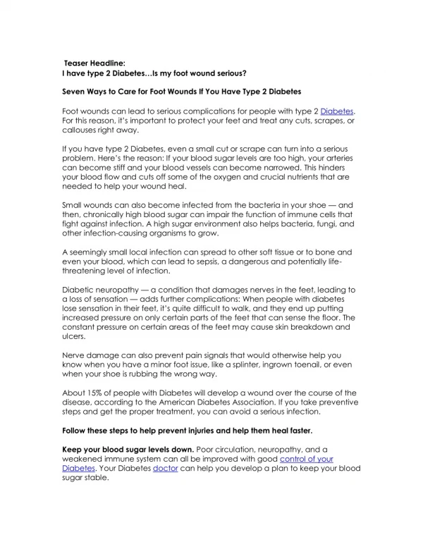
Diabetic Foot
Diabetic Foot . N. Craig Stone April 17, 2003. Introduction. Epidemiology Pathophysiology Classification Treatment. Epidemiology. DM largest cause of neuropathy in N.A. 1 million DM patients in Canada Half don’t know

Diabetic Foot
E N D
Presentation Transcript
Diabetic Foot N. Craig Stone April 17, 2003
Introduction • Epidemiology • Pathophysiology • Classification • Treatment
Epidemiology • DM largest cause of neuropathy in N.A. • 1 million DM patients in Canada • Half don’t know • Foot ulcerations is most common cause of hospital admissions for Diabetics • Expensive to treat, may lead to amputation and need for chronic institutionalized care
Epidemiology • $34,700/year (home care and social services) in amputee • After amputation 30% lose other limb in 3 years • After amputation 2/3rds die in five years • Type II can be worse • 15% of diabetic will develop a foot ulcer
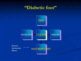
Pathophysiology • ?Vascular disease? • Neuropathy • Sensory • Motor • autonomic
Vascular Disease • 30 times more prevalent in diabetics • Diabetics get arthrosclerosis obliterans or “lead pipe arteries” • Calcification of the media • Often increased blood flow with lack of elastic properties of the arterioles • Not considered to be a primary cause of foot ulcers
Neuropathy • Changes in the vasonervorum with resulting ischemia ? cause • Increased sorbitol in feeding vessels block flow and causes nerve ischemia • Intraneural acculmulation of advanced products of glycosylation • Abnormalities of all three neurologic systems contribute to ulceration
Autonomic Neuropathy • Regulates sweating and perfusion to the limb • Loss of autonomic control inhibits thermoregulatory function and sweating • Result is dry, scaly and stiff skin that is prone to cracking and allows a portal of entry for bacteria
Motor Neuropathy • Mostly affects forefoot ulceration • Intrinsic muscle wasting – claw toes • Equinous contracture
Sensory Neuropathy • Loss of protective sensation • Starts distally and migrates proximally in “stocking” distribution • Large fibre loss – light touch and proprioception • Small fibre loss – pain and temperature • Usually a combination of the two
Sensory Neuropathy • Two mechanisms of Ulceration • Unacceptable stress few times • rock in shoe, glass, burn • Acceptable or moderate stress repeatedly • Improper shoe ware • deformity
Patient Evaluation • Medical • Vascular • Orthopedic • Identification of “Foot at Risk” • ? Our job
Patient Evaluation • Semmes-Weinstein Monofilament Aesthesiometer • 5.07 (10g) seems to be threshold • 90% of ulcer patients can’t feel it • Only helpful as a screening tool
Patient Evaluation • Medical • Optimized glucose control • Decreases by 50% chance of foot problems
Patient Evaluation • Vascular • Assessment of peripheral pulses of paramount importance • If any concern, vascular assessment • ABI (n>0.45) • Sclerotic vessels • Toe pressures (n>40-50mmHg) • TcO2 >30 mmHg • Expensive but helpful in amp. level
Patient Evaluation • Orthopedic • Ulceration • Deformity and prominences • Contractures
Patient Evaluation • X-ray • Lead pipe arteries • Bony destruction (Charcot or osteomyelitis) • Gas, F.B.’s
Patient Evaluation • Nuclear medicine • Overused • Combination Bone scan and Indium scan can be helpful in questionable cases (i.e. Normal X-rays) • Gallium scan useless in these patients • Best screen – indium – and if Positive – bone scan to differentiate between bone and soft tissue infection
Patient Evaluation • CT can be helpful in visualizing bony anatomy for abscess, extent of disease • MRI has a role instead of nuclear medicine scans in uncertain cases of osteomyelitis
Ulcer Classification • Wagner’s Classification 0 – Intact skin (impending ulcer) 1 – superficial 2 – deep to tendon bone or ligament 3- osteomyelitis 4 – gangrene of toes or forefoot 5 – gangrene of entire foot
Treatment • Patient education • Ambulation • Shoe ware • Skin and nail care • Avoiding injury • Hot water • F.B’s
Treatment • Wagner 0-2 • Total contact cast • Distributes pressure and allows patients to continue ambulation • Principles of application • Changes, Padding, removal • Antibiotics if infected
Treatment • Wagner 0-2 • Surgical if deformity present that will reulcerate • Correct deformity • exostectomy
Treatment • Wagner 3 • Excision of infected bone • Wound allowed to granulate • Grafting (skin or bone) not generally effective
Treatment • Wagner 4-5 • Amputation • ? level
Treatment • After ulcer healed • Orthopedic shoes with accommodative (custom made insert) • Education to prevent recurrence
Charcot Foot • More dramatic – less common 1% • Severe non-infective bony collapse with secondary ulceration • Two theories • Neurotraumatic • Neurovascular
Charcot Foot • Neurotraumatic • Decreased sensation + repetitive trauma = joint and bone collapse • Neurovascular • Increased blood flow → increased osteoclast activity → osteopenia → Bony collapse • Glycolization of ligaments → brittle and fail → Joint collapse
Classification • Eichenholtz • 1 – acute inflammatory process • Often mistaken for infection • 2 – coalescing phase • 3 - consolidation
Classification • Location • Forefoot, midfoot (most common) , hindfoot • Atrophic or hypertrophic • Radiographic finding • Little treatment implication
Indications for Amputation • Uncontrollable infection or sepsis • Inability to obtain a plantar grade, dry foot that can tolerate weight bearing • Non-ambulatory patient • Decision not always straightforward
Conclusion • Multi-disciplinary approach needed • Going to be an increasing problem • High morbidity and cost • Solution is probably in prevention • Most feet can be spared…at least for a while