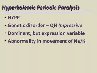
HYPERKALEMIC PERIODIC PARALYSIS
HYPERKALEMIC PERIODIC PARALYSIS. Danielle Swadberg, Brock Roberts, Sameen Singh, Chelle Wheat. Twenty years ago, a quarterhorse named “Impressive” won all of the titles of his class. He was the top-winning, top-producing Quarter Horse stallion of all time.


HYPERKALEMIC PERIODIC PARALYSIS
E N D
Presentation Transcript
HYPERKALEMIC PERIODIC PARALYSIS Danielle Swadberg, Brock Roberts, Sameen Singh, Chelle Wheat
Twenty years ago, a quarterhorse named “Impressive” won all of the titles of his class. He was the top-winning, top-producing Quarter Horse stallion of all time. As a breeding stallion, he proved himself equally deserving of his name, turning out champion after champion. Many of Impressive's offspring bore the same dramatic physical stature as their sire - they too went on to become outstanding and prolific stallions and broodmares. Of the top 15 halter horses in 1992, 13 were descendants of Impressive. Even at the age of 23, Impressive himself was fourth on the list. In 1993, it was estimated that more than 55,000 Quarter Horses, Paints, and Appaloosas bore his pedigree. From the American Quarterhorse Association
Soon his progeny were seen to be affected by a strange muscular twitching that often left them temporarily unable to move. Usually mis-diagnosed as tying-up syndrom or colic, these episodes varied widely in degree and duration....but all had one factor in common, their pedigree. As a result, many people now know HYPP by it‘s more common name: Impressive Syndrome. This turned out to be a genetic mutation that only recently has been implicated in the rare but burgeoning - and sometimes fatal - muscular disorder known as hyperkalemic periodic paralysis. This particular defect is a dominant condition, meaning that at least half of the affected horses' offspring will be affected as well. In the words of one prominent Quarter Horse trainer, this discovery was "one of the most devastating things that ever hit the horse industry."
Hyperkalemic Period Paralysis or HYPP in horses is characterized by sporadic attacks of muscle tremors, weakness and collapsing. These HYPP attacks can also involve loud breathing noises from paralysis of the muscles of the upper airway. Sudden death can occur in these horses from heart failure or respiratory muscle paralysis. Using electromyography (EMG) to measure the electrical activity present in certain muscles, the researchers discovered a wide variety of abnormalities, including spontaneous activity from muscles under no stimulation. Because these muscle tremors happen in other diseases besides HYPP, those results were not enough evidence to give an accurate diagnosis. Eventually, a mutation in a Na channel of the skeletal muscles was found, which was the official diagnosis of HYPP.
Humans also can have HPP (HyperKPP) The disorder involves attacks of muscle weakness or paralysis, alternating with periods of normal muscle function. Attacks usually begin in early childhood. Multiple daily attacks are not uncommon. Attacks typically last only 1 to 2 hours, but can sometimes last as long as a day. They tend to occur while resting after exercise or exertion. Risks include a family history of periodic paralysis. Attacks may be triggered by fasting. Attacks seldom occur during exercise but may be triggered by rest following exercise.Disorders that cause intermittent episodes of paralysis as their primary effect are uncommon. More commonly, an intermittent episode of paralysis or weakness is a symptom of another disorder. Hyperkalemic periodic paralysis occurs in approximately 1 in every 100,000 people. Men are affected more often than women and usually have more severe symptoms.
Human Symptoms • Weakness/paralysis • Most commonly located in the shoulders and hips • Arms and legs may also be involved • Occurs intermittently • May occur on awakening • May be triggered by rest after exercise • May be triggered by fasting • Usually lasts for less than 2 hours • Spontaneous recovery of normal strength between attacks • Normal alertness during attacks
HPP is caused by a flaw in a sodium channel in the muscle membrane. This flaw makes the personwith HyperKPP extremely sensitive to increases in serum potassium that wouldn't bother the average person. Anyone can be made weak by a drastic increase in serum potassium, but the person with HyperKPP gets weak with even a slight elevation in potassium level, and patients with HyperKPP may become profoundly paralyzed while their potassium levels remain well within normal limits, even when their potassium is on the lower end of normal. The human defect is also in Na channels
Weakness most commonly affects the muscles of the arms and legs but may affect the trunk as well. In a few patients the muscles involved in breathing can be affected during severe episodes. An irregular heartbeat can occur during episodes as well. Most patients have normal muscle strength between attacks, but muscle tissue can be damaged by attacks and this damage may eventually cause permanent weakness in some patients once they reach their 50s and 60s. During episodes of muscle weakness the normal flow of sodium ions is interrupted affecting the ability of the cell to contract properly. The potassium level in the blood may not rise during attacks, but many HyperKPP patients have a slightly elevated potassium level between attacks. The human defect is also in Na channels
A tablespoonful of Calcium Gluconate syrup stirred into a glass of Coca Cola or other sweet beverage has proven an effective therapy which aborts mild episodes in the early stages. Calcium Gluconate Syrup is a mineral supplement available off the shelf and is found in most pharmacies. For those who have frequent episodes and whose lives are compromised, more aggressive treatment is advisable, especially since some patients with HyperKPP may develop permanent muscle weakness after years of episodes. The carbonic anhydrase inhibitor 'Diamox' (acetazolomide) is often prescribed for HyperKPP patients. A similar drug called Daranide (diclorphenamide) is far more potent, and often works on patients who respond poorly to Diamox, or on patients who have been on Diamox many years and have become resistant to its effects. About 25% of patients do not respond to Diamox and must be put on other drugs. Treatment is to temporarily increase blood glucose or calcium, or to use K-specific diuretics
Muscles are excitable cells Voltage Gated Na+ channels
The Protein • Purpose of this Study: • EElucidate the Molecular Biology of HyperKPP • LLinker between domains III and IV • SS4-S5 Linker • SS6 segment csbn.concordia.ca/psyc358/ Lectures/voltgate.htm
Functional Processes of the Protein • Activation • This refers to the protein’s ability to open its activation gate (m gate) in response to depolarizing changes in membrane potential • This initiates the action potential when the threshold potential is reached • Studies revolve around quantifying the potential where this response occurs
Functional Processes of the Protein • Fast Inactivation • This is the ability of the protein to close its inactivation gate (h gate) in response to depolarizing changes in membrane potential • Occurs with a slight delay, so that activation and fast inactivation work in tandem to produce action potentials with short duration • Studies are aimed at quantifying the degree of depolarization required to close the h gate
Activation & Fast Inactivation csbn.concordia.ca/psyc358/ Lectures/Nachannel.htm
Functional Processes of the Protein • Slow Inactivation • Refers to the protein’s tendency to inactivate after extended depolarization • Involves unknown molecular mechanism • The potential required to inactivate, as well as the time required to recover from inactivation, are quantifiable and of interest to researchers
Activation -Depolarize to different potentials from a fixed holding potential -Each depolarization results in a quantified current -Current converted to conductance values and normalized -Determines activation of channel as function of potential 20 mV 10 mV 0 mV -10 mV -20 mV -30 mV -40 mV -50 mV -60 mV -70 mV -80 mV -120 mV 25 msec
Activation • In molecular terms, this protocol is designed to establish the potential that is required to open the activation gate • In effect, this determines the threshold potential for this ion channel
Fast Inactivation -Depolarize to fixed potentials from varied holding potentials -Each depolarization results in a quantified current -Current normalized as a fraction of peak current -Determines current as a function of holding potential 20 mV 10 mV 0 mV 0 mV -10 mV 25 msec -20 mV -30 mV -40 mV -50 mV -60 mV -70 mV -80 mV -90 mV -100 mV -110 mV -120 mV 200 msec
Fast Inactivation • This protocol is designed to determine the degree of depolarization required to close the inactivation gate • If the holding potential is sufficiently depolarized, the inactivation gate is closed • Subsequent depolarization to a potential where the activation gate is open generates no current. Why? • Answer: Inactivation gate is already closed
Slow Inactivation -Holding potential is varied, then hyperpolarized to a fixed potential, then depolarized to a fixed test potential -Varied potential is held for a much longer period of time than other protocols, and all membrane changes are sequential 10 mV 0 mV -10 mV -20 mV 20 msec -30 mV -40 mV -50 mV -60 mV -70 mV -80 mV -90 mV -100 mV -110 mV 30 msec -120 mV 50 sec
Slow Inactivation -Emphasis on Sequential Potential Changes Current measured 20 msec 20 msec -10 mV -10 mV -100 mV -100 mV 30 msec 30 msec Change in holding potential 50 sec -130 mV 50 sec
Slow Inactivation • This protocol is designed to determine the degree of depolarization necessary to invoke slow inactivation • Assumption: brief hyperpolarizations designed to remove the effect of fast inactivation have a negligible effect on slow inactivation
Slow Inactivation Recovery -Part one: fast recovery -10mV, 20 msec, current measured >15 min 50 sec 50 sec 50 sec -20 mV -100 mV -100 mV -100 mV -100 mV Increasing recovery time duration Removes fast inactivation, allows recovery
Slow Inactivation Recovery Part two: continuous slow recovery -10mV, 20 msec, current measured 50 sec -20 mV -100 mV 0.5 sec 5 sec 15 sec
Slow Inactivation Recovery • The slow inactivation recovery protocol monitors the amount of time at hyperpolarized potentials required for the slow inactivation gate to open after extended depolarization closes it • This is determined by monitoring the current elicited by rapid depolarization as a function of the amount of recovery time at hyperpolarized potentials
RESULTS • Six different tests were conducted: • Activation • Fast inactivation • Slow Inactivation • Slow Inactivation Recovery • Deactivation • Persistent Current
ACTIVATION & FAST-INACTIVATION • Results: • Slight hyperpolarizing • shift in activation curve • No accountable change • in fast inactivation Fast-inactivation Activation Neurology 2002;58:p.1270
T704 MUTATION Fast-inactivation Activation Cannon, S.C. Neuromuscular Disorders, 1997 p.244
SLOWINACTIVATION • Results: • Slow inactivation does • not occur as easily in • the mutated channel Neurology 2002;58:p.1271
SLOWINACTIVATIONRECOVERY • Results: • Slow inactivation • recovery is easier • in the mutated • channel than the • wild-type Neurology 2002;58:p.1271
DEACTIVATION • Results: • Deactivation was same • for mutated channel • and the wild-type • channel Neurology 2002;58:p.1270
PERSISTENTCURRENT • Results: • L689I mutation showed a slight current after 50ms • Wild-type showed no persistent current Neurology 2002;58:p.1269
SUMMARYOFRESULTS Test Results Conclusion
SUMMARYOFRESULTS (Cont’d) Test Results Conclusion
DISCUSSION AND CONCLUSION • In previous studies on Hyperkalemic Periodic Paralysis (hyperKPP) fast inactivation was thought to be the source of symptoms. • In fast inactivation, Na+ channels open quickly in response to depolarization and then close quickly to a fast inactivated state where openings are rare. • Fast inactivation limits the duration of action potentials and initiates repolarization of muscle fibers. • This results in slower recovery and produces a refractory period in which no action potentials are propagated • It was also found in previous studies that a transversion (bases are switched) mutation is present, which results in the substitution of a conserved amino acid.
SLOW INACTIVATION • In this paper the researchers studied slow inactivation and the mutation that disrupts the process. • Slow inactivation occurs over seconds to minutes and affects the availability of Na+ channels. • In order to study and measure slow inactivation an experimental procedure that includes prolonged depolarization is used. • This depolarization allows slow inactivation to approach steady state. • Then the fraction of channels not slow inactivated is measured as the current that recovers within 20 milliseconds at –100mV.
THE MUTATION • The mutation found is in the SCN4A gene which encodes the human skeletal muscle sodium channel in patients with hyperKPP. • The mutation is a L689I mutation and is close to the mutation I693T (paramyotonia congenita) and the frequent sodium channel mutation (T704M). • The amino acid that is located where this mutation occurs is highly conserved, which means that the same amino acid is seen in many different sodium channels, and that it is important to the function. Figure 1, pg. 1269
THE EXPERIMENT • The experiment used the whole cell patch clamp technique and looked at the expression of the mutation in human embryo kidney 293 cells. • They found a small hyperpolarizing shift in the activation curve, which resulted in the overlap of the activation and inactivation curves. • An impairment of slow inactivation was also discovered. • This hyperpolarizing shift has also been described for other related mutations in similar diseases. FAST INACTIVATION ACTIVATION Figure 3A, pg. 1270
WINDOW CURRENTS • Window current is the area of overlap between the activation and inactivation curves. • In this area, Na+ activation channels are beginning to open, and inactivation channels are beginning to close. • As the activation curve is shifted more to the hyperpolarized region, both inactivation and activation gates are open at the same time. • When this occurs window current gets larger, due to the large influx of Na+ charge down its concentration gradient.
RESULTS • They also found a faster recovery time for the mutants. • The result is that because of the overlap of the activation and inactivation curves, the mutants could produce a generation of persistent currents which they found to be at increased negative potentials. • They did not find a significant change in fast inactivation that could describe the overlap in curves. Figure 4, pg. 1271
RESULTS • The researchers found that approximately 25% of the L689I mutants failed to slow inactivate after a long depolarization (20 min). • This is similar to the behavior of other mutants and in all cases faster recovery from slow inactivation was observed, which allows for more depolarization. • The depolarization was found to prevent the generation of action potentials even by direct galvanic shock, which is what causes the episodic weakness (previous studies). • The experimental data also shows that the linker II S4-S5 is one of the molecular determinants of sodium channel slow inactivation.
DISCUSSION • Normally, in a cell, Na+ channels close at hyperpolarized potentials. When the cell is depolarized the Na+ channels open briefly and then shut to an inactive state so that there are no more openings. • The membrane must then be hyperpolarized to restore the Na+ channels to a closed state from which subsequent openings are elicited. • In hyperKPP, Na+ channels fail to inactivate and prolonged openings and depolarization result. • The result is that persistent Na+ currents are witnessed, the Na+ current is closer to the maximum, and Na+ diffuses down its gradient into the cell which results in a depolarization and a more positive membrane potential. • The current is larger because more Na+ is moving into the cell and the cell is depolarizing. • There is an overlap of the curves because the activation curve is more negative (lower threshold, less voltage required). • Increased extracellular K+ levels worsen the inactivation which in turn causes increased weakness in the patient.
DISCUSSION • Weakness in the patient is caused by the depolarization that results from Na+ moving down its concentration gradient and the positive membrane potential. • The constantly depolarized membrane does not hyperpolarize and thus can not remove inactivation from the Na+ channels and initiate an AP. • There still is a command from the motor neuron to contract the muscle which causes an AP in the motor neuron, and Ach is still secreted into the muscle cell, but the muscle is already so depolarized that it does not fire APs or contract. • K+ causes the symptoms to worsen because it causes depolarization itself. As the K+ concentration rises, the resting potential increases and will open some of the Na+ channels that do not inactivate in turn causing more depolarization. • It is a combination of these two things that make the cell too depolarized to respond to inputs.
CONCLUSIONS • The data shows that the hypothesis that hyperKPP does not require defective fast inactivation is correct and that hyperKPP could be caused by the hyperpolarizing shift in voltage dependence of activation in combination with impairment of slow inactivation. The episodic weakness that results can thus be attributed to a combination of these two defects. • The mutation that was discovered is a transversion mutation that results in the substitution of a highly conserved amino acid, showing its importance in sodium channel function. Due to the fact that slow inactivation is implicated as the cause of HyperKPP, and fast inactivation was proven to not play a role, it can be deduced that because it is a different mutation and different process, it must come from a different part of the molecule. • This study is important because it illustrates how through different functional expression, scientists can begin to discover the structure of a molecule.
CONCLUSIONS Some of the difficulties associated with this study include: It is hard to read and the researchers do not explain why they choose certain durations of time or measurements. It would be helpful if more references to chosen values were included in the paper. Lastly, it is exciting that now researchers can begin to learn about a disease at the molecular level instead of solely through symptom management. This gives hope that in the future, diseases such as Hyperkalemic Periodic Paralysis can be cured.
SOURCES Cannon, Stephen C. “From mutation to myotonia in sodium channel disorders.” Neuromuscular Disorders. 7(1997):241-249. Cannon, Stephen C. “Ion-channel defects and aberrant excitability in myotonia and periodic paralysis.” TINS. 19, #1(1996):3-10.