Understanding Lymphoproliferative Disorders: Myeloma, Leukemias, and Lymphomas
490 likes | 918 Vues
This comprehensive overview explores various lymphoproliferative disorders, focusing on multiple myeloma, plasma cell leukemia, and lymphomas, including Hodgkin’s disease. Key diagnostic features such as the presence of Bence-Jones protein, atypical plasma cells, and the classification of leukemias are analyzed. The significance of specific biomarkers like TRAP in hairy cell leukemia and the morphological classification of lymphoma cells, including Reed-Sternberg cells, is discussed. Our analysis encompasses clinical staging, histology, and recent classification systems relevant to these conditions.

Understanding Lymphoproliferative Disorders: Myeloma, Leukemias, and Lymphomas
E N D
Presentation Transcript
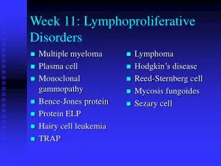
Multiple myeloma Plasma cell Monoclonal gammopathy Bence-Jones protein Protein ELP Hairy cell leukemia TRAP Lymphoma Hodgkin’s disease Reed-Sternberg cell Mycosis fungoides Sezary cell Week 11: Lymphoproliferative Disorders
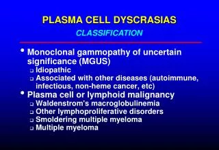
Multiple Myeloma • Plasma cell leukemia • Monoclonal gammopathy ELP pattern with sharp region spike • High ESR, rouleaux, Bence-Jones protein, Platelet dysfunction • Osteolytic (punched out) bone lesions
Other Plasma Cell Disorders • Waldenstrom’s macroglobulinemia • IgM • Soft tissue involvement • CD 20 (B cell) but no CD 21 (mature B) or PC 1 (plasma cell) • Heavy chain disease • Plasmacytoid lymphocyte
Hairy Cell Leukemia • Leukemic reticuloendotheliosis • Probably B cell origin • CD 19, 20, 22, 24 (B cell), 25, 11c (granulocyte), sIg, some CD 5 (pan-T) • Tartrate resistant acid phosphatase (TRAP)
Hairy Cell Leukemia
Acid Phosphatase Naphthol AS-BI phosphate ACP in acid pH > Free naphthol AS-BI + FastGarnet GBC Magnesium > Maroon deposits
Tartrate Resistant ACP: TRAP + Tartrate
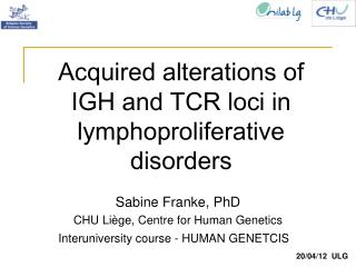
Non-Hodgkin’s Lymphoma • Lymphocytes with clefted nuclei • Sezary cells in mycosis fungoides have many clefts • Rappaport classification based on morphology and differentiation • Lukes-Collin classification with cell types identified • International Working Formulation with tumor aggressiveness and histology • Revised European/American Lymphoma (REAL) classification is most recent
WHO Classification of Acute Lymphoproliferative Syndromes • Precursor B Lymphoblastic Leukemia/Lymphoma (ALL/LBL) -- ALL in children (80-85% of childhood ALL); LBL in young adults and rare; FAB L1 or L2 blast morphology • Precursor T ALL/LBL -- 15% of childhood ALL and 25% of adult ALL • Burkitt Leukemia/Lymphoma (FAB L3)
Skin Biopsy Skin Imprint Sezary Syndrome
Hodgkin’s Lymphoma • Reed-Sternberg cell (RS): lobated nuclei, mutinucleate variants, lacunar cells, lymphocytic-histiocytic cell (LH) • RS: has CD 15 (granulo/mono) and CD 30 (B cell) • Lymphocyte predominant type CD 20 (B cell) • Clinical staging I through IV according to number and location of tumors