Download
1 / 7
70 likes | 82 Vues
<br>Hemperfusion - The Key to Acute Poisoning<br>Jafron HA230 efficiently absorbs toxin in blood and relieves the symptoms of acute poisoning. Clinical Practices prove an ideal efficacy of jafron HA230 in drug poisoning and intoxication caused by fat soluble poision or toxicant that is easily combined with plasma protein.<br>

E N D
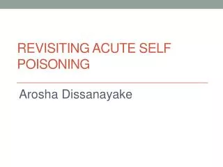
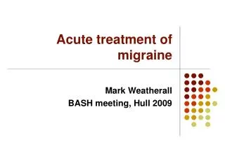
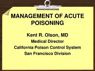
Original Paper Blood Purif 2016;42:93–99 DOI: 10.1159/000445991 Received: November 30, 2015 Accepted: April 4, 2016 Published online: May 18, 2016 Early Stage Blood Purification for Paraquat Poisoning: A Multicenter Retrospective Study An Li a Wenxiong Li b Fengtong Hao a Haishi Wang c a Department of Occupational Diseases and Poisoning Medicine, b Department of SICU, Beijing Chaoyang Hospital, Capital Medical University, Beijing , and c Department of Occupational Medicine and Clinical Toxicology, Shandong Provincial Hospital, Jinan, China Key Words Paraquat · Poisoning · Hemoperfusion · Continuous veno-venous hemofiltration · Conservative treatment SOFA score were independently associated with mortality. HP and HP + CVVH were protective factors. Conclusion: Ear- ly HP or HP + CVVH after PQ poisoning could decrease PQ blood levels, alleviate organ damage, and increase survival. © 2016 S. Karger AG, Basel Abstract Objectives: To evaluate the efficacy of conservative treat- ment vs. hemoperfusion (HP) vs. HP + continuous veno-ve- nous hemofiltration (CVVH) for acute Paraquat (PQ) poison- ing. Methods: This was a multicenter retrospective study of patients with PQ poisoning between January 2013 and June 2014. Clinical data and PQ serum levels were collected at baseline and after 24, 48, and 72 h of treatment. Results: Sev- enty-five, 65, and 43 underwent conservative treatment only (conservative treatment group), conservative treatment + HP (HP group), and conservative treatment + HP + CVVH (HP + CVVH group), respectively. PQ serum levels decreased in all groups after 72 h of treatment (p < 0.001); meanwhile, these values decreased faster in the HP and HP + CVVH groups compared with the conservative treatment group. More importantly, PQ blood levels were significantly lower in the HP + CVVH group compared with the HP group at 24 h (p < 0.05). Sequential organ failure assessment (ΔSOFA) values in the HP and HP + CVVH groups were significantly lower compared with that obtained for the conservative treatment group (p < 0.05). The 60-day survival rates were 21.3, 43.1 and 46.5%, respectively. Multivariate analysis indi- cated that age, PQ dose, admission PQ levels, and admission Introduction Paraquat (PQ) is a water-soluble organic heterocyclic herbicide (1,1-dimethyl-4,4-bipyridine cationic salt) with an apparent distribution volume of 1 liter/kg [1, 2] . PQ is very toxic to human and animals and is without any spe- cific antidote. The oral lethal dose is 1–6 g and the lethal concentration is 3 g/ml [3] . The toxicity of PQ might be due to its accumulation in the alveolar cells resulting in lipid oxidation by free radi- cals of the cell membranes in the lung, kidney, and liver, and manifesting as pulmonary hemorrhage, edema, fi- brosis, and liver and kidney damage [4] . Treatments in- clude emetics, gastric lavage, catharsis, diuretics, reduced glutathione, glucocorticoid, and organ function support [5–7] . The toxicokinetics of PQ in humans is not entirely clear, and most knowledge is obtained from animal studies. The gastrointestinal absorption rate of PQ is low and most of PQ is excreted through the feces. The peak serum concentration appears 2–4 h after oral absorp- 144.82.108.120 - 5/25/2016 1:45:17 AM UCL © 2016 S. Karger AG, Basel 0253–5068/16/0422–0093$39.50/0 Wenxiong Li Department of SICU Beijing Chaoyang Hospital, Capital Medical University Beijing 100000 (China) E-Mail lwx7115 @ sina.com Downloaded by: E-Mail karger@karger.com www.karger.com/bpu
Treatments For conservative treatment, patients received an emetic (so- dium bicarbonate solution or activated carbon suspension of 200 ml injected into the stomach); gastric lavage (15% bleaching clay suspension or 15% activated carbon suspension); catharsis (15% bleaching clay suspension or 15% activated carbon suspension, 300 ml with 20% mannitol, 250 ml); promotion of PQ excretion by fluid infusion and diuresis. Organ function supportive thera- py included oxygen supply, mechanical ventilation, expanding blood volume, and administration of vasoactive drugs to main- tain appropriate tissue perfusion and cell metabolism when nec- essary. For the HP group, conservative treatment was administered as mentioned above; central venous access was achieved by indwell- ing a double lumen catheter within 1 or 2 h after admission. The HP treatment was carried out with the HA330 neutral resin perfu- sion apparatus (Jafron Biomedical Co., Ltd., Zhuhai, China). Blood flow was set at 150–200 ml/min, and each session lasted 3–4 h. The first treatment was conducted on the day of admission, the second treatment 6–8 h later, and subsequent treatments once on days 2 and 3. Heparin was intravenously injected before HP using a load- ing dose of 0.5 mg/kg and continuous intravenous injection using a maintenance dose of 10–20 mg/h. Heparin was stopped 30 min before HP completion. For the HP + CVVH group, conservative treatment was admin- istered as mentioned above; HP lasting 3–4 h was performed on the day of admission, with CVVH immediately conducted for 72 h. Before HP and CVVH, vascular access was obtained using an 11.5F double-lumen catheter (Teleflex, Arrow, USA). The extracorpo- real circulation line and filter (AV600S, Fresenius, Germany) were washed with 100 mg heparin dissolved in 2,000 ml normal saline before CVVH. Postdilution CVVH was performed at a blood flow rate of 150–200 ml/min; ultrafiltration rate was 25–30 ml/kg/h for each new circuit. Circuits were disconnected at high prefilter (>280 mm Hg) or transmembrane (>280 mm Hg) pressure. After discon- nection, a new circuit was immediately initiated until CVVH was completed. A 1.4 m 2 polysulfone membrane filter (AV600S, Fre- senius, Germany) and CRRT device (Multifiltrate, Fresenius, Germany) were used. Replacement fluids were heated to 39 ° C; a combination buffer containing 113 mmol/l Na + , 3.0 mmol/l K + , 0.797 mmol/l Mg 2+ , 118 mmol/l Cl – , 1.5 mmol/l Ca 2+ , 10.6 mmol/l anhydrous dextrose, and bicarbonate-buffered fluids were used post-filtration, adjusted by plasma bicarbonate levels and pH ac- cording to blood gas data. Heparin was intravenously injected at a loading dose of 25–30 IU/kg, and continuous intravenous injec- tion occurred at a maintenance dose of 5–10 IU/kg/h until CVVH was completed. tion. PQ binds weakly to plasma proteins and is not re- absorbed in the renal tubules. The kidney is the organ with the highest concentration of PQ and the main or- gan responsible for PQ excretion. The clearance rate of PQ is associated with the kidney function. The lethal concentration in the lung tissues can be reached within 6 h in case of severe PQ poisoning. PQ levels in muscles are also relatively high. Lung and muscle become a res- ervoir of PQ, releasing it into the blood as the kidneys clear it [8] . Blood purification of PQ poisoning is controversial [8] . Hemoperfusion (HP) can be used to treat PQ poison- ing at the early stage [8–10] , but HP is more suitable for poisons with a large molecular mass, that are lipid soluble, and with a high protein binding rate [11] , which is not the case for PQ. On the other hand, continuous veno-venous hemofiltration (CVVH) is more efficient for clearing wa- ter soluble, small molecular poisons with a low protein binding rate [11] , but there is lack of data for the use of CVVH in PQ poisoning. Therefore, the aim of this retrospective study was to examine the efficacy of conservative treatment, HP, and HP + CVVH for the early treatment of PQ poisoning. Materials and Methods Patients This was a retrospective study of consecutive patients with acute PQ poisoning treated at the Beijing Chaoyang Hospital, The Second Affiliated Hospital of Hebei Medical University, Cangzhou People’s Hospital of Hebei Province, and Shangdong Province- Owned Hospital between January 2013 and June 2014. Inclusion criteria were as follows: (1) PQ poisoning by oral in- take; (2) aged >14; (3) admission within 24 h of PQ poisoning; and (4) urine concentration of PQ was 5–200 μg/ml by the semi-quan- titative sodium bisulfite method. Exclusion criteria were as fol- lows: (1) combined with the other poisonings; (2) hypersensitivity to heparin; (3) history of heparin-induced thrombocytopenia; (4) significant bleeding tendency; (5) history of severe diseases of the heart, lung, liver, kidney, or hematological system; (6) pregnant or lactating; (7) multiple organ failure; or (8) refusal of active therapy, for example, due to economic reasons. This study was approved by the Ethics Committees of each par- ticipating hospital. The need for individual consent was waived off by the committees because of the retrospective nature of the study. Data Collection Baseline patient data were collected including age, gender, dose of PQ, blood levels of PQ at admission, serum levels of superoxide dismutase (SOD), white blood cell counts, platelets counts, blood glucose level, serum levels of creatinine (Cr), alanine aminotrans- ferase, and amylase. The score of acute physiology and chronic health status (APACHE-II) was assessed as well. The serum levels of PQ were monitored and the score of sequential organ failure as- sessment (SOFA) was calculated at 24, 48, and 72 h after admission [8, 12] . ΔSOFA was defined as the differences between the SOFA scores at 24 and 72 h. The patients were followed up for 60 days. Mortality was recorded. Grouping The patients were classified according to the treatment they received: conservative treatment (conservative treatment group), conservative treatment + HP (HP group), or conservative treat- ment + HP + CVVH (HP + CVVH group). The selection of the treatment was made by the attending physician after discussion with the patients and their families after considering the condition of the patients and their financial status. 144.82.108.120 - 5/25/2016 1:45:17 AM UCL 94 Li/Li/Hao/Wang Blood Purif 2016;42:93–99 DOI: 10.1159/000445991 Downloaded by:
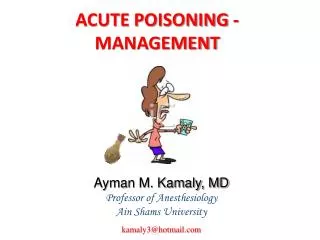
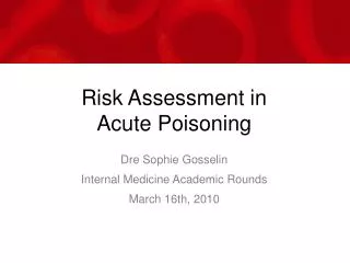
Table 1. Baseline characteristics of the patients with acute PQ poisoning according to subsequent treatments Parameters Conservative treatment (n = 75) HP treatment (n = 65) HP + CVVH treatment (n = 43) p value Gender, n (%) Man Woman Age, years Time to admission, h Dose, ml PQ blood levels at admission, μg/ml WBC, ×109/l SOD, U/g APACHE-II score SOFA score at 24 h after admission Respiratory score Coagulation score Liver function score Blood circulation score CNS function score Kidney function score Total score 35 (46.7) 40 (53.3) 36 (18–56) 12.2 (0.5–22.0) 96 (5,250) 21.6 (0.3–100.2) 14.2±6.8 177.1±51.7 3 (0–8) 36 (55.4) 29 (44.6) 36 (16–52) 7.5 (0.5–20.5) 87 (2,200) 23.0 (0.6–124.5) 16.1±9.9 200.0±77.9 3 (0–11) 25 (58.1) 18 (44.6) 38 (18–60) 7.8 (0.5–19.0) 66 (5,200) 20.8 (0.9–200.6) 15.3±5.9 177.3±23.6 3 (0–9) 0.409 0.782 0.106 0.176 0.071 0.073 0.106 0.208 1.5 (0–3) 0.1 (0–1) 0.4 (0–3) 0.9 (0–3) 0.1 (0–2) 0.4 (0–3) 3.0 (0–9) 1.7 (0–4) 0.5 (0–2) 0.5 (0–3) 1.0 (0–3) 0.1 (0–2) 0.5 (0–3) 2.9 (0–7) 0.9 (0–3) 0.6 (0–3) 0.7 (0–3) 0.9 (0–3) 0.1 (0–2) 0.2 (0–3) 3.1 (0–10) 0.732 0.021 0.414 0.732 0.526 0.927 0.212 WBC = White blood cells. Data are presented as median (minimum–maximum) or as mean ± SD. CVVH groups, respectively. There were no differences among the 3 groups in terms of gender distribution, age, time to admission (time elapsed from PQ poisoning to hospital admission), dose of PQ, blood levels of PQ at ad- mission, serum levels of SOD, APACHE-II score, white blood cell counts, and SOFA score at admission (all p > 0.05; table 1 ). Serum Levels of PQ Total blood samples were obtained with heparin anti-coagula- tion, stored at –80 ° C, and sent on ice for detection within 72 h. Quantitative and qualitative analysis of PQ in blood was per- formed according to previously published methods [13] . Statistical Analysis Continuous data were tested for normality using the Kol- mogorov–Smirnov test. Normally distributed continuous data are presented as mean ± SD and were analyzed using analysis of vari- ance (ANOVA) with the Student–Newman–Keuls post hoc test or repeated measures ANOVA with Bonferroni post hoc test, as ap- propriate. Non-normally distributed continuous data are presented as me- dian (range) and were analyzed using the Kruskal–Wallis test. Cat- egorical data are presented as frequencies and were analyzed using the chi-square test. The risk factors for death and prognosis were assessed by univariate and multivariate non-conditional logistic re- gression. The survival curves were made by the Kaplan–Meier method and compared using the log-rank test. Statistical analysis was performed using SPSS 18.0 (IBM, Armonk, N.Y., USA). Two- sided p values <0.05 were considered statistically significant. Blood Levels of PQ There were no significant differences among the 3 groups at admission (p > 0.05; table 2 ). Blood levels of PQ were significantly lower in the HP and HP + CVVH groups at 24, 48, and 72 h after admission in comparison with the conservative treatment group (p < 0.05); in addition, PQ blood levels were significantly lower in the HP + CVVH group compared with the HP group at 24 h (p < 0.05). The blood levels of PQ were decreased in all 3 groups after 3 days of treatment, though the blood levels of PQ were lower in the HP and HP + CVVH groups 72 h after admission com- pared with the conservative treatment group (p < 0.05). SOFA Scores SOFA scores at admission and at 24, 48, and 72 h after admission are presented in table 3 . There were no signif- icant differences among the 3 groups regarding SOFA scores at 24 h (p > 0.05). ΔSOFA values in the HP and Results Characteristics of the Patients Seventy-five (41.0%), 65 (35.5%), and 43 (23.5%) pa- tients were in the conservative treatment, HP, and HP + 144.82.108.120 - 5/25/2016 1:45:17 AM UCL Blood Purification for PQ Poisoning 95 Blood Purif 2016;42:93–99 DOI: 10.1159/000445991 Downloaded by:
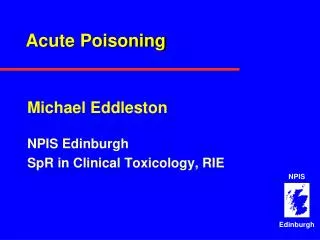
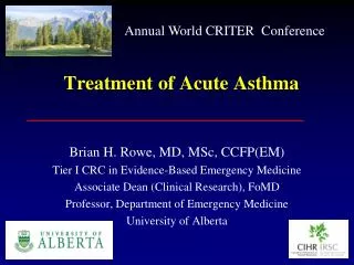
Table 2. Blood levels of PQ comparison at baseline and during treatment Group n Baseline 24 h 48 h 72 h p value 16.71±8.35a 7.84±3.63*, a 4.95±2.81*, #, a <0.001 10.33±6.67a, b 4.54±2.58*, a, b 3.91±1.89*, a, b <0.001 6.02±3.29a–c 2.50±1.34a–c 2.11±1.67a–c <0.001 Conservative HP HP + CVVH p value 75 65 43 – 21.56±11.17 22.95±10.41 20.82±9.26 0.553 <0.001 <0.001 <0.001 All data are presented as mean ± SD, μg/ml. * p < 0.05 vs. conservative treatment; # p < 0.05 vs. HP; a p < 0.05 vs. before baseline; b p < 0.05 vs. 24 h; c p < 0.05 vs. 48 h. Table 3. SOFA score during treatment Conservative treatment (n = 75) HP treatment (n = 65) HP + CVVH (n = 43) p value 24 h 48 h 72 h ΔSOFA 3.03±2.06 4.07±2.72 5.76±3.29 3.34±1.69 2.97±1.82 4.54±3.22 4.43±3.1* 1.72±1.05* 3.06±2.04 4.81±3.70 4.93±3.57* 2.11±1.28* 0.763 0.759 <0.001 <0.001 All data are presented as mean ± SD, score. * p < 0.05 vs. conservative treatment. HP + CVVH groups were significantly lower than in the conservative treatment group (p < 0.05), but there were no significant differences between the HP and HP + CVVH groups (p > 0.05). with the conservative treatment group, but without dif- ference between the HP and HP + CVVH groups (p = 0.535). Analysis of Risk Factors for Death Tables 4 and 5 present univariate and multivariate analyses of factors associated with 60-day mortality, re- spectively. Factors significantly associated with mortality in univariate analyses ( table 4 ) were included in the mul- tivariate model. Results showed that age (OR 1.128, 95% CI 1.030–1.235), PQ dose (OR 1.076, 95% CI 1.020– 1.136), PQ serum levels at admission (OR 1.539, 95% CI 1.142–2.073), and SOFA score at admission (OR 8.073, 95% CI 1.515–43.006) were independently associated with mortality, while HP (OR 0.316, 95% CI 0.119–0.838) and HP + CVVH (OR 0.297, 95% CI 0.111–0.795) were protective factors ( table 5 ). Survival The 3-day survival rates were 86.0 and 65.1% for the HP and HP + CVVH groups, and were significantly high- er than those of the conservative treatment group (56.0%) (p < 0.05). The 7-day survival rates were 56.9 and 65.1% for the HP and HP + CVVH groups, and were signifi- cantly higher than those of the conservative treatment group (41.3%) (p < 0.05). The 60-day survival rates were 43.1 and 46.5% for the HP and HP + CVVH groups, and were significantly higher than those of the conservative treatment group (21.3%, p < 0.05). However, there were no significant differences between the HP and HP + CVVH groups at any time point (p > 0.05). The 60-day survival curve is shown in figure 1 . Median survival times were 3, 15, and 40 days for the conservative treatment, HP, and HP + CVVH groups, suggesting that blood purification could significantly improve survival. Median survival was significantly longer in the HP (p = 0.003) and HP + CVVH (p = 0.001) groups compared Discussion This study showed that conservative, HP, and HP + CVVH treatments all decreased PQ serum levels and im- proved SOFA scores. HP and HP + CVVH treatment 144.82.108.120 - 5/25/2016 1:45:17 AM UCL 96 Li/Li/Hao/Wang Blood Purif 2016;42:93–99 DOI: 10.1159/000445991 Downloaded by:
Table 4. Univariate analysis of factors involved in mortality after acute PQ poisoning HP + CVVH 1.0 HP Parameters OR 95% CI lowe r p value Conservative 0.8 upper Gender Age Time to admission Dose Times of blood perfusion CVVH times PQ blood levels at admission 1.240 SOD Blood purification (control) HP HP + CVVH SOFA score at 1st day WBC Platelets Neutrophils Blood glucose Blood Cr ALT Hemodlastase 1.347 1.055 0.955 1.031 0.879 0.823 0.741 1.032 0.921 1.020 0.691 0.441 1.135 0.986 2.446 1.079 0.990 1.043 1.119 1.536 1.354 1.001 0.328 <0.001 0.012 <0.001 0.295 0.541 <0.001 0.099 0.006 0.006 0.005 <0.001 <0.001 0.509 0.536 <0.001 0.776 0.176 0.889 0.6 Survival (%) 0.4 0.2 0.994 0.358 0.312 2.015 0.908 0.999 1.008 0.817 0.999 0.994 1.000 0.171 0.138 1.545 0.864 0.995 0.982 0.737 0.995 0.985 0.998 0.750 0.705 2.629 0.953 1.003 1.035 0.905 1.003 1.003 1.002 0 0 10 20 30 40 50 60 Time (days) Fig. 1. Sixty-day survival according to treatment after acute PQ poisoning. were better than the conservative therapy; HP and HP + CVVH groups were similar in most parameters studies. However, PQ blood levels were significantly lower in the HP + CVVH group compared with the HP group at 24 h. Multivariate analysis indicated that age, PQ dose, PQ se- rum levels at admission, and SOFA score at admission were independently associated with mortality, while HP and HP + CVVH were protective factors. These results suggest that early HP or HP + CVVH after PQ poisoning could decrease PQ blood levels, alleviate organ damage, and improve survival. PQ undergoes almost no biological transformation in vivo and 90% is excreted through the kidneys. PQ uptake by alveolar epithelial cells is an energy-dependent active process, and the peak concentration in lungs can be reached within 15 h, up to concentrations as high as 10– 90 times the serum levels [8, 12] . Therefore, lung and muscle tissues are regarded as reservoirs for PQ, which is released into the blood once the plateau is reached. Acute respiratory distress syndrome was reported to occur in patients with acute PQ poisoning, and the mortality rate can be as high as 50–80% [6] . Hemodialysis (HD), HP, and CVVH are commonly used blood purification meth- ods for PQ poisoning. It is widely accepted that HP is more effective than HD for PQ poisoning; indeed, the clearance rate of HP is about 5–7 times that of HD [1] , and HP is still effective when blood levels of PQ are low- ered to <0.2 mg/l [14] . WBC = White blood cells; ALT = alanine aminotransferase. Table 5. Multivariate analysis of factors involved in mortality after acute PQ poisoning Parameters OR 95% CI low er upper p value Age Time to admission Dose PQ blood levels at admission 1.539 Blood purification (control) HP HP + CVVH SOFA score at 1st day WBC Blood glucose 1.128 0.923 1.076 1.030 0.772 1.020 1.142 1.235 1.104 1.136 2.073 0.009 0.380 0.007 0.005 0.025 0.021 0.016 0.014 0.451 0.868 0.316 0.297 8.073 1.076 1.023 0.119 0.111 1.515 0.889 0.783 0.838 0.795 43.006 1.304 1.337 WBC = White blood cells. However, PQ is a small water soluble molecule with a low protein binding rate, and secondary distribution can be observed. Theoretically, clearance effectiveness of CVVH is superior to that of HP. Therefore, in this study, results indicated that the blood levels of PQ were signifi- cantly decreased within the first 3 days after admission, 144.82.108.120 - 5/25/2016 1:45:17 AM UCL Blood Purification for PQ Poisoning 97 Blood Purif 2016;42:93–99 DOI: 10.1159/000445991 Downloaded by:
and that the blood levels of PQ were significantly lower in the blood purification groups (HP and HP + CVVH) compared to the conservative treatment group at 24, 48, and 72 h after admission, suggesting that blood purifica- tion could rapidly reduce the blood levels of PQ, which is supported by Pond et al. [12] . Multivariate analysis showed that HP and HP + CVVH treatments were associ- ated with lower mortality. Nevertheless, no matter the blood purification method (HP, CVVH, or HD), the sooner the blood is purified, the better the outcome. It was reported that though HD and HP could decrease the severity of PQ poisoning and pro- long survival, mortality was not decreased [8, 15] . This might be ascribed to the fact that lethal amounts of PQ had started entering into the alveolar epithelial cells and major organs, with blood purification not able to alter the toxicokinetics of PQ in these conditions [8, 12] . The sec- ondary distribution of PQ from lung and muscle tissues to blood circulation could be observed once the blood levels started to decrease; therefore, the timing and dura- tion of blood purification may be very important [16, 17] . Generally, blood purification is recommended to be per- formed as soon as 4–12 h after PQ poisoning, and the earlier the better [16, 17] . Since it takes time for PQ to diffuse from tissues to blood, it might be necessary to con- tinue blood purification [1] . HP was conducted only once on the day of admission in the HP + CVVH group, and then CVVH was immedi- ately performed for 72 h, while HP was carried out once a day within the first 3 days of admission in the HP group. Finally, this study indicated that the PQ clearance rate was equivalent between HP and HP + CVVH, except for the first 24 h, where a faster rate was obtained in the HP + CVVH group; in addition, no significant differences in 60-day survival were observed. Compared to HP, HP + CVVH has some merits [17] : (1) PQ is removed continu- ously to maintain hemodynamics; (2) CVVH corrects the imbalances in water, electrolytes, and acid-base equilib- rium; and (3) CVVH continuously removes metabolites such as urea and Cr. Since HP cannot correct water, elec- trolyte, and acid-base equilibrium imbalances, HP + CVVH should be prioritized for treating PQ poisoning. However, randomized control studies are needed to eval- uate the effects of CVVH alone on PQ poisoning. tients, a bias could have been introduced leading to more wealthy and healthy patients to undergo HP + CVVH. Fourth, urine concentrations of PQ in the study popula- tion were 5–200 μg/ml; therefore, the study results could not be extrapolated for urine concentrations of PQ >200 μg/ml observed in severe PQ poisoning. Fifth, time to ad- mission was higher in the conservative treatment group compared with values obtained for the HP and HP + CVVH groups, although the differences were not statisti- cally significant; whether this has contributed to our re- sults merits further assessment. Finally, the toxicokinetics of PQ is still poorly understood, limiting the application of treatments. Additional studies are still necessary to de- termine the best course of action for acute PQ poisoning. Conclusion HP or HP + CVVH could rapidly decrease the blood levels of PQ at the early stage, alleviate organ injuries, and improve survival. The therapeutic effect was mostly equivalent between HP and HP + CVVH treatment re- garding to the acute PQ poisoning, especially after the first day of treatment. Age, dose, blood levels of PQ at admission, and SOFA score at 24 h after admission were independent risk factors for mortality, while HP and HP + CVVH were independent protective factors. Acknowledgments The authors acknowledge the dedicated help of Professor W. Li, director of Surgical ICU, Chaoyang Hospital, Beijing, China, for his guidance and efforts on the study design, implementation, and manuscript preparation; the funding support from Dr. F. Hao, director of Occupational Diseases and Poisoning Medicine; and the considerable assistance of Mr. H. Wang, lecture in Shangdong Province-owned hospital during the 2-year information collection for the study. Funding The study was funded by the special research project funding (No. 201202006-06) for public welfare from Division of Science and Education, National Health and Family Planning Commis- sion of the People’s Republic of China and National Science and Technology major projects and significant new drug creation (2014ZX09J15104002) Limitations This study does have some limitations. First, the sample size was relatively small. Second, this was a retrospective study with all the inherent biases and limitations. Third, given the limitation of financial resources of some pa- Disclosure Statement The authors declare that they have no conflict of interests. 144.82.108.120 - 5/25/2016 1:45:17 AM UCL 98 Li/Li/Hao/Wang Blood Purif 2016;42:93–99 DOI: 10.1159/000445991 Downloaded by:
References 1 Feinfeld DA, Rosenberg JW, Winchester JF: Three controversial issues in extracorporeal toxin removal. Semin Dial 2006; 2 Goldfarb DS: Goldfrank’s Toxicologic Emer- gencies, ed 9. New York, McGraw-Hill, 2010. 3 Hwang KY, Lee EY, Hong SY: Paraquat in- toxication in Korea. Arch Environ Health 2002; 4 Kang MS, Gil HW, Yang JO, Lee EY, Hong SY: Comparison between kidney and hemo- perfusion for paraquat elimination. J Korean Med Sci 2009; 5 Sabzghabaee AM, Eizadi-Mood N, Montaz- eri K, Yaraghi A, Golabi M: Fatality in para- quat poisoning. Singapore Med J 2010; 496–500. 6 Hagiwara S, Iwasaka H, Matsumoto S, No- guchi T: An antisense oligonucleotide to HSP47 inhibits paraquat-induced pulmo- nary fibrosis in rats. Toxicology 2007; 199–207. 7 Dinis-Oliveira RJ, de Pinho PG, Santos L, Teixeira H, Magalhães T, Santos A, de Lourdes Bastos M, Remião F, Duarte JA, Carvalho F: Postmortem analyses unveil the poor efficacy of decontamination, anti-inflammatory and immunosuppressive therapies in paraquat human intoxications. PLoS One 2009; e7149. 8 Wunnapuk K, Mohammed F, Gawarammana I, Liu X, Verbeeck RK, Buckley NA, Roberts MS, Musuamba FT: Prediction of paraquat exposure and toxicity in clinically ill poisoned patients: a model based approach. Br J Clin Pharmacol 2014; 9 Ghannoum M, Nolin TD, Lavergne V, Hoff- man RS; EXTRIP Workgroup: Blood purifi- cation in toxicology: nephrology’s ugly duck- ling. Adv Chronic Kidney Dis 2011; 166. 10 Yang TS, Chang YL, Yen CK: Haemoperfu- sion treatment in pigs experimentally intoxi- cated by paraquat. Hum Exp Toxicol 1997; 709–715. 11 Ahmad S: Manual of Clinical Dialysis, ed 2. New York, Springer Science+Business, 2009. 12 Pond SM, Rivory LP, Hampson EC, Roberts MS: Kinetics of toxic doses of paraquat and the effects of hemoperfusion in the dog. J Tox- icol Clin Toxicol 1993; 13 de Almeida RM, Yonamine M: Gas chro- matographic-mass spectrometric method for the determination of the herbicides paraquat and diquat in plasma and urine samples. J Chromatogr B Analyt Technol Biomed Life Sci 2007; 14 Hsu CW, Lin JL, Lin-Tan DT, Chen KH, Yen TH, Wu MS, Lin SC: Early hemoperfusion may improve survival of severely paraquat- poisoned patients. PLoS One 2012; 15 Hampson EC, Effeney DJ, Pond SM: Efficacy of single or repeated hemoperfusion in a ca- nine model of paraquat poisoning. J Pharma- col Exp Ther 1990; 16 Senarathna L, Eddleston M, Wilks MF, Wool- len BH, Tomenson JA, Roberts DM, Buckley NA: Prediction of outcome after paraquat poisoning by measurement of the plasma paraquat concentration. QJM 2009; 259. 17 Koo JR, Kim JC, Yoon JW, Kim GH, Jeon RW, Kim HJ, Chae DW, Noh JW: Failure of con- tinuous venovenous hemofiltration to pre- vent death in paraquat poisoning. Am J Kid- ney Dis 2002; 19: 358–362. 4: 853: 260–264. 57: 162–166. 78: 855–866. 7:e48397. 24(suppl):S156–S160. 18: 160– 254: 732–740. 51: 16: 102: 251– 236: 31: 229–246. 39: 55–59. 144.82.108.120 - 5/25/2016 1:45:17 AM UCL Blood Purification for PQ Poisoning 99 Blood Purif 2016;42:93–99 DOI: 10.1159/000445991 Downloaded by: