
Abstract
AlphaLISA ® for the Quantitative Detection of Histone Acetylation Upon Inhibitor Treatment Steven Blasutti, Ian Jaworski, Marjolaine Roy, Sara Howland, Gabriella Szekely-Klepser 1 , Sophie Dahan, and Francesco Lipari 1 Assay Designs Inc. Abstract. AlphaLISA Assay Principle.

Abstract
E N D
Presentation Transcript
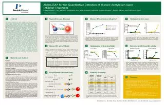
AlphaLISA® for the Quantitative Detection of Histone Acetylation Upon Inhibitor Treatment Steven Blasutti, Ian Jaworski, Marjolaine Roy, Sara Howland, Gabriella Szekely-Klepser1, Sophie Dahan, and Francesco Lipari 1 Assay Designs Inc. Abstract AlphaLISA Assay Principle Optimized in vitro Assay Histone H3 Acetylation with pCAF 1 3 9 6 The dynamic equilibrium of lysine acetylation of histones in vivo is governed by the opposing activity of acetyltransferases and deacetylases. Certain disease processes have been linked to abnormalities in the acetylating/deacetylating events and the corresponding enzymes have become important therapeutic targets in recent years. Specific histone modification inhibitors can also help biologists in studying the complex role of histone post-translational modifications. There are no tools available for high-throughput screening of potential histone acetylation/deacetylation inhibitors. The objective was to develop a high-throughput assay to quantitate the level of acetylation upon treatment with histone modification inhibitors. Acetylation of histone H3 lysine (Lys) 9 was chosen as a model system. Initial experiments with purified histone and AlphaLISA® immunoassay technology demonstrated that the histone was not stable and interacted non-specifically with the antibody and/or bead reagents. The assay involved two steps: 1. acetylation of the histone with pCAF and 2. detection using the immunoassay. First, the reaction with pCAF was optimized so that the histone was exposed to low salt conditions for a minimal time to decrease denaturation of the histone. Next, the buffer conditions for the immunoassay were optimized to eliminate non-specific interactions. The assay conditions were changed to pH 8.5 and 0.5 M NaCl to obtain a specific signal. Using the optimized acetylation and detection conditions, an assay was developed to measure histone acetylation at Lys9 using as little as 0.3 nM histone. The assay was used to detect acetylated histone H3 derived from HeLa cells that were treated with sodium butyrate. Therefore, the assay was used to monitor histone acetylation in vitro or to detect acetylated histone H3 in nuclear extracts. Future experiments will aim at developing an all-in-one well assay, whereby cells can be treated with histone modification inhibitors, lysed, and then histone modification is quantitatively detected all in the same well. These tools will speed up the discovery of histone acetylation inhibitors. The biotinylated anti-analyte antibody binds to the Streptavidin-coated Donor beads while another anti-analyte antibody is conjugated to AlphaLISA Acceptor beads. In the presence of the analyte, the beads come into close proximity. The excitation of the Donor beads provokes the release of singlet oxygen molecules that triggers a cascade of energy transfer to the Acceptor beads resulting in a sharp peak of light emission at 615 nm. The reaction of pCAF and histone H3 were optimized using the Assay Designs acetyl transferase kit, which measures the release of CoA. Different buffers, as well as enzyme and substrate concentrations, were tested. A time course of the optimized reaction is illustrated. A very high maximum signal and a good signal to background (S/B) ratio were obtained at the 30 nM concentration. At 0.3 nM histone, there was a significant difference between the control and acetylated histone signal. Histone H3 : pCAF Model Optimization of Detection Buffer Detecting Ac-H3 from HeLa Cells 4 7 10 Low NaCl High NaCl Sodium butyrate Trichostatin A pCAF (p300/CBP-associated factor) belongs to the GCN5 family of acetyltransferases. It is a transcriptional coactivator and specifically acetylates histone H3 N-terminal tail in vitro at Lys9 and 14 (Schiltz, 1999; Sterner, 2000). The enzyme associates with other proteins such as p300/CBP to modulate transcription. The in vitro experiments herein were performed using a purified recombinant pCAF enzyme and purified recombinant human histone H3. Materials and Methods 2 pCAF pCAF Materials: Acetyltransferase Activity Kits and anti-Histone H3 Acetyl-Lys9 were from Assay Designs. Anti-Histone H3 (C-terminus) was from CST. Biotin-acetyl-histone H3 peptide, pCAF, and histones acid extracted from HeLa cells were from Millipore. Human Histone H3.1 was from NEB. Nuclear extraction kit was from Pierce. White OptiPlate-384, TopSeal-A, EnVision® Multilabel Reader, AlphaLISA® Acceptor beads, and Streptavidin Donor Beads were from PerkinElmer Inc. Histone H3 Acetylation: The acetylation reaction contained pCAF, Histone H3, and acetyl CoA at final concentrations of 10 nM, 1.5 µM, and 25 µM, respectively, in 0.1 M HEPES, pH 7.5, and 0.1% Tween-20. The negative control contained equivalent reagents, but lacked cofactor acetyl CoA. The enzyme reaction was incubated at 30°C for 30 min. Assay Designs Fluorescence Assay: The acetylation reaction was monitored using a fluorescence assay. The enzyme reaction was stopped at 0, 15, 30, and 60 min by adding 5 µL of enzyme reaction to 10µL of isopropanol. After 60 min, 10 µL of 1X Assay Designs detection solution was added to each well and the plate was incubated at 23°C for 10 min. The plate was read using the EnVison Multilabel Reader with an excitation wavelength of 380 nm and an emission wavelength of 520 nm. AlphaLISA Competition Assay: To aid in optimizing the AlphaLISA protocol, a competition assay was performed. Biotinylated H3 peptide (Acetylated at Lys9) (6 nM final) was allowed to react with SA Donor beads (40 mg/mL final) and anti-Histone H3 Acetyl-Lys9 antibody (1 nM final) bound to Protein A Acceptor beads (10 mg/mL final). The reaction was competed with recombinant histone H3, with or without previous pCAF acetylation. The signal was detected using the EnVision Multilabel Reader. AlphaLISA Immunoassay Protocol: The optimized AlphaLISA immunoassay was initiated by combining biotinylated anti-Histone H3 Acetyl-Lys9 (1 nM final) and histone sample in detection buffer (50 mM Tris, pH 8.5, 0.5 M NaCl). The mixture was incubated 30 min at RT and then anti-Histone H3 (C-terminus)-conjugated Acceptor beads (10 µg/mL final) were added and then incubated 30 min at RT. Streptavidin coated donor beads (40µg/mL final) were added to give a final volume of 50 µL, and incubated 60 min at RT. The assays were performed in white OptiPlate-384 and the signal was detected using the EnVision Multilabel Reader. ARTKQTARKS TGGKAPRKQL ATKAARKSAP ATGGVKKPHR YRPGTVALRE IRRYQKSTEL LIRKLPFQRL VREIAQDFKT DLRFQSSAVM ALQEACEAYL VGLFEDTNLC AIHAKRVTIM PKDIQLARRI RGERA135 1 Above are illustrated the competition curves before and after buffer optimization. The binding of the anti-acetyl antibody to the acetyl-histone peptide was competed by both acetylated or unmodified histone (low NaCl), indicating that non-specific interactions were occurring. The addition of 0.5 M NaCl to the detection buffer was critical to decrease the non-specific interactions, so that only acetyl-histone competed the binding (high NaCl). Histone hyperacetylation could be measured in inhibitor-treated HeLa cells. HeLa cells were treated with either sodium butyrate or trichostatin A (TSA). In the sodium butyrate experiment, the histones were acid extracted from nucleosomes, whereas for the TSA experiment nuclear fractions were prepared by cell lysis, separation of the cytoplasmic proteins from the nuclei, and lysis of the nuclei. The extracts from control or treated cells were then assayed using the optimized immunoassay. Histone H3 sequence with pCAF acetylation sites. Acetyl-Histone Detection Assay Antibody Screening 5 8 Histone Summary 11 pCAF + AcetylCoA pCAF + CoA Ac-Histone A homogeneous bead-based assay was developed to measure acetylated histone H3 at Lys9. Acetylated histone H3 can be measured using purified recombinant histone or in cellular extracts. Proper optimization of assay buffer conditions allowed for sensitive and specific detection of the acetylated protein. References Kim, T.Y., Bang, Y.J., Robertson KD. (2006) Epigenetics. 1, 14-23. Schiltz, R.L., Mizzen, C.A., Vassilev, A., Cook, R.G., Allis, C.D., Nakatani, Y. (1999) J. Biol. Chem. 274, 1189-92. Sterner, D.E., Berger, S.L. (2000) Microbiol. Mol. Biol. Rev. 64, 435-59. Biotin-anti-Ac-Lys9 b-anti-Ac-Lys9 : Ac-Histone anti-H3 beads b-anti-Ac-Lys9 : Ac-Histone : anti-H3 beads SA Donor beads Five different antibodies (labelled A – E) were screened to find the configuration providing the highest signal and signal to background ratio. SA Donor beads : b-anti-Ac-Lys9 : Ac-Histone : anti-H3 beads


