Guidelines for Effective Sample Handling in Coagulation Analyses
130 likes | 249 Vues
This guide provides essential methodologies for the preparation and handling of laboratory samples for coagulation analyses, as outlined by Anne-Mette Hvas from Aarhus University Hospital. Key steps include patient preparation, specimen collection techniques including venipuncture and vascular access device use, appropriate tube selection with anticoagulants, specimen transportation protocols to prevent hemolysis, and optimal storage conditions. Special considerations for timing, posture, and pre-collection protocols are emphasized to ensure accurate and reliable results in coagulation laboratory testing.

Guidelines for Effective Sample Handling in Coagulation Analyses
E N D
Presentation Transcript
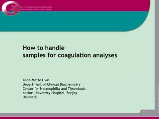
How to handle samples for coagulation analyses Anne-Mette Hvas Department of Clinical Biochemistry Center for Haemophilia and Thrombosis Aarhus University Hospital, Skejby Denmark
For the next 15 minutes • Preparation of the patient • Specimen collection • Venipuncture • Vascular access device • Tubes and anticoagulant • Specimen transport and processing • Sample Storage
Preparation of the patient – the ideal Standardized time of the day Overnight fast or a light meal No alchohol during the No coffee during the past 18-24 h past 1 h No smoking during the 20 minutes rest past 1 h before blood sampling Specified posture (sitting/lying)
Preparation of the patient – possible and acceptable Routine laboratory PT/INR, APTT No preparation of the patient
Preparation of the patient – possible and acceptable • Routine laboratory • PT/INR, APTT No preparation of the patient • Specialised coagulation laboratory • Coagulation factors, natural anticoagulants, No preparation of the patient • von Willebrand factor • Markers of fibrinolysis Standardized time of the day • Tissue-type plasminogen activator (t-PA)Overnight fast or a light meal • Plasminogen activator inhibitor (PAI-1)No alchohol during the past 18-24 h • Plasminogen activityNo coffee during the past 1 h • Plasmin-inhibitorNo smoking during the past 1 h • Specified posture (sitting/lying) • 20 minutes rest before blood sampling
Specimen collection - what is the problem? As soon as the blood vessel is entered: • Tissue factor is released activation of extrinsic pathway • Platelets are activated • Coagulation factors are activated • Natural anticoagulants are activated
Specimen collection • Venipuncture • Needle gauge from 21 to 19 • Max 25 mL (20-gauge needle), max 50 mL (19-gauge needle) • Torniquet application for max 1 min with minimal stasis • Collected directly into the tube containing the anticoagulant • Discard the first 5 mL collected (except for PT/INR or APTT) • The tube must be correctly filled
Specimen collection • Vascular access device • Avoid air leaks • Avoid heparin contamination and specimen dilution • Flush the line with 5 mL saline and discard the first 5 mL or six dead space volumes
Specimen collection • Tubes and anticoagulant • Tubes of non-reactive materials (polypropylene or siliconized glass) • 3.2% trisodium citrate • The proportion of blood:trisodium citrate are usually 9:1
Specimen transport and processing • Transport • Vibration might cause haemolysis and activation of coagulation • Ideally, tubes should be held upright during transport • Processing – centrifugation • To obtain platelet poor plasma (platelet count < 10 x 109/L)
Main documentation • National Committee for Clinical Laboratory Standards. Fourth edition 2003. Document H21-A3 • Woodhams et al. Blood Coagulation and Fibrinolysis 2001;12:229-236 • Laboratory Techniques in Thrombosis. A Manual. 2nd revised edition of ECAT Assay Procedures Edited by J Jespersen, RM Bertina and F Haverkate