CHAPTER 28 Nervous Systems
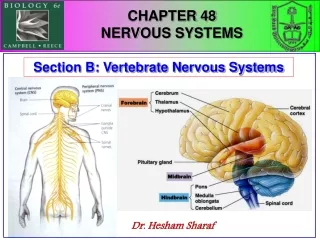
CHAPTER 28 Nervous Systems. Can an Injured Spinal Cord Be Fixed?. The spinal cord is the central communication conduit between the brain and the body It consists of a bundle of nerves. Spinal cord injury disrupts communication between the central nervous system and the rest of the body.

CHAPTER 28 Nervous Systems
E N D
Presentation Transcript
Can an Injured Spinal Cord Be Fixed? • The spinal cord is the central communication conduit between the brain and the body • It consists of a bundle of nerves
Spinal cord injury disrupts communication between the central nervous system and the rest of the body • Paraplegia is paralysis of the lower half of the body • Quadriplegia is paralysis from the neck down • Research on nerve cells is leading to new therapies
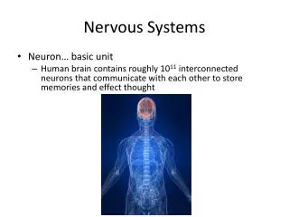
NERVOUS SYSTEM STRUCTURE AND FUNCTION 28.1 Nervous systems receive sensory input, interpret it, and send out appropriate commands • The nervous system has three interconnected functions • Sensory input • Integration • Motor output
SENSORY INPUT INTEGRATION Sensory receptor MOTOR OUTPUT Brain and spinal cord Effector Peripheral nervoussystem (PNS) Central nervoussystem (CNS) Figure 28.1A
The central nervous system (CNS) consists of the brain and, in vertebrates, the spinal cord • The peripheral nervous system (PNS) is made up of nerves and ganglia that carry signals into and out of the CNS • The nervous system can be divided into two main divisions
Sensory neurons convey signals from sensory receptors into the CNS • Interneurons integrate data and relay signals • Motor neurons convey signals to effectors • Three types of neurons correspond to the nervous system’s three main functions
1 Sensoryreceptor 2 Sensory neuron Brain Ganglion 3 Motorneuron Spinalcord 4 Quadricepsmuscles Interneuron CNS Nerve Flexormuscles PNS Figure 28.1B
28.2 Neurons are the functional units of nervous systems • Neurons are cells specialized to transmit nervous impulses • They consist of • a cell body • dendrites (highly branched fibers) • an axon (long fiber)
The myelin sheath is the insulating material in vertebrates • It is composed of a chain of Schwann cells linked by nodes of Ranvier • It speeds up signal transmission • Multiple sclerosis (MS) involves the destruction of myelin sheaths by the immune system • Supporting cells protect, insulate, and reinforce neurons
Dendrites Signal direction Cell body Cellbody Node of Ranvier Myelin sheath Signalpathway Axon Schwann cell Nucleus Nucleus Nodes ofRanvier Schwann cell Synaptic knobs Myelin sheath Figure 28.2
NERVE SIGNALS AND THEIR TRANSMISSION 28.3 A neuron maintains a membrane potential across its membrane • The resting potential of a neuron’s plasma membrane is caused by the cell membrane’s ability to maintain • a positive charge on its outer surface • a negative charge on its inner (cytoplasmic) surface Voltmeter Plasmamembrane Microelectrodeoutside cell –70 mV Microelectrodeinside cell Axon Neuron Figure 28.3A
These pump K+ into the cell and Na+ out of the cell • Resting potential is generated and maintained with help from sodium-potassium pumps OUTSIDE OF CELL K+ K+ Na+ Na+ Na+ Na+ Na+ Na+ Na+ Na+ Na+ Na+ Na+ Na+ channel Na+ Plasmamembrane K+ Na+ - K+pump K+channel Na+ K+ K+ K+ Protein K+ K+ K+ K+ K+ K+ K+ INSIDE OF CELL Figure 28.3B
28.4 A nerve signal begins as a change in the membrane potential • A stimulus alters the permeability of a portion of the plasma membrane • Ions pass through the plasma membrane, changing the membrane’s voltage • It causes a nerve signal to be generated
It is an electrical change in the plasma membrane voltage from the resting potential to a maximum level and back to the resting potential • An action potential is a nerve signal
Na+ K+ Na+ K+ Additional Na+ channels open, K+ channels are closed; interior ofcell becomes more positive. 3 Na+ channels close andinactivate. K+ channelsopen, and K+ rushesout; interior of cell morenegative than outside. 4 Na+ Actionpotential 3 4 2 The K+ channels closerelatively slowly, causinga brief undershoot. 5 Na+ Thresholdpotential A stimulus opens some Na+channels; if threshold is reached,action potential is triggered. 2 1 1 5 Resting potential Neuroninterior Neuroninterior Resting state: voltage gated Na+and K+ channels closed; restingpotential is maintained. 1 Return to resting state. 1 Figure 28.4
28.5 The action potential propagates itself along the neuron Axon Action potential Axonsegment 1 Na+ Action potential K+ 2 Na+ K+ Action potential K+ 3 Na+ K+ Figure 28.5
Its size is not affected by the stimulus strength • However, the frequency changes with the strength of the stimulus • An action potential is an all-or-none event
28.6 Neurons communicate at synapses • The synapse is a key element of nervous systems • It is a junction or relay point between two neurons or between a neuron and an effector cell • Synapses are either electrical or chemical • Action potentials pass between cells at electrical synapses • At chemical synapses, neurotransmitters cross the synaptic cleft to bind to receptors on the surface of the receiving cell
1 SENDINGNEURON Actionpotentialarrives Axon ofsendingneuron Vesicles Synapticknob SYNAPSE 2 3 Vesicle fuses with plasma membrane Neurotransmitteris released intosynaptic cleft SYNAPTICCLEFT 4 Receivingneuron Neuro-transmitterbinds to receptor RECEIVINGNEURON Neurotransmittermolecules Ion channels Neurotransmitter brokendown and released Neurotransmitter Receptor Ions 5 6 Ion channel opens Ion channel closes Figure 28.6
28.7 Chemical synapses make complex information processing possible • Excitatory neurotransmitters trigger action potentials in the receiving cell • Inhibitory neurotransmitters decrease the cell’s ability to develop action potentials • The summation of excitation and inhibition determines whether or not the cell will transmit a nerve signal
Dendrites Synaptic knobs • A neuron may receive input from hundreds of other neurons via thousands of synaptic knobs Myelinsheath Receivingcell body Axon Synapticknobs Figure 28.7
28.8 A variety of small molecules function as neurotransmitters • Most neurotransmitters are small, nitrogen-containing organic molecules • Acetylcholine • Biogenic amines (epinephrine, norepinephrine, serotonin, dopamine) • Amino acids (aspartate, glutamate, glycine, GABA) • Peptides (substance P and endorphins) • Dissolved gases (nitric oxide)
28.9 Connection: Many drugs act at chemical synapses • Drugs act at synapses and may increase or decrease the normal effect of neurotransmitters • Caffeine • Nicotine • Alcohol • Prescription and illegal drugs Figure 28.9
NERVOUS SYSTEMS 28.10 Nervous system organization usually correlates with body symmetry • Radially symmetrical animals have a nervous system arranged in a nerve net • Example: Hydras Nervenet Neuron A. Hydra (cnidarian) Figure 28.10A
cephalization, the concentration of the nervous system in the head end • centralization, the presence of a central nervous system • Most bilaterally symmetrical animals exhibit Eye Brain Brain Brain Brain Ventralnervecord Ventralnervecord Nervecord Giantaxon Transversenerve Ganglia Segmentalganglion B. Planarian (flatworm) C. Leech (annelid) D. Insect (arthropod) E. Squid (mollusk) Figure 28.10B-E
28.11 Vertebrate nervous systems are highly centralized and cephalized CENTRAL NERVOUSSYSTEM (CNS) PERIPHERALNERVOUSSYSTEM (PNS) Brain Cranialnerve Spinal cord GangliaoutsideCNS Spinalnerves Figure 28.11A
The brain and spinal cord contain fluid-filled spaces Dorsal rootganglion(part of PNS) Gray matter Meninges BRAIN White matter Central canal Spinal nerve(part of PNS) Ventricles Central canalof spinal cord SPINAL CORD(cross section) Spinal cord Figure 28.11B
28.12 The peripheral nervous system of vertebrates is a functional hierarchy Peripheralnervous system Sensorydivision Motordivision Sensingexternalenvironment Sensinginternalenvironment Autonomicnervous system(involuntary) Somaticnervous system(voluntary) Sympatheticdivision Parasympatheticdivision Figure 28.12A
This happens because neurons carrying information from the skin and those carrying information from the internal organs synapse with the same neurons in the CNS • Referred pain is when we feel pain from an internal organ on the body surface Heart Lungs and diaphragm Lungs and diaphragm Liver Gallbladder Heart Stomach Liver Pancreas Small intestine Appendix Ovaries Kidney Colon Urinarybladder Ureters Figure 28.12B
The autonomic nervous system exerts involuntary control over the internal organs • The somatic nervous system exerts voluntary control over skeletal muscles • The motor division of the PNS
28.13 Opposing actions of sympathetic and parasympathetic neurons regulate the internal environment • The autonomic nervous system consists of two sets of neurons that function antagonistically on most body organs • The parasympathetic division primes the body for activities that gain and conserve energy • The sympathetic division prepares the body for intense, energy-consuming activities
PARASYMPATHETIC DIVISION SYMPATHETIC DIVISION Eye Brain Constrictspupil Dilatespupil Salivaryglands Stimulatessalivaproduction Inhibitssalivaproduction Lung Relaxesbronchi Constrictsbronchi Acceleratesheart Slowsheart Adrenalgland Heart Stimulatesepinephrineand norepi-nephrine release Liver Spinalcord Stomach Stimulatesstomach,pancreas,and intestines Stimulatesglucoserelease Pancreas Inhibitsstomach,pancreas,and intestines Intestines Bladder Stimulatesurination Inhibitsurination Promoteserection ofgenitals Promotes ejacu-lation and vaginalcontractions Genitals Figure 28.13
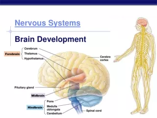
THE HUMAN BRAIN 28.14 The vertebrate brain develops from three anterior bulges of the neural tube • The vertebrate brain evolved by the enlargement and subdivision of three anterior bulges of the neural tube • Forebrain • Midbrain • Hindbrain • Cerebrum size and complexity in birds and mammals correlates with sophisticated behavior
EmbryonicBrain Regions Brain StructuresPresent in Adult Cerebrum (cerebral hemispheres; includescerebral cortex, white matter, basal ganglia) Forebrain Diencephalon (thalamus, hypothalamus,posterior pituitary, pineal gland) Midbrain Midbrain (part of brainstem) Pons (part of brainstem), cerebellum Hindbrain Medulla oblongata (part of brainstem) Diencephalon Cerebralhemisphere Midbrain Midbrain Pons Hindbrain Cerebellum Medullaoblongata Spinal cord Forebrain Embryo one month old Fetus three months old Figure 28.14
28.15 The structure of a living supercomputer: The human brain Table 28.15
Cerebrum Forebrain Thalamus Cerebralcortex Hypothalamus Pituitary gland Midbrain Pons Medullaoblongata Hindbrain Spinal cord Cerebellum Figure 28.15A
Most of the cerebrum’s integrative power resides in the cerebral cortex of the two cerebral hemispheres Left cerebralhemisphere Right cerebralhemisphere Corpuscallosum Basalganglia Figure 28.15B
28.16 The cerebral cortex is a mosaic of specialized, interactive regions • The motor cortex sends commands to skeletal muscles • The somatosensory cortex receives information about pain, pressure, and temperature • Several regions receive and process sensory information (vision, hearing, taste, smell)
Frontal association area (judgment, planning) • Auditory association area • Somatosensory association area (reading, speech) • Visual association area • The association areas are the sites of higher mental activities (thinking)
FRONTAL LOBE PARIETAL LOBE Somatosensoryassociationarea Somatosensory cortex Speech Motor cortex Frontalassociationarea Taste Reading Speech Hearing Smell Visualassociationarea Auditoryassociationarea Vision OCCIPITAL LOBE TEMPORAL LOBE Figure 28.16
“Right-brained” vs. “left-brained” • In lateralization, areas in the two hemispheres become specialized for different functions
28.17 Connection: Injuries and brain operations have provided insight into brain function • Much knowledge about the brain has come from individuals whose brains were altered through injury, illness, or surgery • The rod that pierced Phineas Gage’s skull left his intellect intact but altered his personality and behavior Figure 28.17A
A radical surgery called hemispherectomy removes almost half of the brain • It demonstrates the brain’s remarkable plasticity Figure 28.17B
28.18 Several parts of the brain regulate sleep and arousal • Sleep and arousal are controlled by • the hypothalamus • the medulla oblongata • the pons • neurons of reticular formation Input from ears Eye Motoroutput tospinal cord Reticular formation Input from touch,pain, and temperaturereceptors Figure 28.18A
Two types of deep sleep alternate • Slow-wave (delta waves) and REM sleep • An electroencephalogram (EEG) measures brain waves during sleep and arousal Awake but quiet (alpha waves) Awake during intense mental activity (beta waves) Delta waves REM sleep Delta waves Figure 28.18B, C Asleep
28.19 The limbic system is involved in emotions, memory, and learning • The limbic system is a functional group of integrating centers in the cerebral cortex, thalamus, and hypothalamus • It is involved in emotions, memory (short-term and long-term), and learning • The amygdala is central to the formation of emotional memories • The hippocampus is involved in the formation of memories and their recall
Thalamus CEREBRUM Hypothalamus Prefrontalcortex Smell Olfactorybulb Hippocampus Amygdala Figure 28.19
28.20 The cellular changes underlying memory and learning probably occur at synapses • Memory and learning involve structural and chemical changes at synapses • Long-term depression (LTD) • Long-term potentiation (LTP)
1 Repeatedactionpotentials Sendingneuron Sendingneuron Synapticcleft 2 2 4 Ca2+ Cascade ofchemical changes 3 3 Ca2+ Receiving neuron LTP Figure 28.20