Download

1 / 32
320 likes | 555 Vues
Tobacco - Inside the Body. © Photographs courtesy of Manchester University Pathology Department. This Powerpoint is hosted on www.worldofteaching.com Please visit for 1000+ free powerpoints. Human female chromosome map (XX). Alcohol and nicotine addiction may be in the genes (1).

E N D
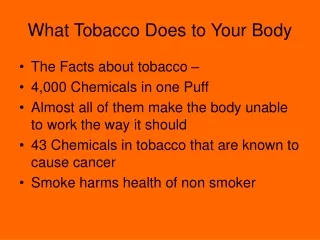
Tobacco - Inside the Body ©Photographs courtesy of Manchester University Pathology Department This Powerpoint is hosted on www.worldofteaching.com Please visit for 1000+ free powerpoints
Human female chromosome map (XX). Alcohol and nicotine addiction may be in the genes (1)
There is accumulation of carbon (smoke) within the walls of these air sacs in the lungs. This makes the air sacs inelastic and it is difficult for air to get out of the lungs (Emphysema) (9)
Clubbing of the fingers is a common condition which may indicate cancers, liver diseases and lung disease (16)
Focal dust emphysema is commonest in cigarette smokers and coal miners.The black colour in this lung is due to cigarette smoke (22)
Cells lining the bronchus- here there are no cilia- they have been damaged by the nicotine (23)
Cells lining the bronchus the large nuclei are in cancerous cells spreading along the bronchus (24)
Lung tissue blackened by smoke and showing large spaces due to emphysema (25)
Emphysema- these large spaces are due to the breakdown of the air sacs in the lungs (26)
Lungs: The picture on the left is of emphysema (large air sacs) and the picture on the right is of normal lung.(27)
The air sacs in the centre of the lung tissue in the picture have broken down. The black dots are smoke particles lodged in the tissues- this condition is emphysema. (28)
A blue colouration occurs when blood circulation is poor such as heart failure, shock and exposure to cold environments.(30)
This man is a heavy smoker (see nicotine stained fingers) and has led to vascular disease which has resulted in a blood clot on one of his fingers on his right hand (31)
Damaged toes due to poor blood circulation- will lead to amputation (32)
Amputated fingers- often as a result of damage to blood vessels due to smoking (33)
Franks sign - an ear lobe crease thought to be indicative of heart problems (34)
Fluid collecting in the tissues, note the dent where somebody has pressed on top of the hand- a sign of heart failure (36)
Heart:This slice is also through both ventricles (compare it to photo 38) The left ventricle is towards the right of the image as you look at it, and shows a white area of in the heart wall. This is tissue, which has died due to lack of blood supply (37)
Heart: The left ventricle is towards the left of the screen, and shows enormous thickening of the wall, particularly in relation to the small cavity. The heart is almost twice the size it should be, as it has had to work much harder than normal. (38)
Expansion of finger ends- associated with chronic breathing and heart problems (39)
Expansion of finger ends- associated with chronic breathing and heart problems (40)
Cancer of oesophagus- this is where the oesophagus meets the stomach (41)
Cancer of the kidney- the cancer growth is the white tissue (42)
Cervical cancer - the nucleus of this pink cell should be round- instead it is mis-shapen and is an early sign of cancer of the cervix (43)
Bladder cancer - the bladder is a hollow bag for storing urine- here it is cut open showing a large cancerous growth inside (44)
Cross-section of a coronary artery (artery to the heart). The inner wall is very thick (pale pink) and you can see a massive blood clot (red) taking up much of the photo (46)
Stomach ulcers (dark brown) - alcohol and smoking are factors (49)
Stomach ulcers (dark brown)- alcohol and smoking are factors. The stomach is cut open. (50)