Download
1 / 66
690 likes | 2.68k Vues
Abnormal Liver enzymes and/or LFTs: work-up and diagnosis. What you need to know. Differential Diagnosis. Population based survey in US 1999-2002 estimated abnormal ALT in 8.9% of population Symptomatic vs Asymptomatic? Acute vs Chronic? Hepatitic vs Cholestatic?. Hepatitic:

E N D
Abnormal Liver enzymes and/or LFTs: work-up and diagnosis What you need to know
Differential Diagnosis • Population based survey in US 1999-2002 estimated abnormal ALT in 8.9% of population • Symptomatic vs Asymptomatic? • Acute vs Chronic? • Hepatitic vs Cholestatic?
Hepatitic: Viral Hepatitis: A, B, C, D, E Alcoholic and Nonalcoholic Steatohepatitis Autoimmune Hemachromatosis Wilson’s disease Alpha-1 antitrypsin Cholestatic: Obstruction Gallstones, malignancy, parasites Primary Biliary Cirrhosis Primary Sclerosing Cholangitis Infiltrative diseases: metastatic cancer, sarcoidosis, amyloidosis Differential Diagnosis Either: • Drugs • Thyroid disorders • Celiac disease • Vascular disease: CHF, Budd Chiari syndrome, Sinusoidal obstructive syndrome
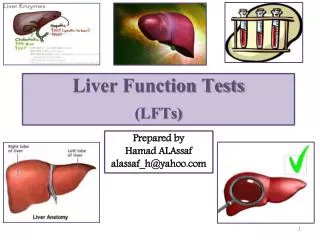
Liver Tests • AST, ALT • Alkaline Phosphatase • GGT • Bilirubin • Albumin • Protime/INR True “liver function tests”
AST, ALT • Aspartate aminotransferase, alanine aminotransferase • Enzymes that are in the hepatocyte and function during gluconeogenesis • Leak out of the hepatocytes in times of injury and can be measured in the serum • Normally present in serum at levels ~30-40 U/L
AST/ALT • AST: • liver > cardiac muscle > skeletal muscle > kidney > brain > pancreas > lung > leukocytes > erythrocytes • Less specific for liver damage • Can increase with strenuous exercise, MI • Located in cytosol and mitochondria of hepatocytes • Cleared more rapidly than ALT • ALT • Mainly from cytosol of hepatocytes • more specific for liver damage
Normal Aminotransferase Levels and Risk of Mortality from Liver Diseases • Study design: Prospective cohort study; Korean Medical Insurance Corporation; 8-year follow-up; 94,533 men and 47,522 women aged 35 to 59 • Results: “...findings indicate that serum aminotransferase concentration is associated with mortality from liver disease, even within the current normal range“; best cut-off value by ROC of ALT for prediction of liver diease in men was 30 U/L • Conclusions: “People with slightly elevated aminotransferase activity, but still within the normal range, should be closely observed and further investigated for liver disease“ Kim HC, et al. BMJ 2004;328:983.
Normal Aminotransferase Levels and Risk of Mortality from Liver DiseasesProspective Cohort Study ALT (IU/L) No (men) RR (95% CI) < 20 37425 1.0 (0.7-1.4) 20-29 36589 2.9 (2.4-3.5) 30-39 11975 9.5 (7.9-11.5) 40-49 4068 19.2 (15.3-24.2) 50-99 3887 30.0 (25.0-36.1) 100 589 59.0 (43.4-80.1) "Normal“ "Elevated“ Kim HC, et al. BMJ 2004;328:983.
Normal Serum AST and ALT and Risk of Mortality from Liver Diseases Prospective Cohort Study DESIGN • Prospective, cohort study • Cause of death from death certificates • 94,533 males and 47,522 females ages 35-59 with 8 years follow-up Number with CHB unknown OBJECTIVE • Determine liver mortality in individuals with normal ALT (<40 IU/L) OUTCOMES • 690 deaths from liver disease (LD) • Death from LD associated with baseline age, AST, BP, FHx of LD, elevated glucose or cholesterol • ALT >20 increased risk for death from LD Risk of death from liver disease based on ALT and AST Kim HC, et al. BMJ 2004;328:983.
The 6 TESTS • HBsAg • HCV Aby • Fe Sat • BMI • Alc history • Medication and herbal history
Alkaline Phosphatase • Exists in liver in membrane of hepatocyte where it lines the canaliculus • Liver > bone > intestine • Placenta • Normally changes with age
Other cholestatic enzymes • GGT: gamma-glutamyltransferase • Found in hepatocytes and biliary epithelial cells • 5’ nucleotidase • Both these enzymes can be used to confirm alk phos elevation is coming from liver • GGT is also sensitive to alcohol ingestion
Bilirubin • Breakdown product of heme • 70-80% of normal production is from breakdown of hemoglobin in senescent RBC • Conjugation of bilirubin occurs in ER of hepatocyte, and conjugated bilirubin is then transported into bile (rate limiting step) • Almost 100% of bilirubin in healthy people is indirect
Bilirubin • Increased bilirubin can occur from: • Overproduction of bilirubin • Uncomplicated hemolysis (rarely levels >5 mg/dL) • Impaired uptake, conjugation, or excretion • Blockage of bile duct • Regurgitation from damaged hepatocytes of bile ducts • Hepatocellular damage • Urinary bilirubin is only conjugated
Albumin • Important plasma protein synthesized by the liver • Half-life 20 days • Levels <3 mg/dL should raise the suspicion of chronic liver disease • ***not specific for liver disease • Also reduced in heavy alcohol consumption, chronic inflammation, protein malnutrition
Protime/INR • Liver synthesizes major coagulation proteins: I, II, V, VII, IX, X, XII, XII • Protime measured II, VII, IX, X: vitamin K dependent factors • Can be elevated in liver disease or Vitamin K deficiency • ***most important abnormality to signify development of fulminant hepatic failure in course of acute liver disease • Degree of elevation is a prognostic factor in many liver diseases
Evaluation of LTs • First step is to repeat the test!! • Many many things can cause a one-time elevation of liver tests • Mild elevations should be monitored for at least 6 months before a full serologic work-up is done • Exception is viral hepatitis serologies in high risk people
Evaluation of LTs • Evaluate how high the test is • Normal values are calculated from “normal” people • Normal = 2 SD above and below the mean • B y definition, this makes 2.5% of normal people have abnormal test! • Think of situations where a high test is normal • Alk phos in pregnancy • AST in marathon runner
Evaluation of LTs • History • How long has elevation been present? • Any symptoms: pruritis, fatigue, RUQ pain, arthralgias, myalgias, rash, anorexia, fever, weight loss, changes in urine/stool • Symptoms of more severe liver disease: jaundice, ascites, LE edema, GI bleeding, confusion or slowed thinking • Other Medical Problems? • Family history of liver disease? • Personal or family history of autoimmune disease or thyroid disease?
Medication History • What medications do you take? • When did you start them? • Any change in doses? • Over the counter medications? • Herbals? – ask specifically!!
Social HistoryEssential in Eval of liver disease • How much do you drink? – be specific!!! • Any recent travel? Born abroad? • Have you had any blood transfusions? • Have you ever had hemodialysis? • Do you work in healthcare? Any needlesticks? • Any tattoos? • Have you ever injected drugs, even once? • Have you ever snorted drugs, even once? • Any recent mushroom ingestion? • Any unprotected sex? Multiple sex partners? • Are you a Vietnam veteran?
Physical Examination • Look for signs of chronic liver disease • Spider angiomata • Firm liver edge • Splenomegaly • Leukonychia • Ascites • LE edema • Abdominal wall collateral vessels • Proximal and temporal muscle wasting • Look for signs of other diseases: acanthosis nigricans, signs of thyroid disease, xanthomas, LAD, etc • Check cardiac exam for JVD, hepatojugular reflex
Most of the time, with the history and physical you should have a good idea what’s the likely culprit of the elevated liver tests!
Case #1 • 48 year old man is found to have abnormal liver tests on routine physical examination • No significant PMH • On no medications • Tried IV drugs “once in college” • PE normal • Labs: CBC, BMP normal • AST 62, ALT 88, Alk phos 75, Tbil 0.7, INR 1.0, Albumin 4.1
Case #1, continued • Hepatitis C: Antibody positive • What now? • Check HCV RNA, genotype, refer to hepatology • Oh yeah, and check other viral serologies too
Hepatitis C • HCV-Ab (hepatitis C antibody) • (+) in chronic or previous infection • Sensitive screening test with elevated ALT • Detectable 8-16 weeks after exposure to virus • False positives in autoimmune dz or hypergammaglobulinemia • False negatives in immunosuppressed
Hepatitis C • Hepatitis C viral level in copies/ml or IU/ml • Qualitative by PCR or TMA (transcription-mediated amplification) to < 10 IU/ml • HCV-RNA detectable 2-4 weeks after exposure • Level is factor in response to Rx but NOT severity of disease
Case #245 year old South African man • Presents for routine physical and found to have elevated liver tests • PMH: hyperlipidemia • Soc: drinks 2 beers/week, no Hx drug use • Born in South Africa, came to US in 1985 • Family history: none (but he mentions that they don’t go to the doctor) • PE: normal
Case #245 year old South African man • Labs: CBC, BMP normal • AST 50, ALT 60, Alk phos 70, Tbili 0.5 • INR, Albumin normal • US normal • What now?
Case #245 year old South African man • Hepatitis B s Ag: positive • HBsAb: negative • HBcAb: positive • What now? • HBV DNA 1.5 billion, HBeAg pos, HbeAb neg • Diagnosis? • Chronic Hepatitis B • What if HBV DNA was 2000?
HBsAg Prevalence (%) 8: High 2-7: Intermediate <2: Low Global Distribution of Chronic HBV Infection • 350 million chronic carriers worldwide • Ninth leading cause of death • Nearly 75% of HBV chronic carriers are Asian
HBV Serologies Acute infection Chronic infection Immunized Past Exposure False pos or Past Exp
HBV Serologies • HBe Ag: only applicable in patients who are chronically infected or carriers • Positive: increased infectivity • Negative: precore mutant of virus, still can be infective, still has advancing disease • Levels of HBV DNA important to decide if patient active or inactive
Case #319 year old female college student • c/o severe fatigue of new onset, jaundice, mild pruritis of few days duration • No EtOH • Meds: minocycline, multivitamin • No Hx of contacts with viral hepatitis, travel • PE: heent: mild scleral icterus abd: nl bs, no organomegaly, no tenderness or palpable mass
Case #3, continued • Labs: alb 4.2, t bili 4.2, alk phos 248, • AST 180, ALT 252; • CBC normal, BMP normal • Acute viral hepatitis serologies neg. What is the most likely diagnosis?
Case #319 year old female college student Drug induced cholestasis secondary to minocycline. Symptoms resolved within 2 weeks of drug d/c, liver profile normalized in 8 weeks.
Drug Induced Liver Injury • Any drug can cause any injury!!!! • Higher risks in women, older age • Can be at start of medication, or in some cases at *any time* during therapy • Should rule out other causes of liver injury
Drug Induced Liver Injury • A note: Tylenol is the most common cause of DILI, and also the most common cause of acute liver failure in US • Normal dose for Tylenol toxicity is 10-12 grams/d, but toxicity can occur in much lower doses in certain circumstances • Alcohol use • Fasting state
Case #456 year old woman • Presents with fatigue, myalgias • PMH: hypothyroidism, HTN • Meds: Synthroid, Atenolol, MVI, Ca • Soc: no T/E/D • FHx: father with vitiligo • PE: appears fatigued, Abd with mild RUQ tenderness to deep palpation
Case #456 year old woman • CBC, BMP normal • AST 245, ALT 280, TBili 2.0, Alk phos 207 • Alb 3.8, INR 1.2 • US: mild hepatomegaly, otherwise normal • Differential diagnosis? • What further labs?
Case #456 year old woman • ANA >1:640 • ASMA >1:380 • Quantitative Ig: Elevated IgG • Viral Markers negative • Diagnosis: Autoimmune Hepatitis
Autoimmune Hepatitis • Middle-aged (or teenage) woman, non-drinker without viral hepatitis • Fatigue, arthralgias/myalgias, oligomenorrhea, jaundice • Increased AST/ALT, gamma globulins • Positive ANA and SMA • Interface hepatitis with lymphoplasmacytic infiltrate • Responds to corticosteroids
Case #5:60 year old man • Presents with new onset diabetes, found to have elevated LTs • PMH: DM II newly diagnosed, OA in hips • Fhx: none • Soc: drinks 3 beers/day, no drug use • PE: normal
Case #5:60 year old man • CBC, BMP normal • AST 80, ALT 65, Alk phos 125, TBili 0.6 • Alb 3.6, INR 1.0 • US: increased echogenicity of the liver • Differential? • Tests?
Case #5:60 year old man • Fe % sat 70%, Ferritin 800 • Viral studies negative • Differential: ETOH vs Hemochromatosis • HFE gene test: C282Y homozygote
Hemochromatosis • Inherited abnormality of iron absorption • Affects 0.5% of Caucasian people • Rare in other races • C282Y/H63D gene abnormalities • Iron overload seen in C282Y homozygotes and sometimes compound heterozygotes (C282Y/H63D) • No role for gene testing without elevated iron tests!! • Iron tests can be falsely elevated in alcohol, acute inflammation, non fasting state
Alcoholic Liver Disease • Seen in 25% of heavy drinkers • >5 drinks/day in men, much lower in women • AST>ALT • AST in mitochondria, and alcohol is a mitochondrial toxin • Also seen when cirrhosis develops in other diseases • Cirrhosis can develop without LT abnormalities! • Alcohol hepatitis rarely has AST>300s
Case #6 A 47 year old Caucasian female presents with complaints of itching, dry mouth, and RUQ abdominal pain. She also notices some pigmentation changes on her eyelids. Her medical history includes frequent UTI’s and osteopenia. What labs are you most interested in seeing for this patient?