Download
1 / 25
260 likes | 850 Vues
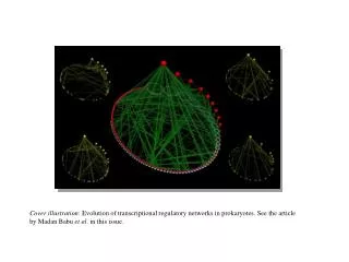
Bacterial Source Tracking by DNA Fingerprinting of Escherichia coli. Bacterial Source Tracking (BST). Determination of the sources of fecal bacteria from an environmental water sample (1995) Several different techniques are currently being used for BST. BST Technology. Genotypic

E N D
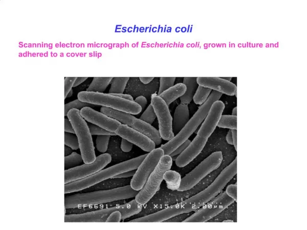
Bacterial Source Tracking by DNAFingerprinting of Escherichia coli
Bacterial Source Tracking (BST) • Determination of the sources of fecal bacteria from an environmental water sample (1995) • Several different techniques are currently being used for BST
BST Technology • Genotypic • Pulsed-Field Gel Electrophoresis (Simmons/Herbein/Hagedorn – Va. Tech.) • Ribotyping (Samadpour – U. of Wash.) • Randomly Amplified Polymorphic DNA (RAPD)/PCR (Sadowsky – U. of Minn.)
BST Technology • Biochemical (Phenotypic) • Multiple Antibiotic Resistance (MAR) (Hagedorn - Va. Tech/ Kator – VIMS/ Wiggins – James Madison) • Coliphage (Geoff Scott – NOAA) • Carbon Source Profiles (Hagedorn – Va. Tech) • Sterols or Fatty Acid Analysis • Chemical (Non-Bacterial) • Optical Brighteners • Caffeine Detection
Fecal Indicators (Microbial) • Fecal Coliforms • Facultative, Gram negative, non-spore forming, rod –shaped, ferment lactose producing gas within 48 hours. Sources: animals, soil (44.50 C is selective) • Fecal Streptococci (Enterococci) • Facultative, Gram positive, tolerate high salt and are used as indicators particularly in brackish or marine waters. Sources: warm-blooded animals • Escherichia coli • A sub-set of the fecal coliform. Sources: mammals and birds
Elevated levels of E. coli • Indicator of potential pathogens • Other bacteria • Viruses • O157:H7 • Beach closings • Shellfish bed closings
Fecal Coliform Standards • Recreational Water • < 200 MPN/100 ml • Shellfish Beds • < 14 MPN/100 ml • Public Drinking Water Supply • < 20 MPN/100 ml • Treated Drinking Water • < 1% positive/month
Sources of Contamination • Treatment plants • Septic systems • Improper disposal from boats • Agricultural runoff • Pets • Wildlife
Project Overview • Study site selection • Monitor study sites for elevated levels of E. coli • Isolate E. coli from known sources • Isolate E. coli from water • Verify as E. coli • DNA analysis (Pulsed-Field Gel Electrophoresis) • Match water sources to known sources • Possible remediation plan
Determining Fate and TransportSample E. coli levels monthly for one year Use GPS and GIS to map and analyze levels Determine principal inputs of contamination
Scat Collection • Locations within drainage area • Identifiable samples
Water Samples • Select rivulets or Drainage areas • Monitor E. coli levels by MPN • Monitor pH, dissolved oxygen, temp., salinity, conductivity • Focus on MPN “hot spots” for collecting isolates for DNA analysis
Isolation of E. coli • A-1 Broth with MUG (4-methylumbelliferyl-B-D-Glucuronide) • E. coli-specific enzyme glucuronidase • Fluorogenic metabolite • Presumptive E. coli : • Turbidity • Gas • Fluorescence
Isolation of E. coli • API 20-E Strips (Biochemical tests for gram negative bacteria) • E. coli verified
DNA Sample Preparation: Enzyme Digestions • Plugs restricted with Not I • Restriction sites recognized: • DNA cut • Bands generated 5’..GC*GGCC GC..3’ 3’..CG CCGG*CG..5’
Gel Preparation • Plugs “glued” to comb • Gel formed around DNA plugs
Pulsed-Field Gel Electrophoresis • Fragments separated by electric current • Multi-directional current over 20 hours • Downward net flow
Gel Analysis • Gels stained with Ethidium Bromide • Gels photographed under UV light • Banding patterns analyzed
Bacterial Source Identification • E.coli banding patterns from scat added to library • E. coli banding patterns from water matched to library
DNA Match: Water Sample to Deer Scat Sample Shiles Creek water sample Shiles Creek deer scat sample
DNA Match: Water Sample to Muskrat Scat Sample Shiles Creek water sample Shiles Creek muskrat scat sample
Conclusions: • Molecular Biology advances provide a means to identify nonpoint sources of bacterial contamination. • Lab results identified sources consistent with rural and urban settings. • This technology adds to our knowledge base for better decision-making regarding potential remediation.
Students/Technical Help • The Described Project: • Jon Neville, Amanda Zych, Julie Wood • Currently: • Natasha Patterson, Gayle Raynor, Rebecca Scott, Gry Wesenberg • Current Technical Help: • Lesley Frana • -
Acknowledgements • Maryland Department of Natural Resources • This project was funded in part by the U.S. Environmental Protection Agency Section 319 Nonpoint Source Program through the Maryland Department of Natural Resources. • Maryland Department of the Environment • Technical and Regulatory Services Administration • R.A. Henson School of Science and Technology