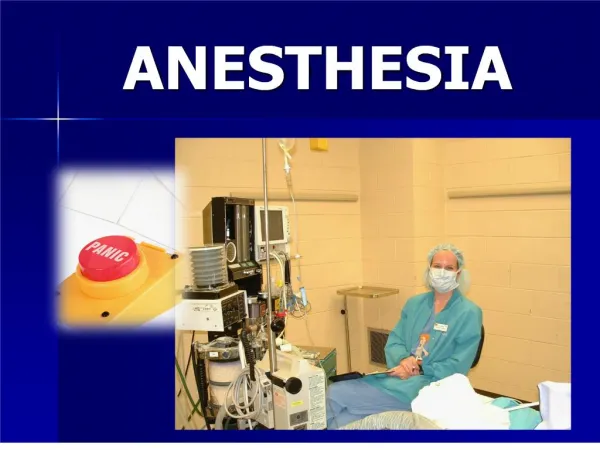
Anesthesia
Anesthesia. Dr. Aidah Abu Elsoud Alkaissi An-Najah National University Faculty of Nursing. History of Anaesthesia. 1842: Ether as an aneasthetic 1844: Nitrous oxide for anaesthesia 1844: sulfuric ether- tooth surgery- for pain 1900: alcohol or opium. Anesthesia.

Anesthesia
E N D
Presentation Transcript
Anesthesia Dr. Aidah Abu Elsoud Alkaissi An-Najah National University Faculty of Nursing Dr. Aidah Abu Elsoud Alkaissi Division of Intensive Care and Anaesthesiology University of Linköping Sweden
History of Anaesthesia • 1842: Ether as an aneasthetic • 1844: Nitrous oxide for anaesthesia • 1844: sulfuric ether- tooth surgery- for pain • 1900: alcohol or opium Dr. Aidah Abu Elsoud Alkaissi Division of Intensive Care and Anaesthesiology University of Linköping Sweden
Anesthesia • From Greekanaisthesismeans ”not sensation” • Listed in Bailey´s English Dictionary 1721 • When the effect of ether was discovered”anesthesia” used as a name for the new phenomenon • Death of pain (Werr Kitchell) Dr. Aidah Abu Elsoud Alkaissi Division of Intensive Care and Anaesthesiology University of Linköping Sweden
Anesthesia Providers • Anesthesiologist ( aphysician with 4 or more yearsof speciality training in anesthesiology after medical school) • Certified registered nurse anesthetist (CRNA), working under the direction and supervision of an anesthesiologist or a physician • CRNA must have 2 years of training in anesthesia Dr. Aidah Abu Elsoud Alkaissi Division of Intensive Care and Anaesthesiology University of Linköping Sweden
Patient Safety • Patient risk and safety are concerns during surgery and anesthesia • Data from a number of studies of death caused by anesthesia indicate a death rate ranging from 1 per 20,000-35,000 • A fourfoulded decline over the last 30 years even though surgical procedures are undertaken on increasingly sicker and much higher risk patients than in the past • Awareness of potential problems and constant vigilance (the process of paying close and continuous attention) are crucial to good patient care Dr. Aidah Abu Elsoud Alkaissi Division of Intensive Care and Anaesthesiology University of Linköping Sweden
Preoperative preparationpatient evaluation • Anaesthesiologist: • reviews the patient´s chart, • evaluate the laboratory data and diagnostic studies such as electrocardiogram and chest x-ray, • verify the surgical procedure, • examins the patient, • discuss the options for anesthesia and the attendant risks and • ordered premedication if appropriate Dr. Aidah Abu Elsoud Alkaissi Division of Intensive Care and Anaesthesiology University of Linköping Sweden
The physical status classification • Developed by the American Society of Anesthesiologist (ASA) to provide uniform guidelines for anesthesiologists • It is an evaluation of anesthetic morbidity and mortality related to the extent of systemic diseases, physiological dysfunction, and anatomic abnormalities • Intraoperative difficulties occur more frequently with patients who have a poor physical status classification • Please read table 9-1, page 148. Dr. Aidah Abu Elsoud Alkaissi Division of Intensive Care and Anaesthesiology University of Linköping Sweden
Choice of anesthesia • The patient´s understanding and wishes regarding the type of anesthesia that could be used • The type and duration of the surgical procedure • The patients´s physiologic status and stability • The presence and severity of coexisting disease • The patient´s mental and psychologic status • The postoperative recovery from various kinds of anesthesia • Options for management pf postoperative pain • Any particular requiremets of the surgeon • There is major and minor surgery but only major anesthesia Dr. Aidah Abu Elsoud Alkaissi Division of Intensive Care and Anaesthesiology University of Linköping Sweden
Types of anesthesia careGeneral Anesthesia • Reversible, unconscious state is characterised by amnesia (sleep, hypnosis or basal narcosis), analgesia (freedom from pain) depression of reflexes, muscle relaxation • Put to sleep Dr. Aidah Abu Elsoud Alkaissi Division of Intensive Care and Anaesthesiology University of Linköping Sweden
Types of anesthesia careRegional Anesthesia • A local anethetic is injected to block or ansthetize a nerve or nerve fibers • Implies a major nerve block administered by an anesthesiologist (such as spinal, epidural, caudal, or major peripheral block) Dr. Aidah Abu Elsoud Alkaissi Division of Intensive Care and Anaesthesiology University of Linköping Sweden
Types of anesthesia caremonitered anesthesia care • Infiltration of the surgical site with a local anesthesia is performed by the surgeon • The anasthesiologist may supplement the local anesthesia with intravenous drugs that prof´vide systemic analgesia and sedation and depress the response of the patient´s autonomic nervous system Dr. Aidah Abu Elsoud Alkaissi Division of Intensive Care and Anaesthesiology University of Linköping Sweden
Types of anesthesia carelocal anesthesia • Employed for minor procedures in which the surgical site is infiltrated with a local anesthetic such as lidocaine or bupivacaine • A perioperative nurse usually monitors the patient´s vital signs • May inject intravenous sedatives or analgesic drugs Dr. Aidah Abu Elsoud Alkaissi Division of Intensive Care and Anaesthesiology University of Linköping Sweden
Premedication • Purpose: to sedate the pat and reduce anxiety • Classified as sedatives and hypnotics, tranquilizers, analgesic or narcotics and anticholinergics • Antiacid or an H2receptor-blockingdrug such as cimitidine (tagamet) or ranitidine (Zantac) to decrease gastric acid production and make the gastric contents less acidic • If aspiration occur this premedication decreases the resultant pulmonary damage • Given 60-90 minutes before surgery, or may be given i.v. After the pat arrives in the surgical suite • NPO for a minimum of 6 hours before elective surgery • Not given to elderly people or ambulatory patients because residual effects of the drugs are present long after the pat have been discharged and gone home Dr. Aidah Abu Elsoud Alkaissi Division of Intensive Care and Anaesthesiology University of Linköping Sweden
Perioperative monitoring • Undergeneral anesthesia: monitoring • Inspired oxygen analyzer(FiO2) which calibrated to room air and 100% oxygen on a daily basis • Low pressure disconnect alarm, which senses pressure in the expiratory limb of the patient circuit • Inspiratory pressure • Respirometer (these four devices are an integral part of most modern anesthesia machine • ECG • BP-automated uni • Heart rate • Precordial or esophagel stethoscope • Temp Dr. Aidah Abu Elsoud Alkaissi Division of Intensive Care and Anaesthesiology University of Linköping Sweden
Perioperative monitoring • Pulse oximeters • End tidal carbon dioxide (ECO2) • Peripheral nerve stimulatorif muscle relaxants are used • Foly catheter • For selected patintwith a potential risk of venous air embolism a doppler probe may placed over the right atrium • Invasive: arterial pressure mesurements, central venous pressure • Pulmonary artery catheter and continous mixed venous oxygen saturation measured Dr. Aidah Abu Elsoud Alkaissi Division of Intensive Care and Anaesthesiology University of Linköping Sweden
Perioperative monitoring • For special conditions other monitors as transcutaneous O2 and CO2 transesophageal echocardiography • Electroencephalogram • Cereral or neurological may be used Dr. Aidah Abu Elsoud Alkaissi Division of Intensive Care and Anaesthesiology University of Linköping Sweden
Pulse oximetry • Please read 149, 151 • Based on principles of spectrometric (equipped with scales for measuring wavelengths ) oximetry, plethysmography, and Lambert-Beer law (The intensity of a beam of monochromatic radiation), which relates the concentration of solute (a substance dissolved in a solvent) in the suspension of the intensity of light transmitted through the solution • It provides a a continous noninvasive indication of the arterial oxygen saturation of functional hemogloin and the pulse rate and provides an early warning of hypoxemia Dr. Aidah Abu Elsoud Alkaissi Division of Intensive Care and Anaesthesiology University of Linköping Sweden
Pulse oximetry • The sensor combines two low-intensity light – emitting diodes (LED) as light sources and photodiode as a receiver or light detector • One LED emits (expel ) red light and the other LED emits infrared light • When transmitted through blood and tissue, the two frequencies are absorbed differently by the tissue componentsand by thr reduced HB and oxyhemoglobin Dr. Aidah Abu Elsoud Alkaissi Division of Intensive Care and Anaesthesiology University of Linköping Sweden
Pulse oximetry • The internal microprocessor analyzes the variations in the absorbtion of light emited from both LEDs and provides a readout of the percent saturation of Hb with O2 • Adverselly affected • Hypothermia, hypotension, hypovolemia, vasoconstriction, hypothermia • Electrosurgery,motion or ambient (of the surrounding area or environment) light may decrease the readout Dr. Aidah Abu Elsoud Alkaissi Division of Intensive Care and Anaesthesiology University of Linköping Sweden
Pulse oximetry • Sensor is usually placed on the third or forh finger • Ear lobe, bridge of nose’care; prevent localized neurovascular or ischemic damage Dr. Aidah Abu Elsoud Alkaissi Division of Intensive Care and Anaesthesiology University of Linköping Sweden
Capnograph • A capnometer measures carbon dioxide • Has a waveform display • It evaluates the alveolar ventilation • There is only a small gradient from the rterial to the alveolar Co2and with forced expiration the ETCO2 privide a close approximation of the arterial CO2 (PaCO2) • Because anesthetized pat only passively expires and the point of measures is near the connection between the pat circuit and the endotrachel tube, there is5-8 torr gradient between ETCO2 an PaCO2 • ETCO2 can be measured by a mass spectrometer or an infrared analyzer Dr. Aidah Abu Elsoud Alkaissi Division of Intensive Care and Anaesthesiology University of Linköping Sweden
Capnograph • Measure the amount of infrared light absorbed by CO2 in the sample of gas • In the mainstream unit all respired gas passes through the detector, whereas with the sidestream unit a portion of the gas is aspirated at a constant rate (50-250)ml/min through a small bore tubing into the unit • Most units display a waveform of the expiratory CO2 partial pressure versus time after a short sampling and processing delay • Waveform is important for correctly interpreting the output data • Digitl readout gives ETCO2 and respiratory rate Dr. Aidah Abu Elsoud Alkaissi Division of Intensive Care and Anaesthesiology University of Linköping Sweden
Capnograph • Clinically, the unit is useful to: • Verify endotracheal intubation • Circuit disconnection • Alveolar ventilation • Early return of muscle function after muscle relaxants are used • Acute alterations in metabolic functions such as malignant hyperthermia or thyrotoxicosid Dr. Aidah Abu Elsoud Alkaissi Division of Intensive Care and Anaesthesiology University of Linköping Sweden
General Anesthesia Dr. Aidah Abu Elsoud Alkaissi Division of Intensive Care and Anaesthesiology University of Linköping Sweden
Dr. Aidah Abu Elsoud Alkaissi Division of Intensive Care and Anaesthesiology University of Linköping Sweden