Download
1 / 24
390 likes | 1.34k Vues
AMOEBIASIS. HISTORY. 2yr 10mo girl Main Complaint: 7 days of loose, bloody diarrhoea and vomiting Lethargic and doesn’t feed well Previous history: Uneventful perinatal history No previous admissions to hospital Numerous clinic visits in preceding months for diarrhoea RVD status unknown

E N D
HISTORY • 2yr 10mo girl • Main Complaint: • 7 days of loose, bloody diarrhoea and vomiting • Lethargic and doesn’t feed well • Previous history: • Uneventful perinatal history • No previous admissions to hospital • Numerous clinic visits in preceding months for diarrhoea • RVD status unknown • No TB or other infectious contacts • No recent travel history / No significant family history • DIET: Never breastfed , Feeds on a family diet that includes mainly veg protein and carbohydrates. Denies traditional medicine ingestion.
EXAMINATION • Growth paramoeters: • Weight: 11kg - 73% of expected for age • Length: 86cm – Below 3rd centile • Head Circ: 48cm – On 50th centile • Weight for height: Just below 3rd centile • Temp: 37.1 HR:135bpm RR:42/m BP:70mmHg syst • Distressed, ill-looking, pale, 7.5% dehydrated • Chest, CVS & CNS: Normal
EXAMINATION • Abdomen: • Soft, distended abdomen with decreased bowel sounds • 3cm hepatomegaly – Firm, sharp edge, smooth surface non-displaced and not tender. • Fullness possibly a mass lesion extending from R flank across midline & tender to touch. • Normal hernial orifices and female genitalia • PR: No exterior abnormalities noted Irregular rectal mucosa Bloody, foul-smelling diarrhoea mixed with pus noted
FBC: WCC: 3.12 Hb: 9.5 MCV: 80.1 Platelets: 127 Diff: Neut: 42.7% Mono: 17.8% Lymph: 35.7% Eosino: 1.8% Smear: Left shift + toxic granulation. CRP: 337 U&E: 129 / 2.6 / 91 / 24 / 3.4 / 38 LFT: Tbili: 13 Cbili: 1 TP: 46 Alb: 17 Glob: 29 ALP: 54 GGT: 32 ALT: 68 AST:81 Urine culture: Negative Stool culture: Negative Blood culture: Negative RVD Elisa: Positive CD4: 188 (4.62%) TB W/Up: Negative SPECIAL INVESTIGATIONS
AXR: Distended loops of large bowel with air fluid levels No free air noted Abd. Sonar: Aperistaltic thickened loops of bowel in RFI No mass seen CT Abdomen: Fluid-filled large & small bowel loops Closely related bowel loops in RFI – bowel wall thickening No definite mass seen Sigmoidoscopy: Mass seen at recto-sigmoid junction – biopsy taken Histology: Rectal biopsy with extensive mucosal ulceration. Marked inflammatory cell infiltrate composed predominantly of chronic inflamm. cells and fibrin deposition Amoebae noted and confirmed with PAS stain. SPECIAL INVESTIGATIONS
SUMMARY & MANAGEMENT • 2yr 10mo girl with • Immunosuppression • Amoebic proctitis and a chronic inflammatory mass at the recto-sigmoid junction consistent with an amoeboma. • Treated with oral Metronidazole • Optimized general condition in terms of nutrition • Referred for initiation of HAART
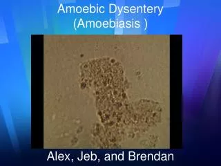
The Organism • 4 species of Entamoeba: • Nonpathogenic: E. dispar, E. coli, E. hartmanni • Pathogenic: E. histolytica • amoebiasis = A Parasitic infection caused by the protozoon Entamoeba histolytica • 2nd to Malaria as protozoan cause of death worldwide • 10% of world’s population infected – Increased prevalence in developing countries (up to 25%) • In SA – More common in KZN • Factors contributing to faecal-oral spread: • Poor education • Poverty and overcrowding • Unsanitary conditions • HIV infection
1. Cyst Stage Infective stage Survive from –4 to 40 Celcius Size – 12mm Quadrinucleated Ingested by contact with fecally contaminated food Passes through stomach, excysts in lower small bowel. Metacystic amoeba with four cystic nuclei from each cyst 8 Small trophozoites from each metacystic amoeba Trophozoites carried to cecum The Life Cycle
The Trophozoite Stage: 10-40 qm, fragile Uninucleate Erythrophagocytosis Reside, feed and multiply by binary fission in lumen of colon May invade – Lytic & physical mechanisms and metastasize to liver and other extra-intestinal sites Galactose-containing molecules & receptors regulate cyst formation Precyst – Cyst – Uninucleate to Quadrinucleate and passed in stool The Life Cycle
The Pathogenesis • 10% of infected individuals develop invasive disease • Factors contributing to developing invasive disease: • Pathogenicity of infecting Entamoeba species • Dose of inoculum • Host factors: Impaired cell-mediated immunity, on steroids • Virulence of infecting species: • Presence of surface adhesion factors • Release of proteolytic enzymes • Release of cytotoxins and inflicting of cytolysis
The Pathogenesis • Trophozoites adhere to colonic mucosal glycoproteins via a galactose and N-acetyl-D-galactosamine-specific lectin. • (Gal/GalNac) – Lectin is 260kD-surface protein consisting of a 225kD subunit and a 35kD subunit. • Adherence results in cell lysis (apoptosis) and PMN invasion • PMN’s are then lysed releasing lytic enzymes, causing more tissue destruction • Small foci of necrosis in the intestinal wall coalesce to form ulcers (Flask-shaped ulcers) • Parasites resist destruction by complement arm of immune system via Gal/GalNac mediated inhibition of the membrane attack complex • Cell-mediated immunity is important in clearing infection through generating Ұ-INF and TNF-α to activate macrophages and neutrophils to kill the trophozoite
Area most commonly involved = Cecum, then Recto-sigmoid area May invade blood vessels causing thrombosis, infarction and dissemination via portal circulation to liver and extra-intestinal sites eg. brain, pleura, pericardium and genito-urinary system. Flask-shaped ulcers The Pathogenesis
The Clinical Features • Many infections = Asymptomatic ‘cyst passers’ • Symptomatic infections may have a gradual, acute or rapid, fulminant course. • Clinical incubation period = 4 days to several months • Oftengradual development of symptoms = Irregular bouts of diarrhoea, abdominal pain, weight loss, nausea, loss of appetite (amoebic proctocolitis) • Less often sudden onset of copious diarrhoea containing mucus and blood. • Findings may include low-grade fever, tenderness on palpation of the abdominal wall overlying involved large bowel.
The Complications • Complications of Intestinal amoebiasis: • Fulminant Amoebic Colitis with Perforation • May have a mortality rate of up to 50% • Children less than 2 yrs at increased risk of perforation • Massive Haemorrhage • Due to vasculitis of large arteries or multiple ulcers leading to small arterial leaks • amoebomas • A granulomatous thickening of the colon resulting from lytic necrosis followed by secondary pyogenic inflammation, leading to fibrosis and proliferative granulation tissue. Lesions are firm, hard, may resemble a carcinoma. • amoebic Stricture • Resulting from fibrosis of intestinal wall. Can involve rectum, anus or sigmoid.
The Complications • Complications of Extra-Intestinal Amoebiasis: • Amoebic Liver Abcess • Most frequent complication of amoebiasis • Male:Female Ratio = 1 in Children and infants • In adulthood = More common in young males • Third to Half may have no history of diarrhoea • Commonly found in Right Lobe of liver • Presents acutely with high fever, RUQ tenderness • Jaundice an unusual finding • Have marked leucocytosis and may have XR abnormalities in 25 to 90% of patients
The Complications • Complications of Extra-Intestinal amoebiasis: • amoebic Peritonitis • As a complication of a ruptured hepatic abcess • Pleuropulmonary amoebiasis • Caused by rupture of Rt. Lobe Liver abcess in 10% of patients • Has cough, pleuritic chest pain & dyspnoea • amoebic Pericarditis • Rare, but most serious complication in 3% of pts. with liver involvement • Rupture of Left Lobe liver abcess • Cerebral amoebiasis • Rare, has altered consciousness and focal neuro signs • CT – Irregular lesions without surrounding capsule or enhancement • Genito-Urinary Involvement • Painful genital ulcers – Punched out appearance & profuse discharge
The Diagnosis • Light Microscopy of Stool • Identification of trophozoites / cysts in fresh stool • Disadvantages: • Not sensitive (miss up to two thirds of infections) • Cannot distinguish between E.histolytica and E. dispar • Serology: • Anti-amoebic antibodies (IgM) 70% sensitive for amoebiccolitis and 90% sensitive for amoebic liver abcess • Stool antigen-detection test or PCR • Sensitive and Specific • Disadvantages: • Antigen detection test (EIA) only available from Blacksburg VA
The Diagnosis • Colonoscopy / Sigmoidoscopy • Colonoscopy preferable • Wet preps of material from ulcer-base can show trophozoites • Biopsies should be taken from edge of ulcers • Recommended to evaluate for amoebic colitis even when Ulcerative Colitis considered
The Diagnosis • amoebic Liver Abcess: • Diagnosis relies on: • Detection of risk factors for E.histolytica infection • Positive Serology • Lesion in Liver : • Abdominal USS • Abdominal CT: Well-rounded, wall enhances • Aspiration may yield “anchovy-paste” material • More often yellow / gray-green • Often odourless and sterile – Highly suggestive of amoebicabcess
The Management • Asymptomatic infections • Luminal agent only recommended but ?not available in SA • In general, not treated in endemic areas • Symptomatic infections • Oral Metronidazole for 10 days • Effective in eradicating amoebae in bowel lumen and wall • Effective in eradicating extra-intestinal disease • Additional luminal agent not necessary • E. dispar infection doesn’t require treatment
Prevention • Improved sanitationandclean water supplyreducefecal-oral transmission • Boiling water, Washing veg with vinegar • Vaccination: • None available currently • Prototype subunit vaccines based on the Gal/GalNAc-lectin under study