Download
1 / 55
570 likes | 2.3k Vues
Outline. History AnatomyIndications TechniquesComplicationsCanal wall up vs. Canal wall down. History. Louis Petit was credit with first describing the procedure in the 1736 with a trochar, although trephination was done since prehistoric times.A chisel and gouge where used extensively throughout the 1800'sSchwartze popularized mastoidectomy in 1870 with detailed drawings. He described the cortical mastoidectomy, which was used extensively in preantibiotic era. Bondy described a techn9455

E N D
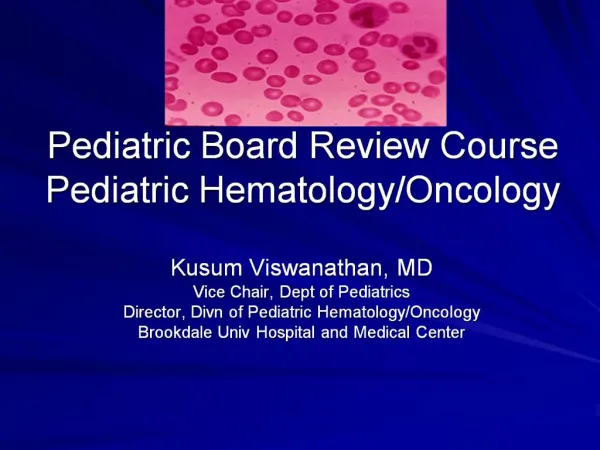
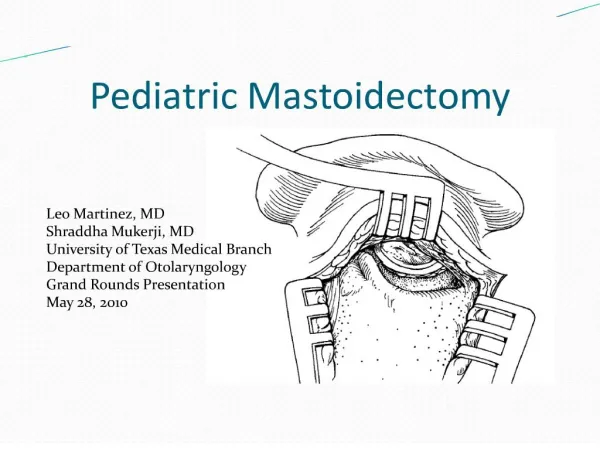
1. Pediatric Mastoidectomy
2. Outline History
Anatomy
Indications
Techniques
Complications
Canal wall up vs. Canal wall down
3. History Louis Petit was credit with first describing the procedure in the 1736 with a trochar, although trephination was done since prehistoric times.
A chisel and gouge where used extensively throughout the 1800�s
Schwartze popularized mastoidectomy in 1870 with detailed drawings. He described the cortical mastoidectomy, which was used extensively in preantibiotic era.
Bondy described a technique in 1910 in which mastoidectomy was performed and the posterior canal wall removed while leaving the pars tensa and ossicular chain intact
4. History 1922 Lempert introduced electrically driven drills in ear surgery, which were already used in dentistry
1930�s Wullstein introduced the operating microscope
1958, the canal wall up mastoid was then popularized by House. He also introduced the suction irrigation system and retractors in mastoid surgery.
5. Anatomy There are four parts to the temporal bone: petrous, tympanic, mastoid, and squamous
A transmastoid procedure allows access to the facial nerve, internal carotid, jugular, and internal auditory canal
6. Anatomy Adult Infant- have poorly developed mastoid and tympanic rings
7. Anatomy- axial mastoid
VII, seventh cranial nerve;
VIII, eighth cranial nerve;
APA, anterior petrous apex;
Ca, carotid artery;
CT, chorda tympani;
EAC, external auditory canal;
ET, Eustachian tube; Fn, facial nerve;
IAC, internal auditory canal;
KS, K�rner septum; LSC,
lateral semicircular canal;
PPA, posterior petrous apex;
PSC, posterior semicircular canal
8. Indications for Mastoidectomy Most common indication for mastoidectomy in children are
Cholesteatoma
Mastoiditis acute and chronic
Coexistence of the diseases
Less common indicators are
Neoplasm of temporal bone
Fracture of temporal bone, CSF leak
Facial nerve decompression
9. The Classical Procedures Canal Wall Up:
Simple Mastoidectomy
Complete Mastoidectomy (facial recess)
Canal Wall Down:
Modified Radical mastoidectomy
Radical mastoidectomy
Combination procedures (tympanomastoidectomy, neurotologic approaches)
10. Simple Mastoidectomy Indicate for acute surgical mastoiditis, commonly called �coalescent mastoid� or acute mastoid osteitis.
11. Simple Mastoidectomy Indicated also for :
Nonsurgical medical management failure of chronic suppurative otitis media/mastoiditis
Cholesteatoma when the cholesteatoma extends into the mastoid cells
Cochlear implant, in which a posterior tympanotomy is part of the procedure
Other uncommon indications in infants and children:
facial nerve decompression, translabyrinthine, labyrinthectomy, neoplasm, and mastoid trauma
12. Preoperative evaluation Preoperative audiometry. One should not operate on an ear in which hearing status is unknown.
Image study, high resolution CT scan of the Temporal bone. Key for assessment of pneumatization, and position of the tegmen and the sigmoid sinus.
Muscle relaxants should be avoided. Nerve monitoring is not essential, but useful especially in revision surgeries.
13. Surgery preparation Supine position with head turned away from affected ear
Hair may be shaven if it is in the operating field, or taped to keep it out of the field.
Injection with lidocaine with epinephrine.
Microscope should be balanced and at 225-300 mm
14. Simple Mastoidectomy A post-auricular approach is used for mastoidectomy in children. In young children the mastoid tip is not well developed and the stylomastoid foramen is located more superficially, making the facial nerve vulnerable to surgical trauma. The inferior aspect of the incision is more posterior and is not carried down as far to avoid injuring the facial nerve (Children younger than four)
15. Simple Mastoidectomy Carry the incision to the loose areolar tissue over the temporalis facia. This can be identified by pulling the auricle while performing the incision.
16. Simple Mastoidectomy The cortex is exposed by an incision through the linea temporalis, with a vertical cut extended to the posterior mastoid tip, in a T fashion. An elevator is then used to free the cortex off the soft tissue.
17. Simple Mastoidectomy Self retaining retractors are positioned and the surface landmarks are identified, which include the spine of Henle, cribriform area, and linea temporalis
MacEwen�s triangle shows the location of the antrum.
18. Simple Mastoidectomy
19. Simple Mastoidectomy
20. Simple Mastoidectomy
21. Simple Mastoidectomy Various drills are available and there are common principles related to bur selection
Larger bur preferred over smaller ones when possible
A bur with a cutting surface is selected for cortical bone, were diamond grain surface is for removing the last layer of bone over facial nerve or sigmoid sinus
Suction irrigation is critical to prevent excessive heat transfer to underlying structures.
Also, it is important to �saucerize� the edges of the mastoid cavity to provide visualization.
22. Simple Mastoidectomy Mastoid cortex is removed and the air cells are exposed.
23. Simple Mastoidectomy Next, identification of the tegmen, as a pink color in the bone superiorly is made. Vessels signal that you are close to the dura
The drilling is along a wide plane to avoid drilling in a hole
The deepest point of the dissection should be over the antrum
24. Simple Mastoidectomy
25. Simple Mastoidectomy Dissection is complete when the anterior epitympanum, zygomatic cells, body of incus and head of malleus are identified.
Cultures can then be taken from the mastoid mucosa, if needed.
A typanostomy tube is placed when acute mastoid osteitis is present.
26. Simple Mastoidectomy
27. Complete mastoidectomy This is an extension of the simple mastoidectomy with greater access to the attic, labyrinth, endolyphatic sac, antrum and facial nerve.
Some authors describe the complete mastoidectomy as a simple mastoidectomy with a facial recess approach.
Opening of the aditus ad antrum allows access to the epitympanum, and the incus and malleus may be removed for greater access
The canal wall remains up.
28. Complete Mastoidectomy The indications are the same for the simple mastoidectomy, with need for greater access to the mastoid cavity, as usually seen with cholesteatomas.
29. Complete Mastoidectomy The complete mastoidectomy starts with a simple mastoidectomy. After discovering the incus, HSSC, and the facial nerve, the facial recess can then be found.
First, the EAC is thinned laterally to medially. The medial portion will uncover the facial recess .
The recess is bound laterally by the chorda tympani, medial by the facial nerve and superiorly by the fossa incus. Opening this will allow access to the middle ear.
30. Complete mastoidectomy
31. Facial Recess
32. Complete Mastoidectomy
33. Complete Mastoidectomy Gaining access to the epitympanum may be necessary in cholesteatoma surgery as the cholesteatoma may track medial to the ossicles or into the anterior epitympanum space.
Also, the decision to remove the incus is made secondary to any erosion of the long process of the incus, in which the malleus head is also remove.
Be aware of dehiscence of the facial nerve, which is in 50% of temporal bones in the tympanic segment, superior to the oval window.
34. Complete Mastoidectomy
35. Modified radical mastoidectomy A modified radical mastoidectomy is more commonly used with cholesteatomas with or without chronic suppurative otitis media.
The epitympanum, the external canal and the mastoid cavity are formed into one common cavity, but the tympanic membrane is maintained.
Indications for use with cholesteatoma and chronic suppurative otitis media w/ mastoiditis is when the disease extends to the mastoid air cells and has failed canal wall up surgery
36. Modified radical mastoidectomy Also, during a surgery , when there appears to be a persistent obstruction between the middle ear and mastoid cavity, i.e. the irrigation fluid fails to flow between the two areas, then a simple mastoidectomy must be converted to a modified radical mastoidectomy.
However, removal of the posterior canal wall or the incus is undesirable in children, therefore every attempt should be made to remove the disease and while promoting adequate drainage from the aditus ad antrum.
37. Modified radical Mastoidectomy
With chronic suppurative disease w/ or w/out cholesteatoma, perioperative antimicrobial therapy is administered, an agent against pseudomonas aeruginosa is recommended as it is the most isolated organism
38. Modified radical mastoidectomy With the modified radical mastoidectomy, a simple mastoidectomy is performed first, then the posterior canal wall is taken down.
39. Modified radical mastoidectomy Care must be made to saucerize the bony edges superiorly and posteriorly so that the surrounding soft tissue may ultimately collapse into the defect and lesson the cavity.
40. Modified radical mastoidectomy The epitympanum and mastoid cavity are exteriorized and the tympanic membrane is replaced.
41. Modified radical mastoidectomy Children have more aerated cavities, and therefore they are not grafted or obliterated. Any residual disease may become obscured when grafted or obliterated and the mastoid cavity becomes smaller with age
A drain is usually not necessary since the mastoid and the external canal are connected.
42. Radical mastoidectomy A radical mastoidectomy consists of the mastoid cavity, the external canal and the middle ear with the epitympanum.
Usually not performed in since the onset of antibiotics, but may be performed with extensive cholesteatoma, such as in children.
43. Radical mastoidectomy Also, radical mastoidectomy was advocated frequently in the past with suppurative intracranial complications developed.
However, this extensive surgery is now not needed due to lesser procedures being as effective and more safe.
Even when a cholesteatoma is present with suppurative disease, a canal wall up tympanomastoidectomy is used in conjunction with a telescope instead of radical mastoidectomy
44. Radical mastoidectomy Indications include
Extensive congenital or acquired cholesteatoma in which lesser procedures are not adequate
Extensive suppurative intracranial complication when canal wall up procedures are not likely to control the disease
Tumors of the ear canal (glomus tumors, SCCA) which are uncommon in children
45. Radical mastoidectomy The radical mastoidectomy is started with a simple mastoidectomy with the posterior external auditory canal is taken down just like the modified radical mastoidectomy
However, now the tympanic membrane is removed with the malleus and incus included
Also, a meatoplasty, which is the removal of soft tissue and conchal cartilage, is performed
46. Radical Mastoidectomy
47. Complications Perioperative complications���Facial nerve injury���Sensorineural hearing loss���Postoperative infection���Brain herniation���Cerebrospinal fluid leakage���Bleeding
48. Complications Delayed complications���Posterior canal breakdown���Perichondritis���Mucosalization of mastoid bowl���Stenosis of external canal
49. Canal wall up vs. canal wall down Controversy over whether to perform a CWU vs.. a CWD procedure has existed since the 1950�s
In infants and children, EVERY effort should be made to avoid a canal wall down mastoidectomy.
Why? Having a life-long mastoid requires periodic cleaning and in children this usually requires general anesthesia.
Swimming is a common activity with children, which predisposes them to infection with an open mastoid.
50. Canal wall up vs. canal wall down However, with a canal wall up mastoidectomy, a second look operation is performed due to the middle ear not being completely visible.
This is performed at 6 months in children as opposed to 12 months in adults, due to the aggressive nature of cholesteatomas in children
If residual disease is found, it is removed if possible, and the mastoid is reexplored in 6 months, otherwise the procedure is then converted to a canal wall down.
51. Canal wall up vs. canal wall down Bluestone found in a study of 244 Pediatric Mastoidectomy surgical procedures that residual or recurrent cholesteatoma developed in 38% of case, in which 23% were detected at the second look procedure.
52. Canal wall up vs. canal wall down In children, a canal wall down is performed when:
1. Intratemporal or intracranial suppurative complications
2. Cholesteatoma in inaccessible areas
3. When a second look operation is not possible due to medical conditions ( congenital heart disease) or accessibility (surgery in developing country)
4. Second look procedures reveals aggressive residual disease
53. Conclusion It is important to understand the anatomy and know and understand the different techniques for mastoid surgery.
The type of surgery chosen to manage these diseases in children should be based on the site, the extent of disease, the presence or absence of otitis media, Eustachian-tube dysfunction, and availability of healthcare.
Each operation should be tailored for each child.
54. Conclusion Every step possible should be made to retain the canal wall up in children.
Follow up and re-exploration is key to prevent and control reoccurrence of the disease.
55. References Rosenfeld RM, Moura RL, Bluestone CD. Predictors of residual-recurrent cholesteatoma in children. Arch Otolaryngol Head Neck Surg 1992;118:384�91
Bluestone CD. Acute and chronic mastoiditis and chronic suppurative otitis media. In: Feigin RD, editor, Wald ER, Dashefsky B, guest editors. Seminars in pediatric infectious diseases. Vol 9. Philadelphia: WB Saunders; 1998;9:12�26.
Bailey BJ, et al, eds.�Head and Neck Surgery - Otolaryngology.�4nd ed.�Philadelphia Pa:�Lippincott-Raven;�2006
Antonelli PJ, Dhanani N, Giannoni CM, et al.�Impact of resistant pneumococcus on rates of acute mastoiditis.�Otolaryngol Head Neck Surg.�Sep�1999;121(3):190-4
Harker LA, Shelton C.�Complications of Temporal Bone Infections.�Cummings Otolaryngology Head and Neck Surgery Fourth Edition.�2005;4:3013-3039
Shambaugh GE, Glasscock ME.�Pathology and clinical course of inflammatory diseases of the middle ear.�Surgery of the Ear.�1967;186-220
Kvestad E, Kvaerner KJ, Mair IW.�Acute mastoiditis: predictors for surgery.�Int J Pediatr Otorhinolaryngol.�Apr 15�2000;52(2):149-55
Shambaugh�GE,�Glasscock�ME:�Surgery of the Ear. �Philadelphia,�Saunders,�1980
Myers, Eugene N, et al.�Operative Otolaryngology-Head and Neck Surgery.�Philadelphia:�WB Saunders;�2008
Cummings CW.�Otolaryngology-Head and Neck Surgery.�5nd ed.�St Louis:�Mosby;�2010