Cytochrome P450
Cytochrome P450. F.G. Guengerich S.D. Black T. Wolff, G. Strobl and H. Greim Sean Ekins F.J. Gonzalez. Lecture. Introduction to Cytochrome P450. Cytochrome P450 reactions Cytochrome P450 Structure P450 Substrates and Inhibitors prediction and design. Introduction.

Cytochrome P450
E N D
Presentation Transcript
Cytochrome P450 F.G. Guengerich S.D. Black T. Wolff, G. Strobl and H. Greim Sean Ekins F.J. Gonzalez
Lecture • Introduction to Cytochrome P450. • Cytochrome P450 reactions • Cytochrome P450 Structure • P450 Substrates and Inhibitors prediction and design
Introduction • R.T. Williams - in vivo, 1947. Brodie – in vitro, from late 40s till the 60s. • Cytochrome P450 enzymes (hemoproteins) play an important role in the intra-cellular metabolism. • Exist in prokaryotic and eukaryotic (plants insects fish and mammal, as well as microorganisms) • Different P450 enzymes can be found in almost any tissue: liver, kidney, lungs and even brain. • Plays important role in drugs metabolism and xenobiotics.
P450 Reactions • Cytochrome P450 enzymes catalyze thousands of different reaction. • Oxidative reactions. SH + O2 + NADPH + H+ SOH + H2O + NADPH+ • The protein structure is believed to determines the catalytic specificity through complementarity to the transition state.
General Features of Cytochrome P450 Catalysis • Substrate binding (presumably near the site of the heme ligand) • 1-electorn reduction of the iron by flavprotein NADPH cytochrome P450 reductase • Reaction of ferrous iron with O2 to yield an unstable FeO2 complex • Addition of the second electron from NADPH or cytochrome b5 • Heterolytic scission of the FeO-O(H) bond to generate a formal (FeO)3+ • Oxidation of the substrate. • Formal abstraction of hydrogen atom or electron • Radical recombination • Release of the product.
Oxidative Reactions • Carbon Hydroxylation • Heteroatom Hydroxylation • Heteroatom Release • Rearangement Related to Heteroatom Oxidations • Oxidation of π-System • Hypervalent Oxygen substrate • Reductive Reactions
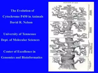
Humans CYP450 -18 families, 43 subfamilies • CYP1 drug metabolism (3 subfamilies, 3 genes, 1 pseudogene) • CYP2 drug and steroid metabolism (13 subfamilies, 16 genes, 16 pseudogenes) • CYP3 drug metabolism (1 subfamily, 4 genes, 2 pseudogenes) • CYP4 arachidonic acid or fatty acid metabolism (5 subfamilies, 11 genes, 10 • pseudogenes) • CYP5 Thromboxane A2 synthase (1 subfamily, 1 gene) • CYP7A bile acid biosynthesis 7-alpha hydroxylase of steroid nucleus (1 • subfamily member) • CYP7B brain specific form of 7-alpha hydroxylase (1 subfamily member) • CYP8A prostacyclin synthase (1 subfamily member) • CYP8B bile acid biosynthesis (1 subfamily member) • CYP11 steroid biosynthesis (2 subfamilies, 3 genes) • CYP17 steroid biosynthesis (1 subfamily, 1 gene) 17-alpha hydroxylase • CYP19 steroid biosynthesis (1 subfamily, 1 gene) aromatase forms estrogen • CYP20 Unknown function (1 subfamily, 1 gene) • CYP21 steroid biosynthesis (1 subfamily, 1 gene, 1 pseudogene) • CYP24 vitamin D degradation (1 subfamily, 1 gene) • CYP26A retinoic acid hydroxylase important in development (1 subfamily member) • CYP26B probable retinoic acid hydroxylase (1 subfamily member) • CYP26C probabvle retinoic acid hydroxylase (1 subfamily member) • CYP27A bile acid biosynthesis (1 subfamily member) • CYP27B Vitamin D3 1-alpha hydroxylase activates vitamin D3 (1 subfamily member) • CYP27C Unknown function (1 subfamily member) • CYP39 unknown function (1 subfamily member) • CYP46 cholesterol 24-hydroxylase (1 subfamily member) • CYP51 cholesterol biosynthesis (1 subfamily, 1 gene, 3 pseudogenes) lanosterol • 14-alpha demethylase
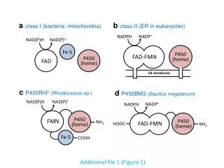
Structure • Till 2001 there was no mammal CYP. • P450cam structure was solved in 1987 • x-ray structure of P450cam with different substrate and inhibitors.
Heme exists in hydrophobic environment, oriented nearly parallel to the surfaces between the L and I helices. Heme-ligating Cys-357 (beginning of L) • Helix-rich on the right side • Beta-sheet-rich on the left side • 14 alpha helices, 5 anti parallel beta-sheets • Compact structure, especially the helical region.
Closed structure, conformational dynamic is essential. • No obvious substrate channel. • The area bounded by B’ F/G and beta 5 identified as the channel. • 6 water molecule fill the substrate active site • Substrate binding loop residues 80-103 • Binding free energy is most likely due to hydrophobic interactions of the substrate and the heme, Leu-244 and Val-295
Structural Model for CYP450 Substrates and inhibitors • Large number of drugs chemical are already known • Systematic attempts to explore substrate and inhibitor specificity of individual cytochrome P40 species
Motivation • Chemical toxicity studies • Predict whether therapeutic effect may be subjected to individual variations. • Predict drugs inhibition.
Elucidate Specificity approaches • Determination of three dimensional structure of the active site. • Design of pharmacophor: • Molecular modeling • Quantitative structure activity relationship
Three dimensional Structure of the Active Site • In P450cam substrate binding, there are three regions of AA flexibility. • One at the substrate binding site • Two are at the assumed substrate access channel • Backbone flexibility of P450cam in case of inhibitor binding.
Conclusions • X-ray structure can serve as an appropriate basis. • Taking to account the degree of flexibility at the active site • Water molecule might accommodate with the active molecule • The development of novel substrate or strong inhibitors might be achieved by docking experiments, energy minimization, molecular dynamics, comparison of electrostatic potential permit.
Design of Substrate Inhibitor Model • Empirical Models • Computer Aided Molecular Design of Pharmacophor Models
Empirical Models • Detect common structural features by: • Comprise stereochemical analysis of metabolites • Binding studies with substrate analogs • Space-filling models • Small number of substrates
Computer Aided Molecular Design of Pharmacophor Models • Quantitative Structure Activity Relationship • Molecular Modeling
Quantitative Structure Activity Relationship • Computational chemistry represents molecular structures as a numerical models and simulates their behavior with the equations of quantum and classical physics. • Available programs enable scientists to easily generate and present molecular data including geometries, energies and associated properties (electronic, spectroscopic and bulk). • The usual paradigm for displaying and manipulating these data is a table in which compounds are defined by individual rows and molecular properties (or descriptors) are defined by the associated columns. • A QSAR attempts to find consistent relationships between the variations in the values of molecular properties and the biological activity for a series of compounds. A QSAR generally takes the form of a linear equation • Biological Activity = Const + (C1 P1) + (C2 P2) + (C3 P3) + ... • where the parameters P1 through Pn are computed for each molecule in the series. • coefficients C1 through Cn are calculated by fitting variations in the parameters and the biological activity.
Molecular Modeling • Utilizing computational techniques to build a pharmacophor by superimposing 3D structures of the ligands. • Identify low-energy conformers. • Identify common electrostatic features. • Structures are superimposed using least squares fit methods.