The Bernard G. Greenberg Lectures
1.27k likes | 1.7k Vues
Breast cancer has served as the disease to illustrate the ideas and apply the results. ... Breast Cancer-Clinical trials The 8 randomized trials have mainly ...

The Bernard G. Greenberg Lectures
E N D
Presentation Transcript
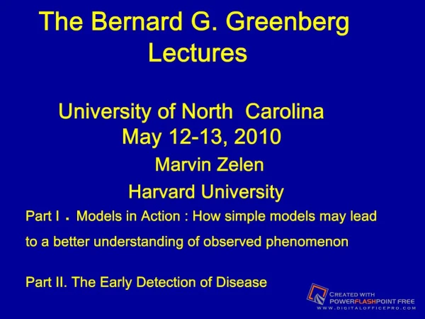
Slide 1:The Bernard G. Greenberg Lectures University of North Carolina May 12-13, 2010
Marvin Zelen Harvard University Part I . Models in Action : How simple models may lead to a better understanding of observed phenomenon Part II. The Early Detection of Disease
My Collaborators Sandra J. Lee, Dana-Farber Cancer Institute and Harvard University Ori Davidov, University of Haifa (now visiting NIEHS) Hui Huang , Dana-Farber Cancer InstituteSlide 3: Part I : Models in Action. How simple models lead to a better understanding of observed phenomenon.
Introduction and general remarks Illustrations 1. Waiting for a bus. Why does it take so long? 2. Family history and disease. Are family members at risk if there is at least one member of family with disease. 3. Cell kinetics. Why do people become refractory to therapy? 4. Prevalence and incidence of disease. What can we learn knowing prevalence and incidence. 5. Data base studies 6. Recurrence time distributions 7. Length biased sampling
Slide 4:Models
Many areas in science and technology cannot seek reliable answers to relevant questions by experiments or empirical studies --- not feasible --- too costly --- takes too long. Models may be developed to describe the phenomenon. The goals of such models are to use known inputs to predict outcomes and to determine how changes in the inputs affect outcomes. Models may serve as a guide for data collection and suggest experiments. inputs model Outputs
Slide 5:Models in Science
Science has many different kinds of models Animal Models : To study human disease Physical models : To demonstrate the apparent truth of a principle. Mathematical Models : To gain insight and understanding of a phenomenon which is being observed.
Slide 6:Models and Their Uses
Nearly all models are simplifications of the phenomenon being observed . However in order for a model to be useful, it must capture the important features of the phenomenon being observed. Models can be used to guide the design of experiments Models may suggest the kinds of data to be collected
Slide 7:Limitations of Models
Models may not entirely reflect features of the observed phenomenon; e.g.: animal disease may not be same as human disease prototype model may not scale up to actual model equations may not properly describe phenomenon Parameters of model may not exist or estimates may be unreliable; e.g. growth of tumor
Slide 8:Probability Models
A probability model is a mathematical model which incorporates the probability aspects of the observed phenomenon when the observed phenomenon has outputs which are subject to probability laws. If the observed process has a time parameter ( or is indexed by something equivalent) we refer to the probability model as a stochastic process.
Slide 9:Data Models
Underlying all statistical analyses is a model of how the data was generated. The most common models relate to linear models where there are unknown parameters and a data structure characterizing the expected values and second moments ( variances and covariances) of the data. Example : Measurement = True value + random error
Slide 10:Mechanistic models
Another kind of model is �mechanistic models�. Such models incorporate the main features of an observed phenomenon and provide a way for predicting outcomes when the input variables are changed. These models attempt to impart an understanding of how outcomes are changed with changing inputs. �They drive our thinking� In this series of lectures we will attempt to illustrate some applications of mechanistic models.
Slide 11:Why Use Mechanistic Models?
Many areas in science and technology cannot seek reliable answers to relevant questions by experiments or empirical studies--not feasible--too costly--takes too long -- may be unethical. Examples : How often should a person visit their physician? Should a therapy be withheld from a man diagnosed with prostate cancer ( no signs or symptoms ) because there is a high probability that it will never be clinical in his lifetime? Model may contain as inputs : Individual health history, risk factors for potential diseases and possible outcomes if disease is discovered early .
Slide 12:Waiting for a Bus
Problem: Why is it when I wait for a bus it always seems to take an unusually long time? It seems that I just missed the bus. Was I just born unlucky? � Or do I have a psychological hang-up? � Or is it really true.
Slide 13:Hypothetical Situation
Observed time between bus arrivals 60 minutes Time 5 Intervals in 60 minutes MBTA = Mean time between arrivals = Total observed time/ No. of intervals = 60/5 = 12 minutes a b c d e f
MBTA = 12 minutes a b c d e f 60 minute clock a b d e Equivalent to placing 5 points at random on clock; i.e. c Flip pointer 5 times 5 intervals Suppose I decide to catch a bus sometime during the observational period. How long do I have to wait for the bus to arrive (on the average)? What is the expected mean waiting time if MBTA = 12 minutes. Waiting time Time when I arrive Find Mean Distance Pe a b c d e P a b c d e f P Informal Analysis Suppose the arrival point is placed on the clock initially P P Or equivalently spin the pointer once and move x to P. Conclusion: Placing a point on a clock is the same as spinning a pointer once. But we have 5 spins and one placed point. This is equivalent to 6 spins and results in 6 intervals. Hence average time between point is 60 minutes = 10 6 Therefore average waiting time = 10 minutes. Reason for unexpected result is that my arrival at the bus stop is equivalent to choosing an inter-arrival interval. The probability of choosing an interval is proportional to the length of the interval. In reality I am sampling possible intervals. The longer the interval the greater the chance of choosing it. This kind of sampling is called Length Biased Sampling. Length Biased Sampling occurs often in modeling biomedical phenomenon.Slide 19:Choosing an interval in proportion to its length
Observed time between bus arrivals 60 minutes Time Choosing a point at random on the line is equivalent to intersecting an interval proportion to its length a b c d e f
Slide 20:What happens to my waiting time if the time between arrivals is smaller?
Suppose the time between bus arrivals is 6 minutes (double the number of buses ). This is equivalent to observing 10 intervals (11 bus arrivals within the hour). Hence by our model, we will have 10 points (buses) and one placed point on the circle. The mean waiting time is 60/11 =5.5 minutes. �Equivalent� to missing the bus by half a minute.
Slide 21:2. Drug Therapy and Cell Kinetics
Life cycle of cell consists of four states G1 : pre-DNA synthesis S : DNA synthesis G2: post-DNA synthesis M : Mitosis G1 S G2 M Two daughter cells Cell maturation Back to cell cycle
Slide 22:Drug Therapy
Some drugs (chemotherapy against cancer) may only destroy tumor cells when they are in the S phase. As a result the longer a tumor cell is in the S phase the more likely it will be destroyed. Consequently the probability of a tumor cell being destroyed may change with additional courses of therapy as the cells with longer S phases tend to be removed from the population. Eventually the tumor cells remaining will have very short S phases and it will be difficult to destroy them . With repeated courses of therapy , the drug will not seem to have any therapeutic effect and the patient will be described as being refractory to treatment with this drug. If therapy can be continually infused, this may increase the probability of destroying tumor cells from a changing population.
Slide 23:Process of Cell Destruction
Idealized process Duration of S Phase Duration of intermitotic times (Cell cycle time) x x X x x x x x x X x x x x x x x x Exposed to therapy Time
Slide 24:3. FAMILY HISTORY AND DISEASE
Some chronic diseases have been found to be associated with family history of disease. Individuals having family members with disease may be at higher risk for disease. Examples: Prostate Cancer, Breast Cancer, BRCA-1/BRCA-2 gene mutations.
Slide 25:Family History and Breast Cancer
Breast cancer risk is higher among women whose close blood relatives have this disease. �Having one first-degree relative (mother, sister, or daughter) with breast cancer approximately doubles a woman's risk. Having 2 first-degree relatives increases her risk about 5-fold�.
Slide 26:STUDY DESIGNS for EVALUATING FAMILY HISTORY
Random Sample of Families from Population Case Control Study With Two Probands
Slide 27:Study Design :Random Sample of Families from Population
Carry out random telephone calls. Determine if probability of having more than one disease case in family is larger than expected. If disease incidence is low, this method may not be feasible. (Breast cancer: disease incidence is 80-100/100,000 per year depending on age). Most of phone calls will not have any disease in family.
Slide 28:Study Design : Case control study
Collect pairs of observations in which one member of pair has disease and the other does not. These are called probands. Then determine if remaining family members have disease for each proband. Calculate observed proportion of having at least one of the remaining family members with disease.
Slide 29:Study Design: Case control study
The proband member of pair with disease can be recruited from hospital . (Referent registry)--- ( R=1) The proband member without disease can be recruited by a random sample. --- (R=0)
Slide 30:Some Calculations
Assume disease incidence is independent of family ? = P{ individual is incident with disease} (Assume ? is very small) S = No. of people in family with disease N = Family Size, P{ N = n | R=0} = p(n) for population (proband without disease) P{ S = 1 | N = n } = 1 � (1- ? )n ~ n? ignoring terms of order ?2. P{S = 1 , N = n ) ~ n? p(n) , ignoring terms of order ?2 P{ N = n | S = 1} = n? p(n) / ?? n p(n) = n p(n) / m ( m= mean family size) P{ N= n | R=1} = n p(n) / m where R= 1 indicates case control proband Note : E[N |R=1] = E[N2 ] / m = (s2 + m2) /m= m [ 1+C2 ] , C2 =s2/m2
Slide 31: P{ N= n | R=1} = n p(n) / m The distribution of family size for a referrent registry is different than the distribution in the population P{ N=n | R=0 } = p(n)
Assume that individuals in family are independent with respect to disease incidence. Then in the family sampled from referent registry having (n-1) remaining members has P{ S=2 |R = 1, N=n} = p{ S = 1| R=1, N=n-1} = 1- (1-? )n-1 ~ (n-1)? ~ n? P{S=2 , N=n | R = 1} ~ ? n2 p(n)/m and P{ S=2 | R = 1} ~ ? ? n2 p(n)/ m =? [ E(N2) ]/ m = ? [ m( 1 + C2) ] Whereas if the population was sampled ,taking into account one family member without disease, P{ S = 1 N=n |R = 0} ~ (n-1) ?~ ? n and P { S = 1 | R=0 } ~ ? ? n p(n) = ? m Hence the ratio is P{S=2 |R = 1 } / P { S = 1| R=0} ~ 1 + C2 Thus assuming independence of family members having disease , the calculations show a bias in which , conditional on having one family member with disease , the probability of having additional family members with disease is greater than a randomly chosen family.
Note: Larger families have a greater probability of having at least one member with disease than smaller families. Conclusion: Referent registries tend to have larger size families then the general population. The probability of having more than one family member with disease is higher for families in referent registry compared to families in general population. This will be true even if family history is not a risk factor for disease. Referent registries yield biased results ConclusionsSlide 33:EXTENT OF BIAS IN FAMILIES FOR RELATIVE RISK
Family Bias (1 +C2 ) Single gender families 1.75 Maternal line 1.5
Slide 34:4. Prevalence and Incidence
Prevalence refers to how many people have disease at some point in time . Incidence refers to how many new cases of disease occurred in a defined time period. Example : In the U.S. in 2007, 2. 6 million women were alive with a diagnosis of breast cancer ; 192,000 new breast cancer cases Since there are approximately 66,000,000 women over the age of 40, we can standardize these numbers by stating in this age group the prevalence is 39 per thousand ; incidence is 2.9 per thousand per year
Slide 35:Prevalence and Incidence
Prevalence may be indicated by P(t) Incidence may be indicated by I(t) t may refer to chronological time or age. In probability terms, Prevalence can refer to the probability of being in a disease state (and alive). Incidence refers to the probability of a transition from no disease to disease over a given period of time.
Slide 36:Models: Stable and Non-stable Disease
Stable disease model : P'(t) = 0 Implies P(t) = P , I(t) = I P = I m where m is mean time with disease. Breast cancer : m = P/I = 39 / 2.9 = 13.5 years Non-stable disease model : P'(t) ? 0 Prevalence and incidence are functions of time. Note : If t refers to chronological time, the stable disease model may be true for some diseases. Origin can be chosen such that P(0) = P
Slide 37:Prevalence , Incidence and Mortality Models
I(t) : Incidence of disease at time t. (t may refer to age or chronological time). M(t) : Mortality of disease at time t P(t) : Prevalence of disease at time t ( Probability of having disease at time t) What are relations between prevalence , mortality and incidence? Can the numbers estimate how long people live with disease? Time Transition from no disease to disease (Incidence) Transition From disease to death (mortality) Disease state Disease free state
Slide 38:Modeling Prevalence and Incidence: Stable Disease Model
where m is the expected value of T (survival)
Slide 39:Non �Stable Disease Model
Suppose P(t) depends on t. Then it can be shown that approximately P(t) ~ m I(t) { 1- � m (C2 - 1) I'(t)/I(t) } where C = s/m. If the time with disease follows the exponential distribution (C = 1) and P(t) = m I(t) exactly. Actually : The N & S condition for P(t) = m I(t) is that T follows an exponential distribution
Slide 40:5. Backwards and Forward Recurrence Times
Backward and Forward Recurrence Times arise in many applications. Examples : Studying the natural history of a disease Early detection of disease Over diagnosis
Slide 41:Definition: Backward and Forward Recurrence Times
Let T be a non-negative random variable and T= t be a possible outcome. Suppose t is randomly divided into two parts: t=U+V U V U = Backward Recurrence Time V = Forward Recurrence Time 0 t
Slide 42:Bus Waiting
A person goes to a bus stop to take a bus. The backward recurrence time is how long the person missed the bus. The forward recurrence time is how long the person has to wait for the next bus to arrive Time U V Arrival at bus stop Bus Arrivals If bus schedule does not change with time, distribution of U and V does not change with time. If bus schedule changes with time, distribution of U and V will be time dependent.
43Slide 43:Application to Chronic Disease Modeling
No disease Alive with Disease Death (S0) (Sa) (Sd) Time S0?Sa Sa?Sd Time: Chronological Time or Age Point Incidence: S0?Sa Point Mortality: Sa?Sd Survival Time
44 Suppose at chronologic time t0 the disease process is beginning to be observed. The process has been going on a long time. Time t0 Some individuals will be in disease free state (S0), others will be alive and in disease state (Sa) at time t0. Backwards Recurrence Time: How long a person has disease up to time t0. Forward Recurrence Time: Time to death with origin at time t0. Only will be observed with additional follow-up time. 45 t0 Length Biased Sampling Individuals in Sa at time t0 are not a random sample of the population with disease. The longer they are alive, the more likely they will be in Sa at time t0. Issues Probability of being in Sa at time t0 ? Does proportion of people in Sa contain information on survival? How long do people in Sa at time t0 live relative to people who become incident with disease after t0? (Forward Recurrence Time) Total time in Sa ? (Survival) Role of disease incidence?Slide 46:Theory: Length Biased Sampling
a(t) = 1 if time with disease is t in sample P{ a(t) = 1| T = t } ~ t ( Length biased sampling) P{ a(t) = 1| T = t } = independent of t ( random sampling) P{ a(t) = 1, t<T = t +dt } ~ t q(t)dt , T has pdf q(t) P{ t<T= t+dt | a(t) = 1} = t q(t) dt / m , m= E(T) Note: E(T | a(t)= 1} = E(T2)/m = m(1+s2/m2) > m P{ t<T= t+dt | a(t) = 1} = t q(t) dt / m , m= E(T)
Slide 47:Theory: length biased sampling and recurrence times
U = Backward recurrence time V = Forward recurrence time Time a(t) = 1 if chosen by length biased sampling and time with disease is t T= U+V is time with disease having pdf q(t) Forward recurence time distribution: f( v | a(t)=1, T=t ) = 1/t , 0 = v < t , f(v , T=t | a(t)=1} = (1/ t ) t q(t)/ m = q(t)/m , 0 = v < t f(t |a(t) = 1 ) = t q(t)/m
Slide 48:Observational Study on a Population
Consider observing a population of individuals. Suppose the observational period begins at time t1 and ends at time t2.. Suppose the individuals in the population are sick or well. Those who are sick have a probability distribution for length of sickness period. Time U U V Those who are ill at time t1 U = how long in sick state before being observed. V = length of time being observed beginning at t1 . Those who are ill at time t2 U = observed period of sick time. (Sick time is right censored) V = residual sick time after observations have ceased. Distribution of U and V depend on whether incidence of sickness is independent or dependent on time t1 Length of line indicates Duration of illness t2 U V
Slide 49: Example: Early Detection of Disease
Suppose a person is in three states with respect to a particular disease ; i.e. S0: Disease free state or if with disease cannot be detected. Sp: Pre-clinical state. Disease may be diagnosed with special examination only. No symptoms. Sc: Clinical state. Disease shows clinical symptoms and is diagnosed. U V S0 Sp Sp Sc Age Disease detected by special exam U: How long person has had disease before diagnosis V: Time gained by earlier diagnosis If incidence of disease (S0 ? Sp) changes with age, then distribution of U and V will depend on age. Lead time
Duration of Pre-clinical State Lead Time Time of Screening Clinical Inception Point diagnosis Of disease |||||||||||||||||||||| Early detection of disease Consider a population of individuals who are screened Screening point Each horizontal line represent duration of pre-clinical disease for an individual OBSERVE: Vertical dotted line has a higher probability of Intersecting horizontal line; i.e.: the screening procedure finds those individuals having longer pre-clinical durations. This kind of sampling is called length biased sampling. Consider a population of individuals who are screened TimeSlide 52:2.9 Overdiagnosis: Prostate Cancer
Background: Prostate Specific Antigen (PSA) test is widely used to diagnose prostate cancer. Nearly all diagnosed cases by PSA are asymptomatic. Question: Would the prostate cancer exhibit clinical symptoms during a man�s lifetime? If no --- PSA diagnosis is an overdiagnosis Time from Diagnosis to death Lead Time S0?Sp PSA diagnosis Death Sp ? Sc Overdiagnosis
Slide 53:Calculations for Over Diagnosis
S0 : disease free state, Sp : pre-clinical state, Sc: clinical state w(x) =P{ transition S0 Sp at age x} , y = age entering clinical state q(y) = pdf in pre-clinical state z = age of diagnosis of disease by early detection program P { T>t } = R(t) where T = survival without disease L = y � z (Lead time) Sequence of events for over diagnosis conditional on being diagnosed at age z Enter Sp at age x, if alive enter Sc at age y , Early diagnosis at age z, Die between (z, z+L) conditional on surviving at least to z where L = y - z. X z death y L
Slide 54:Comparisons of Probability of Over Diagnosis Prostate cancer
Age Model Draisma 55 0.26 0.27 60 .33 .38 65 .40 .47 70 .48 .53 75 .57 .56 80 .65 Draisma et al. jnci (2003)�Based on European Trial; (sojourn time 15.4 yr, relevant cases), Populations : model (U.S. ,all races), Others are white.
The Bernard G. Greenberg Lectures. Part II. The Early Detection of DiseaseSlide 60:Outline :Early detection of disease
Background, Motivation and Rationale 2. Early Detection Clinical Trials 3. The Early Detection Process Models Length and Lead time biases Dynamics
Slide 61:Outline (continued)
4. Applications (breast cancer) Mean lead time vs. disease stage Comparisons of U.S. , U.K. and Nordic public health programs U.S. Preventive Services Task Force recommendations Risk based schedules vs. time based schedules Maximum benefit from screening (?) Is screening women under age 50 and over age 65 beneficial? Screening high risk individuals Over diagnosis (breast cancer) 5. Over diagnosis (prostate cancer)
Slide 62: Motivation
This class of problems has been motivated by the belief that the early detection of disease may lead to higher cure rates and reduced mortality. Breast cancer has served as the disease to illustrate the ideas and apply the results. However the ideas are general and apply to many other cancers as well as possibly to other diseases.
Slide 63:Background and Rationale
Screening Programs : Special exams to diagnose disease when it is asymptomatic. Rationale : Diagnosing and treating the disease early, before signs/symptoms appear, may result in more cures and lower mortality. Caution : If treatment is of little benefit there may be no reason to diagnose disease early.
Slide 64:Examples of Screening Programs
Tuberculosis � Hypertension Diabetes � Coronary Artery Disease Cancer � Thyroid Disease Breast Cancer � Osteoporosis Cervical Cancer � HIV Colorectal Cancer Lung Cancer Prostate Cancer
Slide 65:Some Issues
Public Health Programs : Recommendations Initial age to begin screening, intervals between exams, schedules for high risk individuals. Recommendations should be made by risk status. --- How ?? Costs may be an important consideration. Over diagnosis Disease is diagnosed early but would not be diagnosed by usual care in a person�s lifetime --(Breast and prostate cancer) Maximum benefit What is the maximum benefit that can be achieved from screening by searching for optimal exam schedules ????
Slide 66: Example : Breast Cancer Screening Using Mammography
Breast Cancer-Clinical trials The 8 randomized trials have mainly included women age 50-65. Do younger women ( under age 50 ) and older women (older than age 65) benefit from screening? The U.S. Preventive Service Task Force recommends that beginning at age 50 women have biennial exams. (2009) Nevertheless the American Cancer Society recommends that annual screening begin at age 40 for women at average risk. United Kingdom : The National Health Service offers screening beginning at age 50 with three year intervals for subsequent exams. Nordic countries : The recommendation is that screening begin at age 50 with two year intervals for subsequent exams. What are the trade-offs for the different recommendations?
Slide 67:Scientific Evidence of Screening Benefit
Diagnosing disease early does not necessarily result in benefit; e.g. diagnosing a primary cancer earlier may not be of benefit if the disease has already metastasized. If an effective treatment does not exist, there is no benefit in diagnosing disease early. The general consensus is that randomized clinical trials are the only way to evaluate screening programs for potential benefit.
Slide 68:Early Detection Randomized Clinical Trials
Typical trial consists of two groups . One group (control) receives usual care; the other group (study group) receives invitation to have a finite number of special examinations. Follow up for disease occurrence and death continues after the last exam. Endpoint is death from disease. Randomization may be carried out on an individual basis or by cluster randomization ; e. g. geographical region, physician practice.
Slide 69:Early Detection vs. Therapeutic Trials
Statistical Problem : Design of Early Detection Clinical Trials. -- How many subjects, exams, exam spacing, follow up and optimal analysis time, etc.
Slide 70:Early Detection Clinical Trials
Only subjects who are diagnosed with disease carry information about benefit. Trials need very large number of subjects Relatively low incidence is characteristic of many chronic diseases; e.g. female breast cancer incidence is about 80-100 per 100,000 women per year depending on age. Typical trial will require 10-20 years. During this time the technology for diagnosing disease may have changed. Conclusions may be of limited interest. Is it possible to carry out an early analysis, with limited follow up time?
Slide 71:Need for Models
Issues in the previous slide (optimal schedules, over diagnosis, benefit for women over age 65 and under age 50), high risk individuals, cannot be readily addressed by Randomized Clinical Trials. May not be feasible. Too many variables, takes too long, too costly , ethical concerns. Issues may be addressed by modeling the early detection process
Slide 72:Models
S0: Disease free state : Does not have disease or has disease which cannot be detected by exam. Sp: Pre-clinical state: Has disease but no signs or symptoms; capable of being detected by exam. Individual is asymptomatic. Sc: Clinical state : diagnosis by usual care. S0 ? Sp? Sc : Progressive disease model (Breast cancer) Sp S0 Sc: Progressive disease model : subgroup Sp never goes on to clinical disease -Prostate cancer S0 ? Sp ? Sc : Non-progressive disease model (HPV ,Cervical cancer)
Slide 73:Issues in the interpretation of data
Suppose a group of subjects undergo screening for a particular disease and a number of subjects are diagnosed and treated. The subjects in this screened group have longer survival than a control group (no screening). Is this scientific evidence of the benefit of screening? No ---- Length biased sampling and lead time bias may introduce significant biases
Duration of Pre-clinical State Lead Time (forward recurrence time) Age of Screening Clinical Inception Point Diagnosis Of disease (Early diagnosis) |||||||||||||||||||||| S0 Sp Sp Sc Natural History of Progressive Disease Age S0 = disease free Sp = pre-clinical Sc = clinicalSlide 75:Length biased sampling
Consider a population of cases Time Screening point Horizontal line : duration of time in pre-clinical state Diagnosis : equivalent to placing a random vertical line. Intersection represents case diagnosed. Vertical line is more likely to intersect longer horizontal lines ---people will tend to live longer.
Slide 76: Why would screening result in benefit ?
If screen diagnosed cases are found in an earlier disease stage compared to usual care then there is likely to be benefit. This is referred to as a stage shift . Stage shift can be due to a long lead time ; i.e.cases are diagnosed before they transit to a more advanced prognostic stage. Stage shift may also arise from the length biased sampling. The selection of cases by screening may also be associated with earlier prognostic stages.
Slide 77:Natural History of Disease
S0: Disease Free State or Cannot Be Detected Sp: Pre-clinical State Sc: Clinical State Time (age) Stage II Stage I S0?Sp Sp? Sc Time in stages I and II
Slide 78:Stage Shift and Earlier Diagnosis
S0: Disease Free State or Cannot Be Detected Sp: Pre-clinical State Sc: Clinical State Time (age) Stage II Stage I Early Diagnosis S0?Sp Sp? Sc Note : the longer the mean lead time the greater the probability of diagnosing disease in an earlier stage. Lead time Lead time
Slide 79:Lead Time Bias :Usual care
S0 ? S Sp ? Sc Death Age 50 55 60 Survival from Clinical Diagnosis = 60 � 55 = 5 Years clinical diagnosis S0 = disease free state, Sp = pre-clinical state Sc = clinical state
Slide 80:Early Detection But Survival Is Not Enhanced
Age 50 60 Survival from Screening Diagnosis 60 � 53 = 7 Years 53 Screening Point and Diagnosis S0?Sp Sp?Sc Death Survival (with usual care diagnosis) 60 - 55 = 5 years Diagnosis: usual care 55
Slide 81: Dynamics of the Natural History (1) : Usual care
Age x t y S0 Sp Sp Sc Sc Sd Disease States S0 : Disease free state � disease free or disease state which cannot be detected Sp : Pre-clinical state - asymptomatic with no signs/symptoms Sc : Clinical state � when diagnosed by routine methods Sd : Death state (death due to disease) Usual care : disease is diagnosed and treated at t. . Age x not observed Disease incidence Death from disease Survival (y� t)
Slide 82: Dynamics with Screening (2) : Exam detected case at tS
ts t Sd S0 Sp Sp Sc Ages and x are not observed. Treatment begins at tS Observed survival time (y � ts) (?� ts ) is lead time. Imputed survival = Survival with origin ? = (observed survival) � ( lead time) There may be a number (unknown) of false negative exams x y Exam detected Age Lead Time Lead Time (y � ts) Imputed Survival Observed Survival S0 = disease free Sp = pre-clinical Sc = clinical Sd = death from disease t0 t1 � tj-1 � tj � Exam times Exam times
Slide 83:Dynamics with Screening (3)
t0 t1 tj-1 tj � tr-1 t tr � Survival (y - ) S0 Sp Sp Sc x Interval Case : Case diagnosed between tr-1 and tr y Sc Sd t Time Exams at t0 < t1 < � < tr-1 **
Slide 84:Applications
Breast Cancer Screening (Mammography) Public Health Programs (Breast cancer ) Relation between mean lead time and proportion of negative nodes. Comparison of U.S., U.K. and Nordic countries Choosing screening intervals according to risk. Maximum Potential benefit High Risk Women Do women under 40 and/or over 65 benefit from screening ? Over diagnosis : breast and prostate
Slide 85:Relation of Mean Lead Time and Proportion of Negative Nodes
Most important prognostic variable is whether disease has spread to lymph nodes ; nodes are negative if has not spread , positive otherwise. Distribution of Lead Time derived theoretically � function of exam schedule, risk status and sensitivity. Proportion of negative node diagnosed patients can be found from clinical trials and data bases.
Initial exam (2 county) Subsequent exams (2-county) p = (.080 x mean lead time) + 0.546, p = proportion of negative nodes Mean sojourn time varies by age from 2 years at age 40 to 4 years at age 50 and above. Sensitivity varies from 0.7 at age 40 to 0.9 at age 50 and above. proportion of negative nodes vs mean lead time (2-county, BCSC, HIP, Edinburgh) Initial exam (2 county) Subsequent exams (2-county) Ln(p/1-p) = (.35 x mean lead time) + 0.111, p = proportion of negative nodes Mean sojourn time varies by age from 2 years at age 40 to 4 years at age 50 and above. Sensitivity varies from 0.7 at age 40 to 0.9 at age 50 and above. ln (p/1-p) vs mean lead time (2-county, BCSC, HIP, Canadian I & II)Slide 88:Value of plot : proportion of negative nodes vs. mean lead time
Clinical trials are necessary to evaluate benefit � mortality reduction. If stage distribution is known at diagnosis it is possible to estimate mortality reduction as a function of follow up time. Given : screening schedule, age distribution of population and sensitivity, the mean lead time may be estimated using theoretical distribution. . Hence from plot , the proportion of cases in each stage can be estimated. This allows an estimate of mortality reduction . � No need for clinical trial. Side issues : how does mortality reduction change with changes in sensitivity and scheduling. Methods are very general and may be used for other diseases which satisfy basic assumptions � natural history is progressive and benefit derives form stage shift.
Slide 89:Does length biased sampling of cases account for earlier stage disease ?
Compare stage of interval diagnosed cancers with control group. �Why? The length biased process of early detection selects cases who tend to sojourn longer in pre -clinical state. If longer sojourn times are associated with earlier stage disease , then the interval cancers will tend to have shorter sojourn times and have more advanced staging. If stage of interval cancers is same as control ( no screening group), then length bias does not account for stage shift ; i.e. stage shift is due to diagnosing the disease earlier before it transits to a more advanced stage.
Slide 90:Summary on Stage Shift : Breast cancer
Trial Stage Control Study Screen Interval Detected Detected HIP N+ 52% 41% 30% 50% 2-County II-IV 46% 30% 19% 45% Malmo I * � 59% 39% 23% 56% Edinburgh � 87% 64% 50% 72% ----------------------------------------------------------------- * II-IV : Stages II - IV ( AJCC) The interval cancers tend to have the same stage as a control group. If stage shift was due to length biased sampling, the interval cancers would tend to have more advanced stage than a control group. This is not true.
Slide 91:THRESHOLD METHOD : Application to breast cancer
Background There is general agreement that mammographic screening should begin at 50 years of age. Prevalence at 50 = 1.8 cases/ 1000 Prevalence increases with age First exam at age 50 . Choose subsequent exams so that independent of age, the risk will never be larger than at 50.--- 50, 54, 57, 60, 63, 66, 69, 72, 75, 77
S(t)=Prob(Undiagnosed Pre-Clinical Disease) Exam Schedule with S(50)=.0062 (Threshold Value)Slide 93:Mammogram Exam Schedules for Ages [50, 79]
Annual: U.S.: ACS/NCI Recommendation Every 2 Years: U.S. Preventive Services Task Force and Nordic Recommendation Every 3 Years: U.K. Recommendation * Mortality Reduction = [Mortality (controls) � Mortality (screened )] [ Mortality Controls ] *Threshold exam schedule corresponds to exam schedule whenever risk is the same as at age 50.
Slide 94:Risk Based Screening
Current recommendations for screening are based on time and initial age to initiate screening program. Since goal is to diagnose individual in pre-clinical state � define Risk = Probability individual is in pre-clinical state.
Slide 95:Choosing initial age
Incidence of many chronic diseases increases with age. Hence risk increases with age. The choice of the initial age to begin screening assumes that the risk for lower ages is not high enough to warrant the beginning of a screening program.
S(t): Probability of having undiagnosed pre-clinical disease at age t ( breast cancer) S(40)=0.00164 S(50)=0.0062 S(45) = 0.00352Slide 97:U.S. Preventive Services Task Force (USPSTF) Recommendations for Breast Cancer Screening
November 2009 : Recommends biennial screening mammography only for women ages 50�74. Resulted in much controversy. ACS had long recommended annual screening beginning at age 40. ACS opposed recommendation. December 2009 : USPSTF modified recommendations by stating decision to �start regular biennial screening before the age of 50 should be an individual one and take into account patient�s values regarding individual specific benefits and harms�.
S(t)=Prob (Undiagnosed Pre-clinical Disease) Following Biennial Exam Schedule with =.90 for [50,74] Note: All exams past the age of 50 are at lower risk. S(t)=Prob(Undiagnosed Pre-Clinical Disease) Exam Schedule with S(50)=.0062 (Threshold Value) S(t)=Prob(Undiagnosed Pre-Clinical Disease) Exam Schedule with S(40)=0.00164 (Threshold Value) Note : In order to maintain the same risk level as at age 40, it is necessary to have more than one exam per year after age 56. S(t) with Threshold Method starting at age 40 Note: to maintain threshold , it is necessay to have multiple exams in same year. S(t) with Threshold Method Starting at Age 40 Using Non-Constant Threshold Values .00164 .0042 .00495 .0056 Threshold level changed so that 80% of cases diagnosed in interval.Slide 103:Summary
Recommendations No. of exams Mortality Reduction*** USPSTF* 13 21.8% Threshold* 9 19.0% Mixed threshold** 28 27.9% ACS** (annual) 35 26.8% Threshold** 55 31.2% * Screening ages : 50-74 * * Screening ages : 40-74 *** Deaths counted in age range 40-85
Slide 104:Conclusions
Schedule recommendations based on fixed time periods are illogical as some exams are ultimately given at lower risk levels than the initial exam risk. Risk based schedules are superior to fixed time schedules and result in lower costs and/or higher benefit.
Slide 105:Maximum Benefit from Screening
Maximum benefit occurs if 100% of cases are diagnosed by early detection exam. Schedule Sensitivity = Proportion of cases diagnosed by exam = E / E + I + B E = Exam diagnosed cases I = Interval cases B = Cases diagnosed before first exam
Slide 106:Mortality Reduction vs. Schedule Sensitivity : Ages 50-79
Maximum reduction in mortality is 43.5% if 100% of cases diagnosed by exam.
Slide 107:Screening Younger Women [40, 49]
Screening women in [40, 49] Relatively low chance of developing breast cancer Difficulties in detecting breast cancer Relatively high cost 1997 NIH Consensus Development Panel Review of data from 8 clinical trials �The available data did not warrant a single recommendation for all women in their forties.� Involvement of US Congress Recommendation of ACS and NCI
U.S. Breast Cancer Incidence Rates 1975-2006 Low Incidence Rates for Younger WomenSlide 109:Strategy in Evaluating Benefit for Women Aged 40-49
Clinical trials and recent data indicate a stage shift with early detection for this age group. Node negative ~77%% (screening) vs. 53% (usual care). Compare the mortality of a screened group ( screening only for women in the age group 40-49 ) with a control group (not receiving special exams). Only count deaths for subjects who were in the pre-clinical state during ages 40-9. Note that these subjects may die of disease past the age of 49. The population who were in the pre-clinical state in their 40�s is the target population who can benefit.
Mortality reduction : Screening in 40�s 40, 41, 42, 43, 44, 45, 46, 47, 48, 49 (annual) 40, 42, 44, 46, 48, 49 40, 43, 46, 49 40, 45, 49 40, 49 * Counts only deaths for subjects in pre-clinical state in ages 40-9. ** Counts all deaths for ages 40-79.Slide 111:Conclusion
Screening women in their 40�s does result in some benefit. However the contribution to the overall mortality reduction is 5.3% for 10 annual exams over a 40 year follow-up period. This is too small to pick up in a clinical trial unless there is a huge number of individuals in trial.
Slide 112:Stopping Screening by Age
Should screening be terminated for older women? Suppose breast cancer annual screening is initiated at age 50 and mortality is observed to age 79. Screening is terminated at ages : 64, 69 , 74, 79 Question. How is mortality reduction affected by termination of screening? Deaths are counted in the age interval (50-79)
Slide 113:Mortality reduction vs. Age at which screening is stopped (deaths are counted to age 85)
Slide 114:Screening Schedules for High Risk Population
Subpopulations may be at Elevated Risk No Clear Guidelines for Choosing Screening Schedules Based on Risk Status Risk Factors for Breast Cancer Previous breast cancer, mutations on BRCA1 or BRCA2, First degree relatives with breast cancer, 2+ breast biopsies, 75%+ dense tissue on MM Age at menarche, age at first live birth, no of previous biopsies There are subpopulations who may be at higher risk of developing breast cancer. Although it is intuitive they need to start screening at an earlier age and be followed more frequently, currently there are no clear guidelines available. The risk factors for breast cancer are well established. For example the impact of hereditary factors (BRCA1, BRCA2) for breast cancer is well known. Other non-hereditary risk factors for breast caner are also well established. There are models available (such as Gail et al) which can project the risk of developing breast cancer using the hereditary and non-hereditary risk factors. These models can be adopted to estimate the relative risk of developing breast cancer.There are subpopulations who may be at higher risk of developing breast cancer. Although it is intuitive they need to start screening at an earlier age and be followed more frequently, currently there are no clear guidelines available. The risk factors for breast cancer are well established. For example the impact of hereditary factors (BRCA1, BRCA2) for breast cancer is well known. Other non-hereditary risk factors for breast caner are also well established. There are models available (such as Gail et al) which can project the risk of developing breast cancer using the hereditary and non-hereditary risk factors. These models can be adopted to estimate the relative risk of developing breast cancer.
Slide 115:Initial Age to Begin Screening
Women at normal risk have a probability of 0.0062 of being in pre-clinical state at age 50. Can we use this as a guideline to initiate screening of high risk population at earlier age? If recommendation of normal population is to begin screening at age 50, then using the same probability , a high risk population should begin screening at ages: Application of the model to high-risk population We assume the most beneficial effect of screening begins at age 50. For normal risk women the risk of being in pre-clinical state at age 50 is 0.0062. We used this to propose when to initiate screening based on risk status. Consider RR of 1 � 4. RR=1 (normal population). Women at higher risk will reach the probability of being in pre-clinical state of 0.0062 before age 50. One possible guideline to initiate screening is using the age when the probability of being in pre-clinical state reaches 0.0062. Preliminary calculations - assumed RR applies the same throughout all ages equally. For example RR=2 means twice elevated risk at age 40 as well as age 70. Present the initial age by RR in the table. Application of the model to high-risk population We assume the most beneficial effect of screening begins at age 50. For normal risk women the risk of being in pre-clinical state at age 50 is 0.0062. We used this to propose when to initiate screening based on risk status. Consider RR of 1 � 4. RR=1 (normal population). Women at higher risk will reach the probability of being in pre-clinical state of 0.0062 before age 50. One possible guideline to initiate screening is using the age when the probability of being in pre-clinical state reaches 0.0062. Preliminary calculations - assumed RR applies the same throughout all ages equally. For example RR=2 means twice elevated risk at age 40 as well as age 70. Present the initial age by RR in the table.
Slide 116:Overdiagnosis: Prostate Cancer
Background: Prostate Specific Antigen (PSA) test is widely used to diagnose prostate cancer. A positive result triggers a biopsy Nearly all diagnosed cases by PSA are asymptomatic. Question: Would the prostate cancer exhibit clinical symptoms during a man�s lifetime? If not --- PSA diagnosis is an overdiagnosis S0?Sp PSA Death Sp?Sc Diagnosis Over diagnosis: Lead Time > Residual Survival Age Residual Survival (Time from early diagnosis to death from other causes) Lead Time
Slide 117:Comparisons of Probability of Over Diagnosis (Prostate cancer)
Age Model Draisma 55 0.26 0.27 60 .33 .38 65 .40 .47 70 .48 .53 75 .57 .56 80 .65 Draisma et al. jnci (2003)�Based on European Trial; (sojourn time 15.4 yr, relevant cases) Populations : model (U.S. ,all races), Others are white.
Slide 118:Probability of Over-diagnosis Conditional on Age at Detection: Breast Cancer
Age Probability Conclusion . Over diagnosis not a serious problem
Slide 119:Conclusions
1. Models are necessary in order to answer important issues arising in the screening of chronic diseases. 2. Recommendations for screening schedules should be risk based rather than time based.
THANK YOU FOR COMING The following seven slides may not be used.Slide 122:Initial Age to Begin Screening : High Risk Women
Probability of being in the pre-clinical state for the non-high risk population (RR =1), by age, is: If the high risk women (RR >1 ) were to have the same threshold probability as the non-high risk population, when would screening start?
Slide 123:Age to Start Screening
P(40) = .0016 P(45) = .0035 All high risk women have the same threshold probability as normal population when screening begins
Slide 124:Over Diagnosis
It is possible for some diseases to be diagnosed early which would never have clinical symptoms in a person�s lifetime. Ordinarily the disease is treated when diagnosed; it is not known whether the disease may exhibit clinical symptoms during a person�s lifetime.
Slide 125:Summary on Stage Shift : Breast cancer
Trial Stage Control Study Screen Interval Detected Detected HIP N+ 52% 41% 30% 50% 2-County II-IV 46% 30% 19% 45% Malmo I * � 59% 39% 23% 56% Edinburgh � 87% 64% 50% 72% ----------------------------------------------------------------- II-IV : Stages II - IV ( AJCC) The interval cancers tend to have the same stage as a control group. If stage shift was due to length biased sampling, the interval cancers would tend to have more advanced stage than a control group. This is not true.
Slide 126:Notes on Modeling
Survival begins at point of clinical diagnosis for usual care group (control). In order to make comparisons with control group, all cases in screened group (early diagnosis, interval) must have survival beginning at point of �clinical diagnosis�. This is true for interval cases, but not true for screened diagnosed cases. It is necessary for model to subtract lead time (random variable, not observed) from survival for screened cases so that survival is measured from point of imputed clinical diagnosis (not observed). Screened cases are subject to length biased sampling. This feature must be incorporated in the model.
Slide 127:Stage Shift and Earlier Diagnosis
S0: Disease Free State or Cannot Be Detected Sp: Pre-clinical State Sc: Clinical State Time (age) Stage II Stage I Early Diagnosis S0?Sp Sp? Sc