Download
1 / 60
610 likes | 1.12k Vues
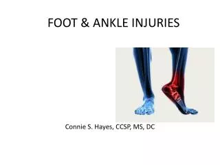
Foot & Ankle Injuries and Treatment. Dr. John R. Sallade Physical Therapist Board Certified – Sports Medicine Fellow – Academy Applied Functional Science. Classes of Conditions. > Traumatic surgical intervention non surgical intervention Insidious onset Congenital.

E N D
Foot & Ankle Injuriesand Treatment Dr. John R. Sallade Physical Therapist Board Certified – Sports Medicine Fellow – Academy Applied Functional Science
Classes of Conditions > Traumatic surgical intervention non surgical intervention • Insidious onset • Congenital
Traumatic • Fractures Ankle Mid foot Forefoot • Tendon tears Achilles (plantaris) Posterior Tibialis Peroneal • Repair, ORIF +/- Immobilization, WB, PT
Post Operative Complications • Stiffness • Weakness • Decreased propioception • Decreased vascularity, edema • Infection • RSD CRIPS • DVT PE
“Non traumatic” InjuriesInsidious Onset • Tendinosis • Stress fractures • Bunions , Hallux Limitus • Hammer toes • Metatarsalgia • Neuromas • Plantar Fascitis • Compartment Syndrome
“Non Traumatic” Injuries • Blisters • Callosities • Sub ungula hematomas • Arthritis • “pump bumps” • Apophositis • Sesmoiditis • Infections
Tendinitis(post. tib., achilles, peroneal) • Usually insidious in onset • Pain with WB – stretch or contraction • Improves with light activity • Latent inflammatory response • TTP, warm • Labs and Radiography not helpful
Treatment • Relative rest • Ice – 15 • Anti inflamatories – dosage and duration • PT - Find the biomechanical cause modalities, stretching, strengthening (hip partner), transverse friction massage, biomechanical control (shoes, inserts, lifts or orthotics)
Ankle Sprains • Account for 25% of all sports injuries • Lateral (ATF+CF)(85%) • Medial (Deltoid)> • “High” (Syndesmosis)> • Mid tarsal Possible causes: • Cavus, poor proprioception, poor rehab, over weight and poorly conditioned • No significant male – female ratio
Treatment • Surgery? • RICE • Progressive WB • Immobilization and Early mobilization • Closed Chain Exercise • Looking for a cause
Causes • Unlocked midtarsal joint at push off phase of gait causing stretch to fascia • Variety of foot types • Tight heelcords for level of function • Tight great toe flexors or fascia • Weakness in control of pronation • Training errors, shoes
Treatment • No correlation to heel spurs • Differentiate from tarsal tunnel • Treat the cause: • Stretch tight heel cords and FHL • Support unstable biomechanics – orthotics, taping or arch strapping • Night splints, morning/first step routine • Analgesic modalities, injections? Surgery?
Bunions • Both medial (1st MTP) and lateral (5th) In medial bunion: • Over pronated foot with abductus (toe out) • Tight heel cords • Forefoot varus
Treatment • Treat the cause • Symptomatic relief with modalities • Heel cord stretching • Fore foot support via orthotic • Strengthening • When is surgery the best option?
Stress Fractures – Micro FracturesMost common sites: metatarsals
Stress FracturesProbable Causes • Increasing the amount or intensity of an activity too quickly (most common) • Hard or uneven running surface • Improper or old shoes • Untreated biomechanical imbalances • Biomechanical limitations of motion (subtalar and midtarsal joints)
Other Risk Factors forStress Fractures Risk Factors • Female, short, thin and caucasian • Certain sports, especially involving plyometric loading: • Distance running • Gymnastics • Dance • Basketball and Tennis • Amenorrhea>decrease hormone support • Poor diet- low in calcium and high in acidity • Osteopenia (Reduced bone thickness or density) • Poor muscle strength or flexibility • Overweight or underweight
Compartment Syndrome • Compartment syndrome is a painful condition that occurs when pressure within the muscles builds to dangerous levels. This pressure can decrease blood flow, which prevents nourishment and oxygen from reaching nerve and muscle cells. • Compartment syndrome can be either acute or chronic. • Acute compartment syndrome is a medical emergency. It is usually caused by a severe injury. Without treatment, it can lead to permanent muscle damage. • Chronic compartment syndrome, also known as exertional compartment syndrome, is usually not a medical emergency. It is most often caused by athletic exertion. • Compartments are groupings of muscles, nerves, and blood vessels in your arms and legs. Covering these tissues is a tough membrane called a fascia. The role of the fascia is to keep the tissues in place, and, therefore, the fascia does not stretch or expand easily.
Symptoms and Diagnosis • Chronic (Exert ional) Compartment Syndrome • Chronic compartment syndrome causes pain or cramping during exercise. This pain subsides when activity stops. It most often occurs in the leg. • Symptoms may also include: • Numbness • Difficulty moving the foot • Visible muscle bulging
Differential Chronic (Exertional) Compartment Syndrome • To diagnose chronic compartment syndrome, other conditions that could also cause pain in the lower leg should be ruled out. Tendonitis can be ruled out but history and physical exam (palpation, passive and resistive tests) . To rules out stress fractures, an x-ray, bone scan or CT scan can be used depending on the duration and location of the injury. • To confirm chronic compartment syndrome, pressure tests of the compartment before and after exercise must be performed . • Treatment may involve a combination of rest, activity modification, change of shoes and orthotics and PT or in more involve cases surgery (fasciotomy).
Reflex Sympathetic DystrophyChronic Regional Pain Syndrome • Hyper reactivity of the sympathetic nervous system causing sustained “fight and flight” response. • The symptoms of RSD/CRPS often progress in three stages—acute, dystrophic, and atrophic. • The acute stage occurs during the first 1–3 months (usually after injury to bone or nerve, surgery and/or immobilization of an extremity) and may include burning pain (not proportionate to the degree of injury), swelling, increased sensitivity to touch, increased hair and nail growth in the affected region, joint pain, and color and temperature changes.
Advanced Symptoms The dystrophic stage may involve constant pain and swelling. The affected limb may feel cool to the touch and appear bluish in color. Muscle stiffness, wasting of the muscles (atrophy), and early bone loss (osteoporosis) also may occur. This stage usually develops 3–6 months after onset of the disorder. • During the atrophic stage, the skin becomes cool and shiny, increased muscle stiffness and weakness occur, and symptoms may spread to another limb. • Characteristic signs and symptoms of sympathetic nervous system involvement include the following: • Burning pain • Extreme sensitivity to touch • Skin color changes (red or bluish) • Skin temperature changes (hot or cold)
Treatment Treatment • The goals of RSD/CRPS treatment are to control pain and promote as much mobilization of the affected limb as possible without increasing symptoms. Treatment must be individualized and will often combine medications, physical therapy, nerve blocks (ganglion blocks with alpha adrenergic antagonist), and psychosocial support. Sympathectomy can be helpful in recalcitrant cases. • Early detection and intervention is paramount.