Hantavirus
Hantavirus . Stephanie Bagley Eugene Khandros Nicholas Bevins. History. Hantaviruses are rodent-borne viruses which may be transmitted to humans in aerosolized urine, feces, or saliva, and occasionally by bite.


Hantavirus
E N D
Presentation Transcript
Hantavirus Stephanie Bagley Eugene Khandros Nicholas Bevins
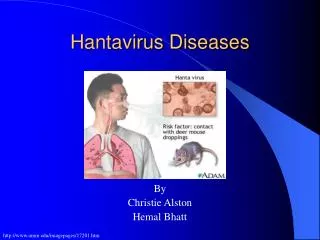
History • Hantaviruses are rodent-borne viruses which may be transmitted to humans in aerosolized urine, feces, or saliva, and occasionally by bite. • Hantaviruses often cause either hemorrhagic fever with renal syndrome (HFRS) or hantavirus pulmonary syndrome (HPS) • Other hantaviruses are not known to be human pathogens.
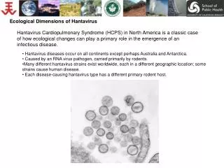
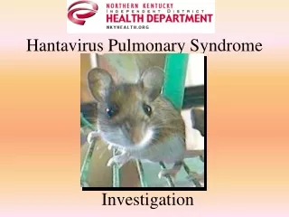
Hantavirus Outbreak in the US • HPS was first described in the United States in May 1993 during the investigation of a cluster of cases of acute adult respiratory distress in the Four Corners region. • HPS was found to be caused by a previously unknown hantavirus, Sin Nombre, detected in deer mice. • Sin Nombre caused approximately 200 confirmed cases of HPS during the outbreak, that led to a 50% mortality rate.
Hantavirus in the US • Prior to the HPS outbreak, the only known hantaviruses were those that caused HFRS • At least three other hantaviruses, New York Virus, Bayou Virus, and Black Creek Canal virus have since been confirmed to cause HPS in the U.S.
HPS in South America • The discovery of HPS in North America led to retrospective studies in South America. • More than 140 cases of HPS confirmed in Argentina. • The Andes hantavirus was discovered in long tailed pygmy rice rats in southern Argentina. • Andes Virus is the only known hantavirus to be transmitted person-to-person.
History of HFRS • While HPS has only been identified since 1993, HFRS has a much longer and complex history. • HFRS may have been recognized in China as early as 1000 years ago. • HFRS described in 1913 Russian records
History of HFRS Outbreaks of HFRS in the 1930’s: • Russia (1932) • Japanese troops in Manchuria (1934) • Sweden (1934)
Characterization of HFRS • 1934 -Japanese outbreak led to studies by Japanese physicians • 1940- Japanese physicians compiled a clinical and pathological description of what was then called “epidemic hemorrhagic fever.” • First to implicate mice in disease transmission. • Physicians injected filtrates of tissue from Apodemus agrarius carrying the hantavirus Hantaan into human subjects.
History of HFRS • 1939-Russian studies also implicated mice in disease transmission. • 1961- Moscow outbreak affecting 113 of 186 workers linked to rodent shipment
Characterization of HFRS 1967-Russian scientists Yankovski and Povalishina provide more insight into disease. • Incubation period up to 6 weeks • Cycles of virus activity parallel population cycles of field mice • Transmission of disease occurred via inhalation of dust contaminated by rodent excretions.
Recent HFRS Cases • 1951- outbreak in Korea during the war affected 3,200 soldiers and resulted in a 7-15% mortality rate. • Korean hemorrhagic fever (KHF) drew widespread international attention and was reclassified as HFRS in 1983 by the WHO. • 1983-Several past outbreaks including KHF, “epidemic hemorrhagic fever” , Russian outbreaks all reclassified as HFRS by the WHO
Family Bunyaviridae 5 genera, 250 species Genus Human disease Bunyavirus LaCrosse encephalitis, others Phlebovirus Rift Valley fever, sandfly fever Nairovirus Crimean-Congo hemorrhagic fever Tospovirus Plant virus, no known human disease HantavirusHemorrhagic fever with renal syndrome Hantavirus pulmonary syndrome
Hantavirus Genus • Hantavirus Similarities • RNA viruses • Lipid membrane • Tri-segmented genome • Hantavirus Differences • Hantavirus transmitted through aerosolized rodent urine, feces and saliva. • Others genera transmitted through arthropod vectors.
Epidemiology and Rodent Hosts • Each strain of hantavirus has a specific rodent host • Hantavirus species appear to have co-evolved with host rodent species • Rodents carrying hantavirus are asymptomatic
Transmission of Hantaviruses Chronically infected rodent Horizontal transmission of infection by intraspecific aggressive behavior Virus also present in throat swab and feces Virus is present in aerosolized excreta, particularly urine Secondary aerosols, mucous membrane contact, and skin breaches are also sources of infection Courtesy of CDC
Virus Strain Rodent Host Hantaan Apodemus agrarius Apodemus flavicollis Dobrava Rattus norvegicus Seoul Bandicota indicus Thailand Microtus pennsylvanicus Prospect Hill Clethrionomys glareolus Puumala Sin Nombre Peromyscus maniculatus New York Peromyscus leucopus Bayou Oryzomys palustris Black Creek Canal Sigmodon hispidus Rodent Hosts Murinae Arvicolinae Sigmodontinae
New York Peromyscus leucopus Sin Nombre Peromyscus maniculatus Prospect Hill Microtus pennsylvanicus Muleshoe Sigmodon hispidus Bloodland Lake Microtus ochrogaster Isla Vista Microtus californicus Bayou Oryzomys palustris Black Creek Canal Sigmodon hispidus El Moro Canyon Reithrodontomys megalotis Rio Segundo Reithrodontomys mexicanus Calabazo Zygodontomys brevicauda Juquitiba Unknown Host Laguna Negra Calomys laucha Caño Delgadito Sigmodon alstoni Choclo Oligoryzomys fulvescens Maciel Necromys benefactus Rio Mamore Oligoryzomys microtis Hu39694 Unknown Host Orán Oligoryzomys longicaudatus Lechiguanas Oligoryzomys flavescens Bermejo Oligoryzomys chacoensis Pergamino Akodon azarae Andes Oligoryzomys longicaudatus New World Hantaviruses Courtesy of CDC
Hantavirus Pulmonary Syndrome Canada (36) Countries with reported cases of HPS (no of cases) Countries with no reported cases of HPS United States (335) Panama (31) Brazil (168) Bolivia (20) Paraguay (74) Uruguay (23) Chile (273) Argentina (404)
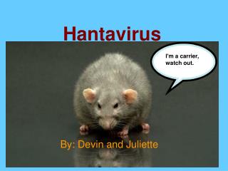
Rodent Hosts in the United States Deer mouse (Peromyscus maniculatus) Carrier of Sin Nombre strain, primary agent of HPS in the US. 250-300 cases since discovery.
Rodent Hosts in the United States White-footed Mouse (Peromyscus leucopus) Carrier of New York strain.
Molecular Biology of Hantavirus • Physical Properties • Structure • Genetics • Replication Cycle • Pathogenesis
Molecular Biology of Hantavirus • Physical Properties • Structure • Genetics • Replication Cycle • Pathogenesis
Virion Properties • Spherical or oval-shaped. • 80-120 nm diameter • Unique grid-like surface pattern, with 7-8 nm projections • Lipid bilayer envelope • Granulofilamentous interior • Survive 12 hours at 4C, high salt concentration and non-physiological pH. • Survives 1-3 days after drying. • Exposure to lipid solvents and nonionic detergents destroys viral envelope
Molecular Biology of Hantavirus • Virion Properties • Structure • Genetics • Replication Cycle • Pathogenesis
Structural Proteins Membrane glycoproteins (G1 and G2) Polymerase (L) Nucleocapsid proteins (N)
Membrane Glycoproteins • G1: 64-67kDa • G2: 54 kDa, highly conserved • Integral membrane proteins • G1-G2 heterodimers form 8 nm projections on virion surface • Cysteine-rich • Contain asparagine-linked sugar groups • Important in cell entry and pathogenesis
Nucleocapsid Protein (N) • 48 kDA • Complexes with genomic vRNA in virus, as well as with cRNA after infection, but not with mRNA • Necessary for virus replication and packaging
Polymerase (L) • 247 kDA • RNA-dependent RNA polymerase (RdRp) • Complexed with ribonucleocapsids in virion • Endonuclease activity to cleave host mRNA • Transcriptase activity for making cRNA and mRNA from vRNA • Helicase activity to unwind vRNA during transcription
Molecular Biology of Hantavirus • Physical Properties • Structure • Genetics • Replication Cycle • Pathogenesis
Genomic Organization • Tripartite negative sense genome • Small (S) segment, 1.7-2.1kb, codes for N nucleocapsid protein • Medium (M) segment, 3.6-3.7kb, codes for G1 and G2 glycoproteins • Large (L) segment, 6.5 kb, codes for L polymerase protein
Panhandle Structure • Conserved repeated complementary sequences at 5’ and 3’ ends form panhandle structures
Transcription • Viral polymerase transcribes negative-strand vRNA to mRNA • Polymerase acts as a endonuclease and cleaves host mRNAs 7-18 nt from the 5’ cap • The capped oligonucleotides act as primers required to initiate transcription • After transcription is primed and the first repeat of the terminal sequence is transcribed, polymerase slips and realigns the nascent RNA, then continues transcription
Replication • Viral polymerase transcribes negative-strand vRNA to sense cRNA • cRNA is used as template to make more negative-strand vRNA • pppG is used to prime cRNA and vRNA synthesis • Same “prime and realign” strategy
Molecular Biology of Hantavirus • Physical Properties • Structure • Genetics • Replication Cycle • Pathogenesis
Attachment Entry Uncoating Release Transcription Replication Assembly Translation
Attachment • Viral G1 and G2 glycoproteins interact with cell surface receptors • Pathogenic hantaviruses bind 3 integrins • Non-pathogenic hantaviruses bind 1 receptors
Entry and Uncoating • Virus particles bound to integrin receptors are taken in by receptor mediated endocytosis • Newly formed vesicles are acidified • Acidic environment changes conformation of G1 and G2 • Viral and cell membrane fuse • Genomic material and polymerase are released into cytoplasm
Primary Transcription • Transcription of negative sense vRNA to mRNA • Viral polymerase (RdRp) transcribes nucleoprotein-coated vRNA • Capped oligonucleotides from cell’s own mRNA are used to prime transcription • Follows “prime and realign” model
Translation • L and S segment mRNA is translated on free ribosomes in cytoplasm • M segment mRNA translated on ER-bound ribosomes • G1 and G2 peptides produced from M mRNA are cleaved cotranslationally • Separate signal sequences for G1 and G2 cause ER attachment and embed the peptides in ER membrane (Signal Hypothesis)
Genome Replication • vRNA is used as a template by viral polymerase to make sense strand cRNA • cRNA is used as a template to make more negative strand vRNA • More genetic material means more virions produced
Secondary Transcription • Extra vRNA synthesized during replication is used as template to make mRNA • Since more template is present after vRNA is replicated, more mRNA can be transcribed, and more viral proteins can be made
Virion Assembly • Membrane-bound G1 and G2 peptides are transported to Golgi and carbohydrates are attached by N-linked glycosylation • vRNA complexes with N nucleopcapsid protein, forms looped panhandle structure, and complexes with L polymerase
Virion Release – Scenario 1 • Nucleocapsid complexes bud into the Golgi membrane with G1 and G2 embedded • Virion particle is formed inside the Golgi • Virions are transported to cell membrane by vesicles and released by exocytosis, just like in secretion • Viruses may prefer different cell surfaces for release