Download
1 / 28
280 likes | 701 Vues
What is OSA . Repeat episodes of partial or complete upper airway obstruction during sleep Result in a disruption of normal ventilation and sleep patterns . Continuum of sleep disordered breathing . . Sleep in children . After 6 months REM sleep and non-REM sleep . REM sleep . Muscle atonia Increased cerebral blood flow Variable HR RR BPIncreased upper airway resistanceDuring REM get bursts of phasic events causing rapid eye movements and myoclonic twitches .

E N D
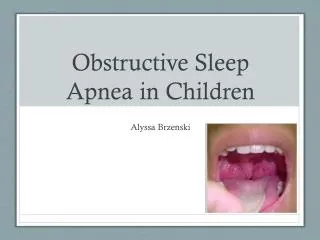
1. Obstructive sleep apnoea in children Joanne Edwards
Senior Paediatric Registrar TCH
2. 4 month old with book over face to assist learning 4 month old with book over face to assist learning
3. What is OSA Repeat episodes of partial or complete upper airway obstruction during sleep
Result in a disruption of normal ventilation and sleep patterns
4. Continuum of sleep disordered breathing
5. Sleep in children After 6 months
REM sleep and non-REM sleep
6. REM sleep Muscle atonia
Increased cerebral blood flow
Variable HR RR BP
Increased upper airway resistance
During REM get bursts of phasic events causing rapid eye movements and myoclonic twitches
7. Non REM sleep Reduced muscle tone
Decreased cerebral blood flow
Regular HR RR BP
Increased upper airway resistance
NREM sleep is divided into stages by EEG criteria which parallel depth of sleep
8. Sleep cycles
9. Respiration during sleep Increased upper airway resistance
Relaxed pharyngeal muscles (dilator)
Probably decreased central respiratory drive
Decrease in lung volumes during REM
11. Sleep disordered breathing Partial or complete collapse at the elvel of extrathoracic airway
Caused by
Small upper airway � smaller in those with OSA
Decreased tone of pharyngeal dilators during sleep
SUbvstantial change in dimensions of airway between inspiration and expiration
12. Predisposing factors Peak age 2-8 years old
Coincides with peak age of lymphoid tissue � ie tonsils and adenoids
Enlarged tonsils and adenoids
Obesity
Mucopolysaccharidoses
Children with airway or facial abnormalities
Midface hypoplasia
Retro or micrognathia Acutely angled skull base
Narrow maxillary arch
Nueromuscular factors � hypotonia or hypertonia
14. Predisposing factors Genetic factors
Both obese and non-obese populations
Drugs
Alcohol
Chloral hydrate
Benzodiazepines
GA
Opioids
15. Pathology Decreased upper airway patency
Adenotonsillar hypertrophy
Allergies causing rhinitis, nasal obstruction
Reduced capacity to maintain airway
Obesity
Neuromuscular disorder
Decreased drive to breathe
Brain stem injury
16. Patterns REM sleep
Hypoventilation
Significant oxygen desaturations
NREM sleep
Relatively protected
17. What are the symptoms and signs?
18. Symptoms � night time Snoring
12% of children snore
Most of children with OSA snore
Pauses in snoring with apnoea
Sleeping
Mouth breathing or unusual positions
Nighttime sweating
Restless or agitated sleep
Parasomnias � sleep terror, sleep walking
Nocturnal enuresis
19. Symptoms � day time Growth deviations
Failure to thrive
Obesity is predisposing factor
Mouth breathing and hyponasal speech
Sleepiness
Daytime napping
Inattention, learning problems, behavioural problems
21. On examination � head and neck Craniofacial anomalies � midface hypoplasia, retrognathia
Obstructive septal deformity
Macroglossia
Hyponasal speech
Mouth breathing � adenoidal hypertrophy
Mucosal or turbinate swelling suggestive of chronic nasal congestion
Suggestive of allergy if dark circles under eyes, swollen eyes, transverse nasal crease
22. Examination Growth
Neuromuscular tone
Mallampati classification of oropharyngeal crowding
BP (hypertension)
23. How is OSA diagnosed Sleep study � polysomnography
What is measured
Airflow � apnoea and hypopnoea
Abdominal and chest wall movements to indicate respiratory effort
End tidal CO2 � adequacy of ventilation
Saturations
EEG � stage of sleep
ECG � cardiac rate and rhythm
EMG � arousals and leg movement
Snore microphone
24. Measurements made Apnoeas
>90% decrease in ariflow that lasts >0% of the duration of 2 normal breaths
Obstructive � continued or increased respiratory effort during period
Central � no respriatory effort during period, event lasts > 20 seconds
Can be mixed
Hypopnoea
Respiratory effort related arousal
25. What is measured Apnoea hyponoea index � total number occurring during 1 hour
Other measures
End tidal CO2
If CO2 exceeds 50 for > 25% of ttoal sleep time � hypoventilation
Hypoexmia < 92%(lowest nadir in normal children)
27. 4 year old girl
Bold bars are REM sleep � most desats durign REM sleep, preserved sleep architecture
CA � central apnoea, CAP � capnography
MA mixed apnoea
OA obstructive apnoea 4 year old girl
Bold bars are REM sleep � most desats durign REM sleep, preserved sleep architecture
CA � central apnoea, CAP � capnography
MA mixed apnoea
OA obstructive apnoea
28. Diagnostic criteria History of snoring, laboured breathing or obstructed breathing during sleep
History of arousals, sweating, neck hyperextension, excessive daytime sleepiness, aggressive or irritable behaviour, slow growth, morning headaches, secondary enuresis
PSG � AHI>1 or frequent arousals with icnreased respriatory effrot, desaturations, hypercapnia
Not explained otherwise
29. Severity Mild
AHI � 1-4, sats nadir 86-91%, CO2 peak > 53
Moderate
AHI 5-10, sats nadir 76-85, CO2 > 60
Severe
AHI > 10, sats nadir < 75, CO2 > 65
31. Management Adenotonsillectomy
Based on clinical experience, difficult to randomize
Known adenotonsillar hypertrophy
CPAP or BiPAP
If adenotonsillectomy too risky or already done
Other
Weight loss, maxillofacial surgery to correct anomalies, nasal steroids, oral appliances
32. Adenotonsillectomy Meta-analysis of 355 children with OSA and adenotonsillar hypertophy
Post adenotonsillectomy 83% had normalized PSG and reduced AHI
If obese, less successful outcomes � AHI>2 persisted in about 76% (compared to 28% lean children
33. Positive airway pressure CPAP
Constant level of positive airway pressure throughout cycle
BiPAP
Higher pressures during inspiration than expiration
Pressures are determined by sleep study
Very poor compliance
34. Oxygen Supplemental oxygen useful in short term if severely hypoxemic until definitive therapy provided
Rarely used
For those who cannot tolerate PPV
Does not improve episodic upper airway obstruction or hypercapnia or sleep fragmentation
May suppress ventilatory drive and worsen hypercapnia