Download
1 / 1
10 likes | 36 Vues
For more details visit our website at - https://cryoviva.in/

E N D
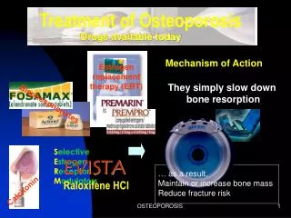
European Cells and Materials Vol. 6. Suppl. 2, 2003 (page 21) ISSN 1473-2262 Stem Cells in the Treatment of Osteoporosis J.A. Gasser Novartis Institutes for Biomedical Research, Basel, Switzerland INTRODUCTION: Osteoporosis may be caused in part by an age-related decline in the number of osteoblast-progenitors (mesenchymal stem cells, MSCs) residing in the bone marrow1. MSCs can be isolated from marrow, cultured and expanded in vitro2,3. It has been hypothesized that such cells from in vitro culture might be infused back to osteopenic subjects in order to replenish their stem cell pool. It is hoped that this procedure would result in a positive bone balance and ultimately the regeneration of the osteopenic skeleton. before declining rapidly in all tissues as the cells were removed almost completely by macrophages within 7 days following their administration. Bone administration of MSCs was unable to prevent OVX-induced bone-loss or regenerate bone tissue in osteopenic rats to any significant degree after systemic infusion. However administration of GFP-tagged MSCs directly into the proximal tibia metaphysis conclusively showed that, injected cells were involved in the injury-related bone formation response triggered by the local injection. Locally administered MSCs also failed to prevent the OVX induced bone loss. effects: In contrast to PTH, i.v.- At present it is unknown how MSCs distribute in a mammalian organism after intravenous infusion and especially whether they have indeed the capacity to preferably ‘home’ to bone marrow from the blood stream and form colonies as proposed by D.J. Prockop4 and collaborators. DISCUSSION & CONCLUSIONS: Single or multiple i.v.-infusion of syngeneic MSCs at cell- doses of 1x10⁷ is well tolerated in Fisher F344 rats (no anaphylaxis, no ectopic bone or cartilage formation). MSCs did not home preferentially to bone tissue but were found to be enriched in lymphoid organs. The cells did not show long-term seeding. In bone, GFP-labelled osteocytes were detected in the mineralised matrix indicating that injected MSCs did differentiate down the osteogenic pathway and participate in local bone formation processes. However, MSCs did not prevent oestrogen-deficiency induced bone loss in rats after local or systemic administration to any significant degree. METHODS: MSCs were labelled with radioactive [3H]thymidine (> 10 dpm/cell) and their organ distribution measured at various time-points for up to 7 days after i.v.-infusion. In order to study seeding, survival and multiplication of cells in various organs including bone, infusion of cells stably expressing green fluorescent protein (GFP) were performed. The therapeutic potential of MSCs was tested under various conditions in rats models of postmenopausal osteoporosis. Syngeneic rat MSCs were harvested and culture expanded from donor rats. Cells were infused one week before or after ovariectomy at a dose of 10 Mio/kg into the tail vein of skeletally mature (8 month old), inbred Fisher F344 rats to prevent bone loss associated with oestrogen deficiency. Injections of parathyroid hormone (PTH), a known bone anabolic principle served as positive control. Non– invasive bone measurements were carried out by pQCT on an XCT-960 Pforzheim, Germany) in the proximal tibia metaphysis at baseline, 4 and 8 weeks. In separate experiments, GFP-labelled MSCs were injected directly into the metaphysis of anaesthetised oestrogen deficient (ovariectomized, OVX) rats. The failure of MSCs in osteoporosis models suggests that, in a situation of oestrogen deficiency, the local ‘catabolic’ environment does not provide the required stimulus to the MSCs for their differentiation down the osteoblast lineage. It may be necessary to either combine MSCs with an osteoinductive carrier material, or provide an autologous stimulus by differentiation-inducing agent such as BMP or TGFβ into the cells (MSC-based gene therapy) in order to achieve therapeutically significant bone regenerative effects. The local environment may be more favourable and chances for success of stem cell therapy higher in situations such as fracture repair and bony ingrowth of implants, where the local injury provides a strong signal for bone repair. (Stratec-Norland, expressing a cell RESULTS: Organ distribution: From the data obtained we calculated organ loads relative to organ wet weight. The values suggest that the lung accumulates the majority of cells at 4h and 24h following infusion. Higher than average values were also seen in duodenum, spleen, thymus. Approximately 800-1900 cells were found in the tibia at 24 hours. Organ loads peaked at 24 hours 1A.J. Friedenstein (1976) Int Rev REFERENCES: Cytol47:327-59; 2B.A. Ashton et al (1980) Clin Orthop 151:294-307. 3A.I. Caplan, (1991) J Orthop Res9:641- 50; 4D.J. Prokop (1997) Science,276:71-74