sdAb as Therapeutic Agent
Full-length monoclonal antibodies have been highly successful as therapeutic agents against various immune diseases and cancers. However, the large size severely limits their applications. As an alternative, single domain antibodies (sdAbs) present great advantages as novel therapeutic agents, such as small size, high expression, improved robustness, and a large number of accessible epitopes. Creative Biolabs is committed to providing customized proposals and solutions to develop novel sdAb-based therapeutics for disease treatment.<br>https://www.creative-biolabs.com/sdab/sdab-as-therapeutic-agen

sdAb as Therapeutic Agent
E N D
Presentation Transcript
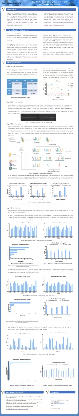
CaseStudy-SdAbDevelopmentfor3Targets via1CamelidImmunization SingleDomainAntibody Introduction CreativeBiolabshasbeenalong-termexpertinthefieldof Singledomainantibody(sdAb),isakindofantibodyfragments single domain antibody (sdAb) development. Our scientists have extensive experience in immunizing camelid animals with the targetofinterestto generatenovelsdAbs.In termsof our advancedHi-Affi™phagedisplayplatform, wecanuse1 immunizedhostanimalto generatehigh-specificsdAbsfor multiple antigens. This is a cost-effective and time-saving option forspecificsdAbdevelopment,especially when youneedto investigate differenttargets with low homology. consisting of a single monomeric variable antibody domain and lackingthelight chainandCH domainof theheavy chainin conventional Fab region. In terms of only 12-15 kDa molecular weight, which ismuch smaller than eitherfull length antibody (150-160kDa)orotherantibodyfragments(Fab~50kDa,scFv ~25kDa), sdAbtakesgreatadvantagesofstability and penetrability, which are essential to the development of several antibody drugsordiagnostic tools. ProjectObjective&Achievement For this case study, THREE different targets were provided as antigens and screening targets. Creative Biolabs is entrusted to immunize only ONE camelid host animal with these targets and thendevelopantigen-specificsingledomainantibodies, respectively. ForTarget1,allthe40cloneswereobservedaspositivethrough monoclonalphage ELISAand7uniqueVHHsequences have beenidentifiedandconfirmedtorecognizethetargetspecifically. ForTarget2,allthe40cloneswereobservedaspositivethrough monoclonalphage ELISAand5uniqueVHHsequences have beenidentifiedandconfirmedtorecognizethetargetspecifically. ForTarget3, 22of the40cloneswereobservedaspositive throughmonoclonalphageELISAand19uniqueVHHsequences have been identified and confirmed to recognize the target specifically. Withtheprovidedantigens(namelyTarget1, Target 2, and Target 3 or T1, T2, T3 for short), one camelid was immunized with mixedantigens. Promising immune response for each antigen wasobservedafter 4injections, whichisqualifiedforlibrary construction. One uniform immune library was then constructed with the capacity of over 109. Three rounds of biopanning were successfully performedagainsteachof thethreetargets respectivelywithsignificant goodenrichment.40cloneswere randomlypickedfromthe3rdroundenrichedpoolofeachtarget forvalidation. Finally, there are 7 unique T1-specific sdAbs, 5 unique T2-specific sdAbs, and 19 unique T3-specific sdAbs be discovered in this project. MilestoneOverview Stage1:AnimalImmunization After the fourth injection, test bleed was collected and 2nd titration wasconductedto monitorthe immuneresponse. The three targets were coated separately and tested in-parallelwith pre- immune sera (negative control) and antisera. As shown in Figure 1, good immune response was observed for all the three targets: thetiterofT1and T3wasover1:128,000,andT2reached 1:32,000. One native (non-immunized before) camelid animal was employed for this project. The immunization process was planned to last 70 days (4 injections with 3-week interval) and performed via multiple sites subcutaneous immunization strategy, which contributes to triggering immuneresponse forall thethreetargets. Table 1.TypicalCamelidImmunization Schedule. Figure1.2ndtitrationresults. Stage2:LibraryConstruction After4thinjection,theantiserawerecollectedandsubjectedtoPBMCisolation, RNA extraction,and cDNApreparation,freshlyonthesameday. TheVHH geneswerethenPCR amplifiedbyusingourspecies-specificprimers. Thephagemidlibrary was constructed with high-quality phagemidvectorsandoptimizedligationstrategiestoachieve100%correctinsertionrate(Figure2).Itwasthendesaltedandsubjectedto electrotransformationwithE.coliTG1asthehoststraintoformtheoriginalbacterialibrary.BasedontheQCcolonyPCRandDNA sequencing analysis, a qualifiedimmune librarywith capacity of over 109hasbeengeneratedsuccessfully. Figure2. QCcolony PCR of randomclones from theend library Stage3:LibraryScreening CreativeBiolabscantailoraseriesoflibraryscreeningstrategiestofindthebest-fitoneofyourproject.Ourscientistsarecommittedtocollecting the most reliable data that contribute to understanding the actual situation of each step. For a typical screening process, pre-absorption will be performedbeforeeachroundofscreeningtoeliminatenon-specificbindersagainsttheplatesurface,correspondingblockingbuffer,and negative target (if exists) as much as possible. From thesecondround,“NoCoating”control is also performed in parallel with the “Target Coating” group. If there is any negative target required by the project, an in-paralleltestof “Negative” control will be involved as well from the secondround. Figure3.Flow diagram of phagedisplay-based screening. Forthiscasestudy,solid-phasescreeningstrategywasperformed,whichthetargetswereimmobilizedontheplatesurfacedirectlyand screened separately. After three rounds of biopanning, good enrichment was observed for all the three targets and clear difference was found betweenthe“TargetCoating”groupand“NoCoating”control(Figure4).Thisindicatedsome specificbindershavebeenselectedforthetargets. Figure4. Processmonitoringoflibrary screening stage. (Enrichment is increasedround by round and presents significantdifference betweennocoating control.) Stage4:BinderValidation After the biopanning, 40 clones were randomly picked from the 3rdround output of each target group. The monoclonal phage ELISA was then performed against the target,respectively. For Target 1, 40 positive clones were observed and then processed for DNA sequencing (Figure 5). 7 unique clones were identified in CDR level (Figure6).Alltheseuniquecloneswerethenpreparedas solubleformat(phage-free)forthevalidationof QC solubleELISA. As showninFigure 7, allof themwere finally confirmed to recognizethe target positively. Figure5. MonoclonalphageELISAof the40randomlypicked clones [Target1]. Figure6.Summary ofDNAsequencingresults [Target1]. (Abundance of each unique clone indicates the number of sequenced clones present thesamesequencinginformation.) Figure7. QC solubleELISAof theuniquesdAbcandidates[Target1]. For Target 2, 40 positive clones were observed and then processed for DNA sequencing (Figure 8). 5 unique clones were identified in CDR level (Figure 9). All these unique clones were then prepared as soluble format (phage-free) for the validation of QC soluble ELISA. As shown in Figure 10, allof themwerefinally confirmed to recognize the target positively. Figure8. MonoclonalphageELISAof the40randomlypicked clones [Target2]. Figure9.Summary ofDNAsequencingresults [Target2]. (Abundance of each unique clone indicates the number of sequenced clones present thesamesequencinginformation.) Figure 10.QC soluble ELISAof the uniquesdAbcandidates [Target2]. ForTarget3,22positivecloneswereobservedandthenprocessedforDNAsequencing(Figure11).19uniquecloneswereidentifiedinCDR level (Figure 12). All these unique clones were then prepared as soluble format (phage-free) for the validation of QC soluble ELISA. As shown in Figure13, allof themwere finally confirmedto recognize the target positively. Figure11.MonoclonalphageELISAofthe40randomlypickedclones [Target3]. Figure 12.Summary ofDNAsequencingresults [Target3]. (Abundance of each unique clone indicates the number of sequenced clones present thesamesequencinginformation.) Figure13.QCsolubleELISAofthe unique sdAb candidates [Target 3]. Conclusion&KeyWords ContactUs • One AnimalImmunization-Multiple antigenswithlowhomology can be immunized together fornovelsdAbdiscovery. • High-Quality SdAb Library - Creative Biolabs’ Hi-Affi™ platform can contribute to generating immunelibrarywith maximizeddiversity andcapacity. • High Fidelity Screening - Solid-phase strategy combined with in-parallel control group, which achievedgreatenrichmentandsupportthereliabilityofthescreeningoutcomes. • Two-Step Validation - Antigen-specific clones were identified and validated through both monoclonalandsolubleELISA,whichcanavoidpotentialfalsepositive. • One-Stop Solution - Extensive experience and integrated procedure enable our scientists to smoothlyadvancetheprojectandmeetallyour objectives. USA 45-1RamseyRoad,Shirley,NY11967,USA Tel:1-631-381-2994 Fax:1-631-207-8356 Email:info@creative-biolabs.com Europe Tel:44-207-097-1828