Download
1 / 47
530 likes | 999 Vues
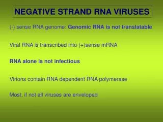
NEGATIVE STRAND RNA VIRUSES. (-) sense RNA genome: Genomic RNA is not translatable. Viral RNA is transcribed into (+)sense mRNA. RNA alone is not infectious. Virions contain RNA dependent RNA polymerase. Most, if not all viruses are enveloped.

E N D
NEGATIVE STRAND RNA VIRUSES (-) sense RNA genome: Genomic RNA is not translatable Viral RNA is transcribed into (+)sense mRNA RNA alone is not infectious Virions contain RNA dependent RNA polymerase Most, if not all viruses are enveloped
A diverse array of negative-strand RNA viruses infect vertebrate hosts Negative-strand RNA viruses are less diverse, but very successful in plant and invertebrate hosts Rhabdoviruses and bunyaviruses infect and are successful in many different vertebrate, invertebrate, and plant hosts All plant-infecting negative strand RNA viruses also infect and replicate in their invertebrate vectors Negative strand RNA viruses with segmented genomes most likely evolved from nonsegmented negative strand RNA viruses Plant Vertebrate Invertebrate
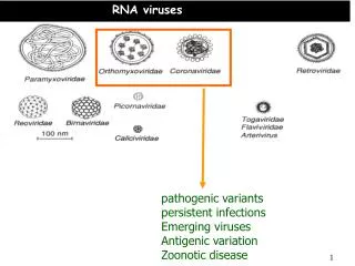
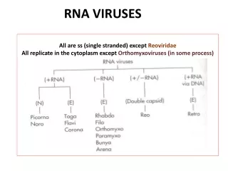
FAMILIES of NEGATIVE STRAND VIRUSES NON-SEGMENTED (-)STRAND VIRUSES RHABDOVIRIDAE Rabies, VSV, & Plant viruses FILOVIRIDAE - Marburg & Ebola viruses PARAMYXOVIRIDAE - Measles, Mumps, RSV, & Distemper BORNAVIRIDAE – Neurological diseases of humans and many animals SEGMENTED (-)STRAND VIRUSES ORTHOMYXOVIRIDAE - Influenza virus SEGMENTED AMBISENSE VIRUSES BUNYAVIRIDAE - Hantavirus, plant Tospovirus and Tenuivirus ARENAVIRIDAE - Lassa fever
NONSEGMENTED NEGATIVE STRAND RNA VIRUSES ORDER MONONEGAVIRALES The family RHABDOVIRIDAE - 45-100 X 100-430 nm bacilliform or bullet-shaped particles - membrane spikes composed of only G protein - helical nucleocapsids unwind to 20 X 700 nm - 1 segment, I0-14kb genome - 5-10 genes encode 5-10 proteins; most have 5-6 genes - 200+ virus species - infect vertebrates, invertebrates and plants - Vesicular stomatitis & Rabies virus; many plant & fish viruses - replicate in both host & insect vectors
G protein M Protein P Protein N Protein L Protein Rhabdovirus structure All 5 proteins encoded by most animal rhabdovirus genomes are part of the virion.
Relationships among rhabdoviruses Fish hosts Animal hosts Plant hosts
FAMILY RHABDOVIRIDAE Genus Vesiculovirus - Vesicular stomatitis virus, type species, is best studied rhabdovirus - causes epizootics in cattle, swine, & horses; insect & tick hosts also - rapid replication leads to many defective-interfering (DI) particles Genus Lyssavirus - Rabies virus - continuous health problem worldwide - thousands of cases per year - in dogs and cattle in Central & South America; skunks, racoons, foxes in North America - vampire bats associated with endemic spread in Central America - prevention by animal immunization; rarely used for humans - treatment shortly after transmission required; natural mortality in untreated humans is 15-25% - particularly unpleasant death makes this one of the most feared viruses Genus Ephemerovirus - Bovine ephemeral virus is most important - low mortality, but fever, lameness, anorexia of livestock make it a serous problem - more complex genome – 10 genes Genus Novirhabdovirus - cause diseases of fish of significant economic importance - similar to VSV in genome structure, but contain 6th gene for nonstructural protein Genus Cytorhabdovirus - Lettuce necrotic yellows virus is type member - different species replicate in and transmitted by different vectors - functional transcriptase can be isolated - bud through membrane in cytoplasm Genus Nucleorhabdovirus - Potato yellow dwarf virus is type member; Sonchus yellow net virus is best studied - different species replicate in and are transmitted by different vectors - functional transcriptase has not been isolated - replicate in nucleus and bud through inner nuclear membrane
Entry of Vesicular Stomatitis Virus into Cells Virus moves to clathrin coated pits and is taken into the cell by receptor mediated endcytosis. Endosome moves into the cytoplasm to a moderately low pH. The G protein has a conformational change and the nucleocapsid is released into the cytoplasm. G protein binds to Sialic Acid receptors on Cell surface. VSV has broad host range and has many potential receptor proteins. The M protein is released to activate mRNA transcription.
Animal rhabdovirus infection cycle 1. Virus binds receptor and enters by endocytosis 2-3. Membrane fusion and core release 4-6. Sequential transcription and translation of mRNAs 7-8. After nucleocapsid protein (N) accumulates, switch from transcription to replication occurs 5,9,10. Glycoprotein (G) enters secretory pathway and is embedded in plasma membrane 11,12. Nucleocapsid with associated phosphoprotein (P) and RdRp (L) are organized into bullet-shaped core particles by matrix protein (M) 13. Cores bud through membrane at sites of G-protein accumulation
VSV is the model rhabdovirus Rhabdovirus transcription and replication • Rhabdovirus genes are transcribed sequentially, in order of greatest requirement and time of requirement • Regulatory leader sequence is transcribed first and binds N protein early and often, resulting in maintenance of transcription mode • Sufficient N-protein accumulation and phosphorylation state of P results in switch from transcription to replication • Full-length antigenome (+ sense RNA) is template for - sense genome replication • SEE AND UNDERSTAND FIGURE ON P. 200-201
Details of genome structure of VSV. Coding regions for the nucleoprotein (N), phosphoprotein (P), matrix protein (M), glycoprotein (G) and polymerase protein (L) are separated by regulatory sequences that contain a transcription stop (gene end) signal, a polyadenylation signal, a nontranscribed intergenic region and a transcription start (gene start) signal. Transcription units are flanked by a leader (Le) and trailer (Tr) region that contain the genomic and antigenomic viral promoters, respectively. Note that the genome sense strand is shown, so by convention, the 3’-end is on the left. From Neumann et al., J. Gen Virol. DOI 10.1099/vir.0.18400-0
Rabies Virus FIG. 1. (A) Organization of the 12-kb genome of RV. Filled boxes represent the open reading frames of the N, P, M (matrixprotein), G, and L genes. c represents the nontranslated 39 sequence of the G cistron (pseudogene). (B) Organization of chimeric viruses. Open reading frames are drawn as shaded boxes, and transcriptional stopypolyadenylylation signals are represented by flags. In SAD VCAT, a DNA fragment composed of the NyP cistron border and the CAT open reading frame was used to replace the major part of the pseudogene sequence of standard RV SAD L16; for construction of SAD XCAT, the fragment was inserted into the StyI site of the pseudogene. The former coding part of the N gene is shown as an open box.
Transcription and replication initiate at separate sites on the vesicular stomatitis virus genome Fig. 6. Model for RNA synthesis: Schematic of the VSV genome depicting the leader region (Le) and the N and P genes. During transcription the RdRP, a complex of L (large oval) and a trimer of P (small oval), binds to specific sequences and initiates synthesis at the N gene start. The products of this reaction are the VSV mRNAs, of which the N and P mRNAs are shown. The 5'-terminal cap is depicted by a black diamond, and the 3' polyadenylate tail by A(n). During replication, the RdRP initiates at the 3' end of the genome. Initiation at the 3' end provides leader RNA (not shown) and the full-length antigenome. Replication requires protein synthesis to supply N protein for encapsidation of nascent RNA. N protein (hatched oval) is kept soluble by interaction with P, in a 2:1 complex. Positions of initiation are indicated by the black triangles. From Whelan and Wertz, 2002, PNAS 99: 9178-9183
Particle structure, genome organization, and expression of Vesicular stomatitis virus, a cytoplasmic rhabdovirus
Nucleorhabdovirus Cytorhabdovirus Two genera of plant-infecting rhabdoviruses: one replicates in cytoplasm, the other replicates in nucleus
Serious and highly contagious Mostly in wild animals; domestic animals now less than 10% of cases Symptoms include insomnia, anxiety, confusion, slight or partial paralysis, excitation, hallucinations, agitation, hypersalivation, difficulty swallowing, and hydrophobia, then death Transmitted by bites of infected animals Bats are commonly associated with rabies Rabies
www.lmb.uni-muenchen.de/groups/ Conzelmann/default.htm Rabies is a virus that attacks the central nervous system of warm-blooded animals. It can be transmitted by the bite of a rabid animal or via the introduction of the saliva of a rabid animal into a fresh (less than 24 hours old) wound. Transmission via other routes is rare.
History of Rabies Virus Causes the most lethal of all infectious diseases. Even the most extreme modern medical interventions are usually not successful. The disease was recognized in Egypt before 2300 B.C. and in ancient Greece, where it was described by Aristotle. The method of transmission of rabies was not recognized until 1804. Rabies is thought to be responsible for the origin of vampire legends (bats, biting, hypersexuality are associated with rabies). Infects all Mammals. In North America, rabies is most common in skunks, foxes, raccoons, bats, dogs, and cats. Occurs worldwide, with the exception of Hawaii, Japan, Great Britain, & smaller islands
Racoon Striped Skunk Red Fox Silvertailed Bat Racoons, Skunks, Foxes and Bats Are Major Rabies Reserviors
Positive Fluorescent Antibody Test Brain tissue showing Negri bodies. Rabies Disease Syndrome • Incubation normally 1-2 months occasionally up to two years. • Multiplies in muscle and connective tissue. • Enters peripheral nerves to CNS • First symptoms of malaise, sore throat, fever. • Increased sweat, hydrophobia, difficulty swallowing, muscle spasms, convulsions • Levels of virus in the blood is not very high. • Invariably fatal. • Death by respiratory paralysis. • Virus can be diagnosed in brain tissue by staining for Negri bodies and by fluorescent antibody tests.
Rabies Vaccines for Humans Inactivated whole virus vaccines are available for humans. • Nervous Tissue Preparations - Associated with the rare complication of demyelinating allergic encephalitis. • Duck Embryo Vaccine is grown in embryonated duck eggs The vaccine has a lower risk of allergic encephalitis but is considerably less immunogenic. • Human Diploid Cell Vaccine (HDCV) - this is currently the best vaccine available with an efficacy rate of nearly 100% and rarely any severe reactions. However it is very expensive. • Other Cell culture Vaccines - because of the expense of HDCV, other cell culture vaccines are being developed for developing countries. However recent data suggests that a much reduced dose of HDCV given intradermally may be just be effective.
World-WideControl of Rabies • Urban - Canine rabies accounts for more than 99% of all worldwide human rabies. Control measures against canine rabies include; • stray dog control. • vaccination of dogs. • quarantine of imported animals. • Wildlife - Rabies in wild animals is much more difficult to control than canine rabies. However, on-going trials in in the USA and Europe using bait containing rabies vaccine given to foxes and raccoons reduces rabies levels.
Rabies Control • Eliminate rabies from hosts/victims • Oral baits w/antivirus. • Has been effective (Europe, Canada). • Slowed potential outbreak in Ohio raccoons. • Used in Texas for coyotes & foxes. • Trap/vaccinate/release (TVR) • Effective in Canada raccoons. • Often combined with oral baits.
Several negative sense RNA viruses infect the central nervous system Neurotropic – infect neural cells Neuroinvasive – can enter CNS (spinal cord or brain) Neurovirulent – can cause disease of nervous tissue Mumps – highly neuroinvasive, but low neurovirulence; most infection leads to infection of CNS, but neurological disease is mild Rabies – highly neuroinvasive, high neurovirulence – readily infects periferal NS and spreads to CNS, with 100% lethality if no antiviral therapy (Herpes) – low neuroinvasiveness, high neurovirulence – always infects periferal NS rarely spreads to CNS, severe consequences when it does From Flint et al., 2004 – your text
FAMILY FILOVIRIDAE - 80 x 800-1000nm or filaments sometimes longer in EMs - very long branched, 6-shaped or U shaped particles - helical nucleocapsids - genome is single negative sense ssRNA, 19Kb -only 2 antigenically unrelated member viruses - Marburg virus & Ebola virus, several strains of the latter - Marburg epidemic of lethal hemorrhagic fever reported in Germany & Bulgaria (1967) - epidemiology linked to green monkey kidney handling - Ebola epidemic in the Sudan area in 1976 up to 88% lethal in the 550 cases identified; most lower (<35% lethal) These viruses appear to be among the least successful of negative strand RNA viruses in that they have a restricted host range and limited distribution. They are among the most feared by humans because of their spectacular symptomatology and often fatal outcome of infection. T
FAMILY PARAMYXOVIRIDAE - pleomorphic, 150-250nm diameter; filamentous forms common - evident spike glycoproteins (1 or 2) and unglycosylated proteins (1 or 2) - filamentous helical nucleocapsids - 15-16 kb ssRNA genome - 7-8 functional genes encode 10-12 polypeptides - found only in vertebrates; no vectors, most airborne; horizontal transmission only - large family of viruses, 3 genera contain serious human & animal pathogens measles, mumps, distemper, respiratory syncytial virus - genome organization similar to rhabdovirus; structure and biology more similar to orthomyxovirus (influenza)
Subfamily Paramyxovirinae • Genus Morbillivirus • - measles virus, canine distemper virus main pathogens • - Members contain hemagglutinin, notneuraminidase activity • Genus Paramyxovirus • - Sendai virus of mice is the model system • - Members contain both hemagglutinin and neuraminidase activity • - Members encode C protein • Genus Rubulavirus • - Mumps virus is major pathogen • - Members contain both hemagglutinin and neuraminidase activity • Members do not encode C protein • Subfamily Pneumovirinae • Genus Pneumovirus • Respiratory syncytial virus is major pathogen • Members lack hemagglutinin or neuraminidase activity
Paramyxoviridae Glycoproteins - do not form such prominent spikes as on influenza virus: HN - haemagglutinin + neuraminidase activities;Measles - referred to as H protein - no neuraminidase activity; RSV - G protein - neither activity. F - consists of 2 disulphide-linked subunits (F1 + F2) - responsible for cell fusion + haemolytic function. Other proteins:The M (matrix) protein lines the inner surface of the envelope. NP - nucleoprotein. L and P - polymerase activity From Alan Cann website
Measles Family – Paramyxoviridae Genus – Morbillivirus Enveloped virions contain a helical nucleocapsid composed of negative-sense ssRNA, nucleocapsid protein, and replication-associated proteins. Virions are roughly spherical ~ 200nm in diameter but may appear larger and more pleomorphic in negative stained EMs. The virus envelope is a lipid bilayer, studded with virus encoded glycoproteins which have properties of haemagglutination and fusion (the F protein). Stain penetrating Images from Linda M Stannard
Measles virus From Flint et al., ASM Press, 2004
Measles virus From Flint et al., ASM Press, 2004
Serious and highly contagious Usually found in non-immunized or partially-immunized (single vaccine, no booster) Most born before 1957 have had measles Measles virus is spread easily Through air by coughs or sneezes By direct contact with nose or throat secretions Symptoms Rash that starts on the face and neck, then spreads High fever Runny nose Red, watery eyes Cough Measles
Symptoms start about 10 days after exposure Average 10 days from exposure to onset of fever Average 14 days from exposure to onset of rash Other symptoms and complications Ear infection Pneumonia CNS/ brain infection (as SSPE, subacute sclerosing panencephalitis) Complications may be lethal More serious in infants and adults, less in children and teens Vaccine Measles (Paramyxoviridae), mumps (Paramyxoviridae), rubella (Togaviridae, + sense) (MMR) vaccine is a live vaccine Has been very effective in limiting spread Links of vaccine to autism have been proposed but not shown Measles
Neural manifestation of measles virus Defective form of virus that is more strongly cell-associated found in SSPE brains Specific mutations result in virus that is more fit in neural cells Usually occurs 6-12 years after measles infection More rarely associated with live measles vaccine Infection rate 1 per million; higher in males & rural Mortality high Recovery often associated with permanent neurological or brain damage Subacute-sclerosing panencephalitis (SSPE)
Many lawsuits trying to link MMR vaccine to autism are still pending
Infectious cDNA clones of negative strand RNA viruses have many uses: Examination of: - 3' and 5' sequences required for replication - minimal sequences for amplification of DI RNAs - sequences responsible for DI RNA interference - effect of changing intergenic dinucleotide sequence - effect of changing virus gene order Expression of: - various reporter genes - structural genes of other (-) strand RNA viruses Will be especially useful for vaccine production
Infectious cDNA clones of nonsegmented negative-strand RNA viruses - Construction of infectious cDNA clones of nonsegmented negative- strand RNA viruses is much more difficult than positive-strand RNA viruses - First done with Rabies virus (Schnell et al., 1994, EMBO J. 13:4195) - Requires: 1. plasmids encoding nucleoprotein (N) and polymerase proteins (L and P), all under control of bacteriophage T7 promoter 2. plasmid containing complete viral sequence (+)strand (the antigenome) under control of phage T7 promoter 3. recombinant Vaccinia virus encoding phage T7 RNA polymerase These three are co-transfected to susceptible cells; once replication cycle begins, it is no longer dependent on initially added components
Infectious cDNA clones of negative-strand RNA viruses Fig. 2. Systems for the generation of negative-sense RNA viruses from cloned cDNA. (A) Schematic diagram for the generation of nonsegmented negative-sense RNA viruses. Cells are cotransfected with protein expression plasmids for the N, P and L proteins and with a plasmid containing a full-length viral cDNA, all under the control of the T7 RNA polymerase promoter. Following infection with recombinant VV encoding T7 RNA polymerase, vRNA is synthesized and the virus replication cycle is initiated. (B) Schematic diagram for the generation of influenza A virus. Cells are cotransfected with plasmids that encode all eight vRNAs under the control of the RNA polymerase I promoter. Cellular RNA polymerase I synthesizes vRNAs that are replicated and transcribed by the viral polymerase and NP proteins, all provided by protein expression plasmids. Neumann et al. 2002 JGV 83, 2635
Infectious cDNA clones of a multipartite negative sense RNA virus such as Influenza virus are especially difficult to make Figure from The Scientist, based on Neumann et al., PNAS 96: 9345