Download
1 / 32
560 likes | 2.59k Vues
The Neonatal Airway and Neonatal Intubation. Matthew L. Paden, MD Pediatric Critical Care Fellow Emory University Children’s Healthcare of Atlanta. Goals of Presentation. Recognize differences between neonatal and adult airway Review neonatal intubation technique and equipment

E N D
The Neonatal Airway and Neonatal Intubation Matthew L. Paden, MD Pediatric Critical Care Fellow Emory University Children’s Healthcare of Atlanta
Goals of Presentation • Recognize differences between neonatal and adult airway • Review neonatal intubation technique and equipment • Review common mistakes and complications of intubation • Examine syndromes commonly associated with difficult neonatal airways
Why do we care? • Prompt intubation of a distressed neonate can be life-saving • Increasingly premature population • Residents are getting less training at this • RRC limitation of intensive care training (1994) • Revision of NRP protocols (2000)
Why do we care? • Database of all neonatal intubations at UCSD from 1992-2002 • 9190 attempts recorded • What did they find? • Successful intubation on each attempt • PGY1 33%, PGY2 40%, PGY3 40% • Total intubations attempted during residency • 1994 – 38(+/- 19), 2002 – 12(+/- 6) • Total intubations successful during residency • 1994 – 24(+/- 14), 2002 – 4(+/-2) • Conclusion • “Pediatric trainees are currently provided inadequate experience to allow development of proficiency at neonatal intubation.” NN Finer, et al. Neonatal Intubation: Success of Pediatric Trainees. J Peds 2005;146:638-41.
The Neonatal Airway • Compared to adults, structures are… • Smaller • More anterior • Epiglottis is floppier • Larger tongue • Larger occiput • Narrowest portion of airway is the cricoid
Airway Anatomy • Embryology • Larynx from 4th and 5th arches • Primitive larynx altered by hypobranchial eminence, epiglottis, arytenoids • Laryngeal lumen obliterated and recanalized
Indications for Intubation • In delivery room • Cardiorespiratory instability • Meconium during birth, with a depressed infant • Prematurity requiring need for surfactant therapy • Congenital malformations
Indications for Intubation • In NICU • Unable to protect airway • Hypercarbic respiratory failure • Hypoxic respiratory failure • Therapeutic indication
What do you need? Monitors - Cardiac and pulse oximetry Suction - Yankauer or catheter Machine - Laryngoscope, ventilator or bag/mask Airway - Endotracheal tube Intravenous - Peripheral or central line Drugs -- Sedation/analgesia/paralysis/atropine
Laryngoscope Blades • Straight blades are placed under the epiglottis and used to lift anteriorly to expose the cords. • Curved blades are placed in the valecula and lifted anteriorly to expose the cords. Miller Macintosh Wisconsin
Endotracheal Tubes • Endotracheal tubes are divided by the size of their internal diameter • For neonates endotracheal tube size roughly corresponds to 1/10th of gestational age rounded down to the nearest size. • For example • A 36 week premie would get a 3.5 ETT • A 28 week premie would get a 2.5 ETT
Intubation Procedure • Proper positioning • Equipment • Bed and patient at comfortable height • Suction and meconium aspirator readily available • Endotracheal tubes not under warmer • All equipment tested and working just prior to use • Patient • Shoulder roll • Head in sniffing position • Too much hyperextension can make visualization difficult
Intubation Procedure • Pre-oxygenate with 100% bag valve mask ventilation • Contraindicated in known congenital diaphragmatic hernia • Apply monitors • Give drugs • Remember minimum atropine dose • Ensure ability to bag/mask ventilate before paralysis
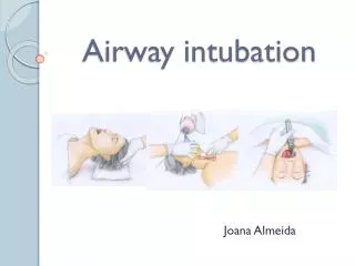
Intubation Procedure • Inserting the laryngoscope blade • Hold laryngoscope in left hand • While standing above the patient, insert the blade in the right side of the mouth WITHOUT trying to visualize the cords.
Intubation Procedure • Take a step back • Lower your head to the level of the label • Slowly advance laryngoscope until you visualize the epiglottis • Use straight or curved blade appropriately
Intubation Procedure • Visualize the vocal cords • Meconium below cords? • Both moving if not paralyzed? • Structurally normal? • Pick up endotracheal tube and pass between vocal cords
Assessing Endotracheal Tube Placement • Direct visualization • End tidal CO2 monitoring • Chest rise • Auscultation • ETT vapor • Less reliable • Chest X-ray
Intubation Procedure • Secure endotracheal tube to lip with tape • Do not let go of tube until secure • Reassess that endotracheal tube is still in place. • Assess the neonate – • Improving? More pink? Heart rate increasing? • Continue resuscitation – proceed to B and C….
Common Problems • Esophageal Intubation • Blade placed too deep, cords not visualized • Tongue obscures visualization • Sweep tongue to one side with blade • More anterior lift • Tape on blade • Cannot see cords • Head is hyper-extended - reposition
Common Problems • Cannot intubate • Most neonates can be bag valve mask ventilated easily • Call early for anesthesiology assistance • “Bag ventilating with oxygen can prolong life for a long time, repeatedly attempting and failing intubation will not.” • Surgical airway
Difficult Neonatal Airways • Must always be prepared for something abnormal • Increasing awareness of problems beforehand because of neonatal ultrasound • “Things you can see” versus “Things you may find”
Difficult Neonatal Airways • Congenital malformations • “Things you can see” • Predictable from looking at the patient • Cleft lip and palate • Pierre Robin syndrome • Treacher Collins syndrome • Goldenhar syndrome • Apert and Crouzon Syndrome
Congenital Malformations • Cleft Lip and Palate • Most common congenital face malformation • Pierre Robin Sequence • Obstruction is usually at the nasopharyngeal level
Congenital Malformations • Apert and Crouzon • Maxillary hypoplasia • Nasopharyngeal airway compromise • Goldenhar syndrome • Unilateral anomalies • Higher incidence of airway anomalies
Congenital Malformations • Treacher Collins • Choanal atresia/stenosis more common • Down’s Syndrome • Subglottic stenosis more common • Remember atlantoaxial instability
Difficult Neonatal Airways • Congenital Malformations • “Things you may find” • Laryngomalacia • Hemangioma or Lymphangioma • Tracheal web • Laryngeal atresia • Subglotic stenosis
Congenital Malformations • Laryngomalacia • A sequence between fully formed to atresia
Congenital Malformations • Laryngeal Web • Tracheal Atresia • Survive only if tracheoesophageal fistula or emergent trach
Congenital Malformations • Hemangioma or Lymphangioma • Only about 30% present at birth
Congenital Malformations • Subglottic Stenosis
In Review • Proper positioning is critical for successful neonatal intubation • Call for help early if unable to intubate or for any congenital anomalies • Continue to provide oxygen with bag valve mask ventilation • Practice makes perfect • It is estimated that you need to perform at least 90 intubations to be able to intubate successfully on the first or second attempt at least 80% of the time