
Celiac
E N D
Presentation Transcript
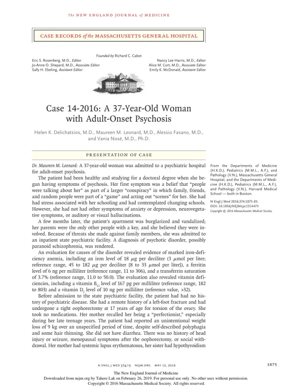
The new england journal of medicine Case Records of the Massachusetts General Hospital Founded by Richard C. Cabot Eric S. Rosenberg, M.D., Editor Jo‑Anne O. Shepard, M.D., Associate Editor Sally H. Ebeling, Assistant Editor Nancy Lee Harris, M.D., Editor Alice M. Cort, M.D., Associate Editor Emily K. McDonald, Assistant Editor Case 14-2016: A 37-Year-Old Woman with Adult-Onset Psychosis Helen K. Delichatsios, M.D., Maureen M. Leonard, M.D., Alessio Fasano, M.D., and Vania Nosé, M.D., Ph.D. Presentation of Case Dr. Maureen M. Leonard: A 37-year-old woman was admitted to a psychiatric hospital for adult-onset psychosis. The patient had been healthy and studying for a doctoral degree when she be- gan having symptoms of psychosis. Her first symptom was a belief that “people were talking about her” as part of a larger “conspiracy” in which family, friends, and random people were part of a “game” and acting out “scenes” for her. She had had stress associated with her schooling and had contemplated changing schools. However, she had not had other symptoms of anxiety or depression, neurovegeta- tive symptoms, or auditory or visual hallucinations. A few months later, the patient’s apartment was burglarized and vandalized; her parents were the only other people with a key, and she believed they were in- volved. Because of threats she made against family members, she was admitted to an inpatient state psychiatric facility. A diagnosis of psychotic disorder, possibly paranoid schizophrenia, was rendered. An evaluation for causes of the disorder revealed evidence of marked iron-defi- ciency anemia, including an iron level of 18 μg per deciliter (3 μmol per liter; reference range, 45 to 182 μg per deciliter [8 to 33 μmol per liter]), a ferritin level of 6 ng per milliliter (reference range, 11 to 306), and a transferrin saturation of 3.7% (reference range, 11.0 to 50.0). The evaluation also revealed vitamin defi- ciencies, including a vitamin B12 level of 167 pg per milliliter (reference range, 182 to 803) and a vitamin D2 level of 10 ng per milliliter (reference value, >32). Before admission to the state psychiatric facility, the patient had had no his- tory of psychiatric disease. She had a remote history of a left-foot fracture and had undergone a right oophorectomy at 17 years of age for torsion of the ovary. She took no medications. Her mother recalled her being a “perfectionist,” especially during her late teenage years. The patient had reported an unintentional weight loss of 9 kg over an unspecified period of time, despite self-described polyphagia and some hair thinning. She did not have diarrhea. There was no history of head injury or seizure, menopausal symptoms after the oophorectomy, or social with- drawal. Her mother had systemic lupus erythematosus, her sister had hypothyroidism From the Departments of Medicine (H.K.D.), Pediatrics (M.M.L., A.F.), and Pathology (V.N.), Massachusetts General Hospital, and the Departments of Medi‑ cine (H.K.D.), Pediatrics (M.M.L., A.F.), and Pathology (V.N.), Harvard Medical School — both in Boston. N Engl J Med 2016;374:1875-83. DOI: 10.1056/NEJMcpc1514473 Copyright © 2016 Massachusetts Medical Society. 1875 n engl j med 374;19 nejm.org May 12, 2016 The New England Journal of Medicine Downloaded from nejm.org by Tahere Lak on February 26, 2019. For personal use only. No other uses without permission. Copyright © 2016 Massachusetts Medical Society. All rights reserved.
The new england journal of medicine and hyperparathyroidism, her maternal grand- father had diabetes, and an aunt had breast cancer. There was no family history of psychiat- ric disease. The patient had been previously employed in human resources and had lived alone. She had traveled to the southern United States, coastal Massachusetts, and New York City but not abroad. She followed a pescatarian diet that in- cluded dairy foods and an egg daily as part of a lunch salad. She drank one alcoholic beverage per month, did not smoke cigarettes but had used marijuana, and drank two caffeinated bev- erages daily. After a 1-month inpatient stay at the state psychiatric facility, the patient was discharged. Her medications included risperidone, sertraline, ferrous sulfate, calcium, vitamin D, vitamin C, and a multivitamin. At a routine follow-up visit 6 weeks after discharge, the patient was evaluated by an internist at her primary care physician’s office, who found her to be excessively thin. On examination, a thyroid nodule was identi- fied, which prompted consultation with an en- docrinologist. A biopsy of the nodule was per- formed, and examination of the biopsy specimen revealed evidence of Hashimoto’s thyroiditis and papillary thyroid carcinoma. Radioactive iodine ablation was suggested, but the patient opted for total thyroidectomy. After the thyroid surgery and despite the administration of escalating doses of oral levothyroxine, the patient’s thyro- tropin level reportedly remained high (value not available) and the free thyroxine level was 0.73 ng per deciliter (9 pmol per liter; reference range, 0.80 to 1.80 ng per deciliter [10 to 23 pmol per liter]). Several weeks later, after further dose escalation of levothyroxine, the free thyroxine level was 0.80 ng per deciliter (10 pmol per liter). A limited follow-up examination that was per- formed by the endocrinologist 6 months after the initial consultation revealed a height of 167.6 cm, a weight of 45 kg, and a body-mass index (the weight in kilograms divided by the square of the height in meters) of 16.1. The patient was afebrile and had a regular pulse of 75 beats per minute and a blood pressure of 90/60 mm Hg. The thy- roidectomy scar was well-healed, and the remain- der of the examination was reportedly normal. However, the patient’s psychiatric symptoms were believed to be poorly controlled by antipsy- chotic medications, and it was unclear whether her thyroid conditions were related to her psy- chosis. Diagnostic tests were performed, and the patient presented to this hospital for further evaluation. Differential Diagnosis Dr. Helen K. Delichatsios: In this previously healthy 37-year-old woman, psychotic symptoms devel- oped over the course of several months. Psychi- atric conditions often develop during early adulthood, and this patient presented with a relatively late onset of psychotic symptoms; there- fore, we first must consider whether her psycho- sis is a primary psychiatric disorder or is due to an underlying medical condition. There is no family history of psychiatric illness, which ar- gues against a diagnosis of a primary psychiatric disorder. This patient also has multiple other dis- orders — including iron-deficiency anemia, defi- ciencies in vitamins B12 and D, papillary thyroid cancer, autoimmune thyroiditis, and clinically significant weight loss — that need to be con- sidered in formulating a differential diagnosis. Iron and Vitamin Deficiencies Do this patient’s iron and vitamin deficiencies help to explain her psychosis? Her diet was de- scribed as pescatarian and included fish, dairy, eggs, and infrequent alcohol consumption. We do not know whether she regularly consumed fruits and vegetables, so she may have had gaps in her nutritional intake. This patient had marked iron-deficiency ane- mia (iron level, 18 μg per deciliter; ferritin level, 6 ng per milliliter), which is not unusual in a menstruating woman. We do not have informa- tion about whether she had heavy menses. De- pending on her fish consumption, her dietary iron intake may not have been adequate. How- ever, iron deficiency alone would not cause her psychosis. This patient also had vitamin B12 deficiency (vitamin B12 level, 167 pg per milliliter). This degree of deficiency can be seen in vegetarians or persons who do not have a regular source of dietary vitamin B12. Despite the patient’s young age, I would consider the diagnosis of pernicious anemia and check an intrinsic-factor level. Al- 1876 n engl j med 374;19 nejm.org May 12, 2016 The New England Journal of Medicine Downloaded from nejm.org by Tahere Lak on February 26, 2019. For personal use only. No other uses without permission. Copyright © 2016 Massachusetts Medical Society. All rights reserved.
Case Records of the Massachusetts General Hospital though vitamin B12 deficiency can cause neuro- logic symptoms such as paresthesias, balance disorders, and confusion, it generally does not cause psychosis.1 There is disagreement about the blood 25- hydroxyvitamin D level that constitutes a vitamin D deficiency, but this patient’s level of 10 ng per milliliter was low according to any criteria.2,3 Low blood 25-hydroxyvitamin D levels are not uncommon in the northeast United States, espe- cially during the winter months. Low vitamin D levels have been associated with psychosis.4 How- ever, vitamin D deficiency has been associated with many other conditions, and it is unclear whether it is the primary cause of these condi- tions. Although I doubt vitamin D deficiency is the sole cause of this patient’s psychosis, it may be a contributing factor and therefore remains a consideration in this case. On occasion, a patient may present with low levels of all three of these micronutrients (iron, vitamin B12, and vitamin D). For example, after bariatric surgery, it is not uncommon for these deficiencies to develop and require lifelong monitoring and supplementation.5 The presence of multiple vitamin and mineral deficiencies raises concern about an impairment of absorp- tion, which may lead to other nutrient deficien- cies. Additional vitamin deficiencies associated with psychiatric and neurologic symptoms in- clude vitamin B1 (thiamine) deficiency, which can cause Wernicke’s encephalopathy; vitamin B3 (niacin) deficiency, which can cause pellagra; and vitamin B6 (pyridoxine) deficiency, which can cause weakness and difficulty walking.6,7 Although we do not know the blood levels of these vitamins in this patient, it is unlikely that any of these vitamin deficiencies would ade- quately explain her psychotic symptoms. head to rule out a primary brain cancer or meta- static tumor. The patient had self-described polyphagia, which makes anorexia nervosa un- likely but bulimia possible. Thyroid Disease The patient reportedly noted hair thinning. Weight loss, polyphagia, and hair thinning are suggestive of an endocrine condition, such as hyperthyroidism, which can cause psychosis. Severe hypothyroidism is also associated with psychosis, but the weight loss and polyphagia argue against this diagnosis. During the course of this patient’s illness, an astute internist found her to be excessively thin and identified a thyroid nodule on palpation. Subsequent examination of a biopsy specimen revealed evidence of Hashimoto’s thyroiditis (autoimmune thyroiditis) and papillary thyroid cancer. These findings are more likely to be in- cidental than part of the underlying process that is causing the psychosis. Thyroid nodules are very common and more likely to be detected on palpation in a thin person than in a person who is not so thin. Papillary thyroid cancer is not uncommon and would not explain the psychosis and weight loss. A separate diagnosis of auto- immune thyroiditis would not be surprising in this case, given the patient’s family history. However, the autoimmune condition should as- sume a prominent role in narrowing the differ- ential diagnosis for psychosis in this case. After the thyroidectomy and despite the ad- ministration of escalating doses of levothyroxine, the patient’s thyrotropin level remained high. The absorption of levothyroxine is nearly 100%,8 so the inability to normalize the thyrotropin level despite the administration of escalating doses of levothyroxine suggests impaired ab- sorption. The patient took iron and calcium sup- plements and sertraline, agents that may delay or impair absorption of levothyroxine.9,10 How- ever, escalating doses of levothyroxine should be able to overcome these medication effects. The patient’s psychiatric symptoms were also poorly controlled despite the appropriate anti- psychotic medications. This is not particularly surprising, because her psychiatric condition is likely to be secondary to an underlying medical condition. As with levothyroxine, the psychiatric medications may not have been properly ab- Weight Loss In addition to having nutritional deficiencies, this patient presented with weight loss. She was described as a perfectionist by her mother, and this trait has been associated with the diagnosis of anorexia nervosa. An unintentional weight loss of 9 kg over an unspecified period of time is worrisome, and cancer should be included in the differential diagnosis. No imaging studies are available in this case, but I would recommend performing magnetic resonance imaging of the 1877 n engl j med 374;19 nejm.org May 12, 2016 The New England Journal of Medicine Downloaded from nejm.org by Tahere Lak on February 26, 2019. For personal use only. No other uses without permission. Copyright © 2016 Massachusetts Medical Society. All rights reserved.
The new england journal of medicine sorbed. Taken together, these clues may point us toward disorders associated with malabsorption. schizophrenia onset at menopause, this patient was not yet menopausal. She did present at the peak age for the onset of a delusional disorder, which is a psychotic disorder with primary delu- sions. This disorder tends to occur at approxi- mately 35 years of age and is fairly common. Many patients with delusional disorder do not seek medical care, and when they do, they often have a poor response to antipsychotic medica- tions. Delusional disorder is characterized by isolated false, fixed beliefs and is not associated with some of the problems of cognitive and ex- ecutive function that are seen in patients with other psychotic disorders, such as schizophrenia and bipolar disorder. The diagnosis of a delu- sional disorder fits well with the features of this patient’s presentation. Some of her delusions involved her going on hunger strikes because she thought the hunger strikes would stop the conspiracy against her. This type of delusion would result in rapid weight loss and perhaps long-term nutritional deficiency. However, a de- lusional disorder is a diagnosis of exclusion; therefore, underlying medical conditions that could cause her psychosis should be ruled out. Thyroid Disease, Autoimmunity, and Malabsorption Although this patient did not have any gastroin- testinal symptoms, we have ample evidence to suggest that she may have had impaired absorp- tion of micronutrients and several medications. She also had thyroid cancer, which I suspect was an incidental finding and probably unrelated to her psychotic symptoms. However, the finding of Hashimoto’s thyroiditis and the subsequent inability to absorb levothyroxine after thyroidec- tomy are pivotal elements in shaping my think- ing about a possible cause of her psychosis. The patient’s family history of autoimmune disorders and development of autoimmune thyroiditis point us toward disorders associated with auto- immunity. The combination of malabsorption and autoimmunity strongly suggests the possi- bility of celiac disease, which is not always as- sociated with gastrointestinal symptoms. Mal- absorption associated with celiac disease would account for most if not all of the key features in this case, including the vitamin deficiencies, poor response to levothyroxine, autoimmune thyroiditis, and weight loss. Finally, although neurologic and psychiatric symptoms of celiac disease are not widely recognized, they have been reported11,12 and would account for the patient’s adult-onset psychosis. In order to establish the diagnosis of celiac disease, I would perform blood testing for IgA tissue transglutaminase antibodies and with a celiac disease–specific panel. I would also con- sider referral to a gastroenterologist for endos- copy and possible biopsy. Dr. Eric S. Rosenberg (Pathology): Dr. Deans, would you tell us your impression when you evaluated this patient at an outpatient psychiat- ric clinic after she had been discharged from the state psychiatric facility? Dr. Emily C. Deans (Psychiatry): When I initially evaluated this patient, I considered the possibil- ity of an affective disorder, major depression, and bipolar disorder with psychosis. She did not present with any mood symptoms, so a primary mood disorder was ruled out. Schizophrenia was considered, but the onset of schizophrenia typi- cally occurs in patients who are younger than this one. Although there is a second peak of Dr. Helen K. Delichatsios’s Diagnosis Celiac disease complicated by psychosis. Clinical Diagnosis Delusional disorder. Discussion of Management Dr. Rosenberg: Dr. Leonard, would you tell us what happened next with this patient? Dr. Leonard: On the basis of testing performed by the patient’s endocrinologist, celiac disease was considered to be the cause of a malabsorp- tion syndrome; it accounted for the patient’s anemia, weight loss, vitamin deficiencies, and poor response to levothyroxine. The diagnostic standard for celiac disease is a biopsy of the small intestine. Serologic tests — including tests for IgA tissue transglutaminase antibodies, en- domysial antibodies, and deamidated gliadin peptide antibodies — help to identify persons who may benefit from duodenal biopsy.13 Gen- eral consensus regarding these studies is that 1878 n engl j med 374;19 nejm.org May 12, 2016 The New England Journal of Medicine Downloaded from nejm.org by Tahere Lak on February 26, 2019. For personal use only. No other uses without permission. Copyright © 2016 Massachusetts Medical Society. All rights reserved.
Case Records of the Massachusetts General Hospital the test for IgA tissue transglutaminase anti- bodies is the most reliable and cost-effective; the test was performed in this patient and was strongly positive at 179 U per milliliter (refer- ence value, <20). Next, the patient was referred to a gastroen- terologist for further evaluation for possible ce- liac disease. The gastroenterologist performed esophagogastroduodenoscopy with small-bowel biopsy. Pathological examination of the biopsy specimen reportedly confirmed the diagnosis of celiac disease. After receiving the diagnosis of celiac disease, the patient thought her practitioners were being deceitful regarding the diagnosis and refused to adhere to a gluten-free diet. Psychotic symptoms and paranoia persisted, and she continued to “find clues” of conspiracy against her. She lost her job, became homeless, and attempted suicide; her family took out a restraining order against her. Eventually, she was rehospitalized at a psychi- atric facility, where she was placed on a gluten- free diet. After nearly 3 months at the psychiatric facil- ity, the patient’s delusions dissipated. She was discharged on a strict gluten-free diet and was taking risperidone daily. At that time, she sought the opinion of the celiac center at this hospital to determine whether her psychiatric symptoms could be related to celiac disease. When we evaluated the patient at this hospi- tal, our goal was to determine whether she had celiac disease, and if so, to determine whether her psychiatric symptoms were related to this diagnosis. Celiac disease should be considered in patients with gastrointestinal symptoms and in patients with extraintestinal problems, such as iron-deficiency anemia that is unresponsive to treatment, arthritis, or elevated levels of liver enzymes; it should also be considered in high- risk patients, including those with a family his- tory of celiac disease, those with type 1 diabetes mellitus, and those with autoimmune thyroid disease.14 This patient had gastrointestinal in- volvement that was manifested by weight loss and had evidence of malabsorption (deficiencies in vitamins D and B12); she also had iron-defi- ciency anemia that was unresponsive to therapy, one of the most common extraintestinal mani- festations seen in adults presenting with celiac disease. Furthermore, her recent diagnosis of Hashimoto’s thyroiditis placed her in a high-risk group for celiac disease, thus indicating that screening should be considered. The first step in her evaluation when she presented to this hospi- tal was to review the biopsy specimen of the small bowel that had been obtained by the gas- troenterologist. Pathological Discussion Dr. Vania Nosé: Review of the duodenal-biopsy specimen obtained by the gastroenterologist showed histologic changes involving the villi, crypts, enterocytes, and lamina propria (Fig. 1A). The duodenal mucosa showed moderate-to- marked villous blunting and atrophy. The sur- face epithelial cells appeared cuboidal or flat- tened, and a few goblet cells, attenuation of the brush borders, cytoplasmic basophilia, loss of polarity, and loss of the basal nuclear orienta- tion were present. There was an increased num- ber of intraepithelial lymphocytes in the surface epithelium and associated lymphoplasmacytosis in the lamina propria. More than 40 intraepithe- lial lymphocytes over 100 surface enterocytes were seen. Immunostaining for CD3 revealed an in- creased number of intraepithelial and lamina propria lymphocytes (Fig. 1B), and immuno- staining for CD8 revealed an increased number of intraepithelial lymphocytes (Fig. 1C). There was marked crypt hyperplasia with elongation of the crypts and an expanded proliferative zone of the crypts, as well as increased mitotic activity and a decreased number of goblet cells (Fig. 1D). Staining for Ki-67 highlighted hyperplastic crypts (Fig. 1E and 1F). The overall findings for this biopsy specimen are consistent with celiac dis- ease (modified Marsh classification 3b). Follow-up Dr. Leonard: On review of the clinical history, positive serologic tests, and duodenal-biopsy specimen with villous blunting, we confirmed that the patient had celiac disease. At the time of the patient’s visit to the celiac center at this hospital, she was following a gluten- free diet; she reported and her psychiatrist con- firmed that the delusions were not present. Therefore, if the symptoms were in fact related to celiac disease, the disease must have been in remission. To assess this, we repeated the sero- 1879 n engl j med 374;19 nejm.org May 12, 2016 The New England Journal of Medicine Downloaded from nejm.org by Tahere Lak on February 26, 2019. For personal use only. No other uses without permission. Copyright © 2016 Massachusetts Medical Society. All rights reserved.
The new england journal of medicine A B D C E F Figure 1. Initial Biopsy Specimen of the Duodenum. Hematoxylin and eosin staining of a biopsy specimen of the duodenum shows damaged surface epithelium, numer‑ ous intraepithelial lymphocytes, and an increased number of lamina propria lymphocytes and plasma cells (Panel A). Immunostaining of the same field for CD3 shows an increased number of intraepithelial and lamina propria lym‑ phocytes (Panel B). Similarly, immunostaining for CD8 shows an increased number of intraepithelial lymphocytes (Panel C). In addition, hematoxylin and eosin staining shows mild‑to‑moderate villous blunting, crypt hyperplasia, and an expanded proliferative zone of the crypts (Panel D). Immunostaining for Ki‑67 shows marked proliferation in the epithelial cells of the crypts that extends to the surface epithelial cells, highlighting the expanded proliferative zone of the crypts and hyperplastic crypts (Panels E and F). Together, these findings are consistent with the diagno‑ sis of celiac disease (modified Marsh classification 3b). 1880 n engl j med 374;19 nejm.org May 12, 2016 The New England Journal of Medicine Downloaded from nejm.org by Tahere Lak on February 26, 2019. For personal use only. No other uses without permission. Copyright © 2016 Massachusetts Medical Society. All rights reserved.
Case Records of the Massachusetts General Hospital logic test for IgA tissue transglutaminase anti- bodies, which was negative, and we scheduled a repeat endoscopy with duodenal biopsy. If the patient’s disease were in remission, we would no longer see active villous blunting (consistent with Marsh classification 3) and would instead see healing mucosa without abnormality (Marsh classification 0) or possibly see a mild increase in the number of intraepithelial lymphocytes (Marsh classification 1) and crypt hyperplasia (Marsh classification 2).15 A Additional Pathological Discussion B Dr. Nosé: Examination of the second duodenal- biopsy specimen, which was obtained at this hospital, showed that the duodenal mucosa had normal villous architecture. There was no villous blunting, villous atrophy, crypt hyperplasia, or increase in the number of intraepithelial lym- phocytes (Fig. 2). The regression of mucosal abnormalities, with normalization of villi to a Marsh classification of 0, is considered to be a histologic remission. Additional Discussion of Management C Dr. Alessio Fasano: Extraintestinal manifestations of celiac disease may be more common than gastrointestinal manifestations, and physicians should have a low threshold to test patients for IgA tissue transglutaminase antibodies in order to establish a diagnosis of celiac disease. In this case, the astute endocrinologist connected the signs of malabsorption, the history of Hash- imoto’s thyroiditis, and the poor response to levothyroxine to make the diagnosis of celiac disease. Patients with abnormal test results for IgA tissue transglutaminase antibodies or those in whom celiac disease is highly suspected must be referred to a gastroenterologist for confirmatory testing with endoscopy and biopsy. The patient must remain on a gluten-containing diet for testing to be accurate. A trial of a gluten-free diet is not appropriate unless celiac disease has been ruled out; once the patient is on a gluten- free diet, a clinician cannot distinguish between Figure 2. Subsequent Biopsy Specimen of the Duodenum (Hematoxylin and Eosin). A second biopsy specimen (Panels A, B, and C) shows the duodenal mucosa with normal villous architecture and no villous blunting or atrophy. There is no increase in the number of intraepithelial lymphocytes, the gob‑ let cells are preserved, and the polarity of the nuclei is maintained. The regression of mucosal abnormalities, with normalization of villi to a Marsh classification of 0, is considered to be a histologic remission. 1881 n engl j med 374;19 nejm.org May 12, 2016 The New England Journal of Medicine Downloaded from nejm.org by Tahere Lak on February 26, 2019. For personal use only. No other uses without permission. Copyright © 2016 Massachusetts Medical Society. All rights reserved.
The new england journal of medicine agnosis of celiac disease? It is not unusual for involvement of the central nervous system to lead to neurologic or psychiatric symptoms, or both. Celiac disease was classically described as a gastrointestinal condition that almost exclu- sively affects white children.16 Now celiac dis- ease is described as an autoimmune disorder that can affect persons of any age or race and can in- volve any tissue or organ of the body (Table 1).17 The typical gastrointestinal symptoms (diarrhea, bloating, failure to thrive, and weight loss) can be easily understood by the underlying intestinal damage caused by the autoimmune attack that occurs after the ingestion of gluten, but the many extraintestinal symptoms that patients with celiac disease often have are more difficult to explain. New insights on the pathogenesis of celiac disease suggest that it is truly a systemic disease that can spread from the intestine to any tissue or organ of the body.12,17 One of the most intriguing yet controversial clinical presentations of celiac disease involves the nervous system as a preferred target.18 Patients with celiac disease often have chronic headache, short-term memory loss, irritability, anxiety, and depression and more rarely have seizures, ataxia, autism, attention- deficit disorders, and psychosis (Table 2).12,18 Although we do not have a definitive explana- tion of the way in which an inflammatory pro- cess in the intestine affects the brain, there is growing evidence of close functional and organic interactions between these two systems, which are typically described as the gut–brain axis.19 A strict gluten-free diet is necessary to control symptoms; however, in patients with psychoses or other neuroinflammatory conditions, adher- ence to a gluten-free diet can be difficult. Patients with celiac disease should be monitored by a gastroenterologist and by a dietician with exper- tise in celiac disease and the gluten-free diet. Table 1. Extraintestinal Manifestations of Celiac Disease. Neurologic: peripheral neuropathy Dental: oral cavities, aphthous ulcers, and enamel defects Cutaneous: dermatitis herpetiformis, eczema, psoriasis, brittle nails, and hair thinning Cardiovascular: associated with myocarditis, blood‑flow alterations, and atrial fibrillation Pulmonary: Lane–Hamilton syndrome Pancreatic: acute pancreatitis Renal: increased risk of glomerulonephritis and end‑ stage renal disease Reproductive: infertility, miscarriage, and delayed puberty Hematologic: anemia Hepatic: hepatitis Musculoskeletal: joint pain, osteopenia, and osteoporosis Table 2. Neuropsychiatric Symptoms Associated with Celiac Disease. Confirmed Loss of short‑term memory Anxiety and depression Psychosis Ataxia Seizures Irritability Chronic headache Possible Autism Attention deficit–hyperactivity disorder Schizophrenia celiac disease and other gluten-related disorders, including nonceliac gluten sensitivity, because both can have extraintestinal manifestations. If a patient has initiated a gluten-free diet without having had the proper testing, a genetic test may be helpful to identify whether there is a need to reintroduce gluten for confirmatory testing for celiac disease. Confirming the diagnosis of ce- liac disease is essential, since the diagnosis of an autoimmune disease alters a patient’s future treatment. Does this patient’s psychosis fit with the di- Additional Follow-up Dr. Leonard: This patient was motivated to dis- cover whether her delusional symptoms were directly related to celiac disease. Under the care of her psychiatrist, she was weaned off the very small dose of psychiatric medication she had been taking, and she remained symptom-free for several months. Unfortunately, during this time, the patient 1882 n engl j med 374;19 nejm.org May 12, 2016 The New England Journal of Medicine Downloaded from nejm.org by Tahere Lak on February 26, 2019. For personal use only. No other uses without permission. Copyright © 2016 Massachusetts Medical Society. All rights reserved.
Case Records of the Massachusetts General Hospital inadvertently ingested gluten. She became delu- sional, was hospitalized, and her level of IgA tissue transglutaminase antibodies, which had previously been normal, was again elevated. Currently, the patient’s level of IgA tissue trans- glutaminase antibodies is persistently elevated, her anemia has returned, and she is not follow- ing a gluten-free diet because of a delusion that the diagnosis of celiac disease is incorrect. Final Diagnosis Celiac disease in histologic remission. This case was presented at Medical Grand Rounds. Dr. Fasano reports receiving consulting fees from General Mills, Crestovo, and Pfizer and lecture fees from Mead Johnson and holding stock in Alba Therapeutics. No other potential con- flict of interest relevant to this article was reported. Disclosure forms provided by the authors are available with the full text of this article at NEJM.org. References 1. Toh BH, van Driel IR, Gleeson PA. Pernicious anemia. N Engl J Med 1997; 337: 1441-8. 2. Ross AC, Taylor CL, Yaktine AL, Del Valle HB, eds. Dietary reference intakes for calcium and vitamin D. Washington, DC: National Academies Press, 2011. 3. Holick MF, Binkley NC, Bischoff-Fer- rari HA, et al. Evaluation, treatment, and prevention of vitamin D deficiency: an Endocrine Society clinical practice guide- line. J Clin Endocrinol Metab 2011; 96: 1911-30. 4. Belvederi Murri M, Respino M, Ma- sotti M, et al. Vitamin D and psychosis: mini meta-analysis. Schizophr Res 2013; 150: 235-9. 5. Mechanick JI, Youdim A, Jones DB, et al. Clinical practice guidelines for the perioperative nutritional, metabolic, and nonsurgical support of the bariatric sur- gery patient — 2013 update: cosponsored by American Association of Clinical Endo- crinologists, the Obesity Society, and American Society for Metabolic & Bariatric Surgery. Surg Obes Relat Dis 2013; 9: 159- 91. 6. Abdou E, Hazell AS. Thiamine defi- ciency: an update of pathophysiologic mechanisms and future therapeutic con- siderations. Neurochem Res 2015; 40: 353- 61. 7. Pfeiffer RF. Neurologic manifestations of malabsorption syndromes. Handb Clin Neurol 2014; 120: 621-32. 8. Fish LH, Schwartz HL, Cavanaugh J, Steffes MW, Bantle JP, Oppenheimer JH. Replacement dose, metabolism, and bio- availability of levothyroxine in the treat- ment of hypothyroidism: role of triiodothy- ronine in pituitary feedback in humans. N Engl J Med 1987; 316: 764-70. 9. Jonklaas J, Bianco AC, Bauer AJ, et al. Guidelines for the treatment of hypothy- roidism: prepared by the American Thy- roid Association task force on thyroid hormone replacement. Thyroid 2014; 24: 1670-751. 10. McCowen KC, Garber JR, Spark R. Elevated serum thyrotropin in thyroxine- treated patients with hypothyroidism given sertraline. N Engl J Med 1997; 337: 1010-1. 11. Jackson JR, Eaton WW, Cascella NG, Fasano A, Kelly DL. Neurologic and psy- chiatric manifestations of celiac disease and gluten sensitivity. Psychiatr Q 2012; 83: 91-102. 12. Leffler DA, Green PH, Fasano A. Ex- traintestinal manifestations of coeliac disease. Nat Rev Gastroenterol Hepatol 2015; 12: 561-71. 13. Rostom A, Dubé C, Cranney A, et al. The diagnostic accuracy of serologic tests for celiac disease: a systematic review. Gastroenterology 2005; 128: Suppl 1: S38- 46. 14. Fasano A, Berti I, Gerarduzzi T, et al. Prevalence of celiac disease in at-risk and not-at-risk groups in the United States: a large multicenter study. Arch Intern Med 2003; 163: 286-92. 15. Marsh MN. Gluten, major histocom- patibility complex, and the small intestine: a molecular and immunobiologic approach to the spectrum of gluten sensitivity (‘ce- liac sprue’). Gastroenterology 1992; 102: 330-54. 16. Auricchio S, Greco L, Troncone R. Gluten-sensitive enteropathy in child- hood. Pediatr Clin North Am 1988; 35: 157-87. 17. Fasano A, Catassi C. Celiac disease. N Engl J Med 2012; 367: 2419-26. 18. Bushara KO. Neurologic presentation of celiac disease. Gastroenterology 2005; 128: Suppl 1: S92-97. 19. Galland L. The gut microbiome and the brain. J Med Food 2014; 17: 1261-72. Copyright © 2016 Massachusetts Medical Society. Lantern SLideS Updated: CompLete powerpoint SLide SetSfromthe CLiniCopathoLogiCaL ConferenCeS Any reader of the Journal who uses the Case Records of the Massachusetts General Hospital as a teaching exercise or reference material is now eligible to receive a complete set of PowerPoint slides, including digital images, with identifying legends, shown at the live Clinicopathological Conference (CPC) that is the basis of the Case Record. This slide set contains all of the images from the CPC, not only those published in the Journal. Radiographic, neurologic, and cardiac studies, gross specimens, and photomicrographs, as well as unpublished text slides, tables, and diagrams, are included. Every year 40 sets are produced, averaging 50-60 slides per set. Each set is supplied on a compact disc and is mailed to coincide with the publication of the Case Record. The cost of an annual subscription is $600, or individual sets may be purchased for $50 each. Application forms for the current subscription year, which began in January, may be obtained from the Lantern Slides Service, Department of Pathology, Massachusetts General Hospital, Boston, MA 02114 (telephone 617-726-2974) or e-mail Pathphotoslides@partners.org. 1883 n engl j med 374;19 nejm.org May 12, 2016 The New England Journal of Medicine Downloaded from nejm.org by Tahere Lak on February 26, 2019. For personal use only. No other uses without permission. Copyright © 2016 Massachusetts Medical Society. All rights reserved.