Download
1 / 27
440 likes | 1.2k Vues
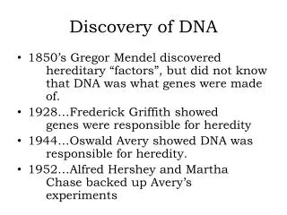
The History of DNA Structure Discovery. 1869 – Friedrich Miescher. Studied the nuclei of white blood cells Isolated the material using HCl (aq) and digestive proteins Named the substance nuclein Found the material was rich in nitrogen and phosphorus. 1919 – Pheobus Levene.

E N D
1869 – Friedrich Miescher • Studied the nuclei of white blood cells • Isolated the material using HCl(aq) and digestive proteins • Named the substance nuclein • Found the material was rich in nitrogen and phosphorus
1919 – Pheobus Levene • Discovered that DNA was made up of chains of nucleotides NITROGEN RICH ACID DEOXYRIBOSE
1920 – DNA vs Protein • thought that 4 nucleotides were connected in the same repeated pattern • protein have 20 amino acids which could be combined in many combinations
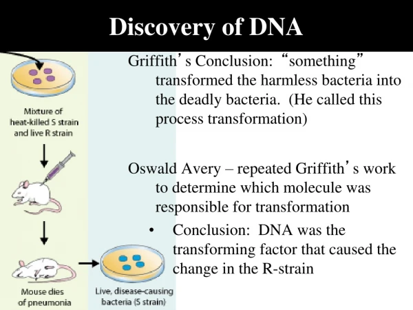
1928 – Frederick Griffith • studied two strains of pneumococcus bacteria • rough strain = nonvirulent • injection into mouse did not result in death • smooth strain = virulent • injection caused mouse to die
Griffith’s Conclusions • some “factor” from the dead, virulent smooth strain “transformed” the living, non-virulent rough strain • non-virulent rough strain picked up DNA to become virulent
nucleus at bottom of stalk 1930 – Joachim Hammerling Acetabularia – type of alga
Hammerling’s Experiment Hereditary information is stored in the nucleus. no regrowth
1940s – Joshua Lederberg • demonstrated bacterial conjugation • bacteria can exchange DNA • bacteria have no nucleus or chromosomes
1940s – Edwin Chargaff • for all organisms A = T and G = C Chargaff’s Rule • organisms with more Gs and Cs tend to be more complex
1952 – Hershey & Chase • conducted experiments to definitively show that DNA is the hereditary material • bacteriophage used to infect bacteria • bacterial virus
Hydrophobic interactions and van der Waalsinteractions CH CH2 CH2 H3C CH3 OH Polypeptidebackbone H3C CH3 Hyrdogenbond CH O HO C CH2 CH2 S S CH2 Disulfide bridge O -O C CH2 CH2 NH3+ Ionic bond
X-ray Crystallography • physics approach to examining biological molecules
Rosalind Franklin’s X-rays The photo indicated: • Backbone of alternating phosphate and sugars • Backbone is a helical structure • Double helix structure (molecule is a uniform helix) • Nitrogenous bases are in the middle of the molecule • Bases are at right angles to the backbone
1953 – James Watson & Francis Crick • inspired by alpha-helix model of proteins • determined howA + T and G + C bonded together • width of purine + pyrimidine bonds fit perfectly between the sugar-phosphate backbone (DNA width = 2 nm) • the double helix model offered an easy method for replication
DNA Backbone alternating sugar-phosphate backbone sugar and phosphate backbone attached by a phosphodiester bond DNA is said to be read from 5’ to 3’ opposing backbones are antiparallel 1’ 4’ 5’ 2’ 1’ 4’ 2’ 3’
Complementary Base Pairing Chargaff’s Rule adenine : thymine (1:1) guanine : cytosine (1:1) the diameter of a base pair is 2 nm 3 H-bonds between G&C 2 H-bonds between A&T
Overall DNA Structure right handed helix 0.34 nm between adjacent base pairs one turn of the helix is 10 base pairs (3.4 nm) 0.34 nm 3.4 nm
Homework • Answer questions pg. 209 #1-4 • Read section 4.2 • Answer questions pg. 216 #1-9