Download
1 / 42
500 likes | 1.45k Vues
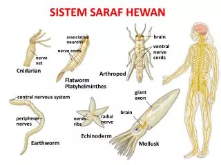
giant axon. central nervous system. associative neurons. brain. brain. ventral nerve cords. nerve cords. peripheral nerves. radial nerve. nerve ribs. nerve net. Mollusk. Earthworm. Arthropod. Flatworm Platyhelminthes. Cnidarian. Echinoderm. SISTEM SARAF HEWAN.

E N D
giant axon central nervous system associative neurons brain brain ventral nerve cords nerve cords peripheral nerves radial nerve nerveribs nerve net Mollusk Earthworm Arthropod FlatwormPlatyhelminthes Cnidarian Echinoderm SISTEM SARAF HEWAN
Kumpulan Neuron Saraf korda Saraf radial Saraf rusuk Saraf jalat Cacing PipihPlatyhelminthes Cnidarian Echinodermata Cefalisasi = EvolusiOtak • Cephalization= clustering of neurons in “brain” at front (anterior) end of bilaterally symmetrical animals where sense organs are More organization but still based on nerve nets; supports more complex movement Simplest, defined central nervous system more complex muscle control Simplest nervous system no control of complex actions
Sistem Saraf Pusat Akson Raksasa Otak Otak ventral nerve cords Sistem Saraf Tepi Molluska Cacing Tanah Arthropoda Cefalisasi = EvolusiOtak • Interneuron bertambahbanyakdidalamOtak More complex brains connected to all other parts of body by peripheral nerves More complex brains in predators most sophisticated invertebrate nervous system Further brain development ganglia = neuron clusters along CNS
Shark Frog Crocodile Cat Human Spinal cord Hind: Medulla oblongata Hind: Serebellum Optic tectum Bird Midbrain Fore: Serebrum Organ Olfaktori EvolusiOtak Vertebrata Otak Depan Otak DepanSerebrum Dominan Otak Belakang Otak Depan
PembagianFungsionalOtak • Otak Belakang (Hind Brain) • Mengendalikan Fungsi Autonomi dan Integratif • Batang Ptak • pons • medulla oblongata • Otak tengah • Serebellum • Thalamus, Hipothalamus
BatangOtak • The “lower brain” • medulla oblongata • pons • midbrain • Fungsi: • homeostasis • Koordinasi Pergerakan • Konduksi Impuls ke Pusat Otak
Medulla oblongata & Pons • Kontrol Fungsi Otonomi Homeostatis • Aktivitas pembuluh darah dan jantung • Pernafasan • Menelan • Muntah • Pencernaan • Relay Informasi dari • dan ke Pusat Otak
Otak Tengah • Terlibat dalam integrasi informasi sensori • pengendalianvisual reflexes • Pengendalian informasiauditory reflexes
Formasi Reticular • Tidur & pola kebugaran aktivitas elektrik di dalam otak • Dilaporkan sebagai ElectroEncephaloGram (EEG) • Umumnya bermimpi terjadi selama REM(rapid eye movement) sleep
satu sentimeter kubik otak manusia mengandung lebih dari 50 juta sel saraf, yang masing-masing bisa berkomunikasi dengan ribuan neuron lain dalam jaringan kerja pengolahan informasi • saraf dikhususkan untuk transmisi impuls dengan cepat, secepat 150 m/detik (lebih dari 330 mil per jam)
Gambaran Umum Sistem Saraf Analog dengan telepon • Berfungsi Sebagai: • Input Sensori • Integrasi • Output Motoris • ‘ SISTEM SARAF Tersusun atas Neuron dan sel-sel pendukung
SISTEM SARAF SelSarafatauNeuronadalah Unit DasarKomunikasipadasistemsaraf Vertebrata
TigaKelas Neuron • Sirkuit Neural Terdiri Atas: • Sensory neurons • Reseptor rangsang • Interneuron (CNS) • Integrasi sinyal • Motor neuron • Transfer sinyal ke efektor (muscle)
Anatomi Neuron • Cell body: Bagian Fungsional • Dendrites: Ekstensi Pendek Penerima sinyal • Axon: Ekstensi Panjang yang menerima impuls
BagaimanaKerja Neuron MemegangdanMenggerakan Info? • Neuron pada waktu istirahat memiliki voltase berbeda dengan neuron menembus membran plasma, disebut “resting voltage potential” • Potensial Aksi(PA) terjadi apabila muatan menembus membran yang berubah secara singkat • Potensial Aksi bergerak ke bawah membran dengan cepat. • Potensial aksi dapat bergerak lebih cepat pada bagian yang bermyelin, disebut “saltatory conduction”
Na+ Na+ Na+ Na+ Na+ Na+ A. Polarisasi K+ K+ K+ K+ ACh Na+ Na+ Na+ Na+ Na+ Na+ B. Depolarisasi K+ K+ K+ K+ K+ K+ K+ K+ C.Repolarisasi Na+ Na+ Na+ Na+ Na+ Na+ K+ K+ Gb. Muatan Elektrik dan Konsentrasi Ion pada Sarcolemma
Table 7–1 State or Event Description Resting Potential Polarization • Sarcolemma has a () charge outside and a () charge inside. •Na� ions are more abundant outside the cell; as they diffuse inward, the sodium pump returns them outside. •K� ions are more abundant Inside the cell; as they diffuse out, the potassium pump returns them inside. Action Potential Depolarization • ACh makes the sarcolemma very permeable to Na� ions, which rush into the cell. • Reversal of charges on the sar- colemma: now () outside and () inside. • The reversal of charges spreads along the entire sarcolemma • Cholinesterase at the sar- colemma inactivates ACh. Repolarization • Sarcolemma becomes very per- meable to K� ions, which rush out of the cell. • Restoration of charges on the sarcolemma: () outside and () inside. • The sodium and potassium pumps return Na� ions outside and K� ions inside. • The muscle fiber is now able to respond to ACh released by another nerve impulse arriving at the axon terminal.
SIFAT SINYAL SARAF DENDRIT AKSON IMPULS Impuls adalah sinyal listrik yg bergantung pada aliran ion yang menembus membran plasma neuron. Potensial Membran disebabkan oleh perbedaan konsentrasi ion antara isi sel dengan cairan ekstraselluler
ImpulsSarafadalahSinyalBioelektrik • Pompa Sodium-Pottasium menggunakan ATP untuk mentransport ion sodium ke luar dan ion pottasium masuk ke dalam membran • Pada waktu neuron beristirahat memiliki muatan negatif relatif terhadap keadaan di luar membran • Potensial aksi reversal dan restorasi muatan berbeda yang menembus membran • Pompa sodium-potassium dapat menyimpan kembali persebaran original ion-ion • Potensial Aksi adalah “all-or-none events”
Resting Membrane Potential, Graded Potentials, and an Action Potential
Transfer Informasidari Neuron ke Target • Synaptic transmission: • Mengeluarkan neurotransmitter: menaikan potential • Pengaruh neurotransmitter: • Excitatory: depolarisasi sel postsynaptic • Inhibitory: hiperpolarisasi sel postsynaptic • Peran postsynaptic neuron: integrasi dan proses informasi
Lokasi Reseptor Akson Neuron Presynaptic Inaktivator (Cholinesterase) Vesicle Neurotransmitter Na+ Na+ Na+ Dendrit Neuron Postsynaptic Mitokondrion Neurotransmitter (Acetylcholin) Neurotransmitter yang teraktivasi Gb. Impuls Transmisi pada Synapse
TipeKimiawi synapse • Acetylcholine: neuromuscular junctions, kelenjar, otak dan spinal cord • Norepinepherine: mempengaruhi bagian otak berkaitan dengan emosi dan mimpi
JalurAliranInformasi • Sinyal di antara Otak dan Spinal Cord bergerak ke bagian tubuh melalui sel saraf • Sensory nerves :sinyal bergerak ke arah Otak dan Spinal Cord • Motor neurons: menggerakkan sinyal dari Otak atau Spinal Cord ke Bagian tubuh
PembagianSistemSaraf • Sistem Saraf Pusat • (Central nervous system) • CNS • Otak dan Spinal Cord • Sistem Saraf Tepi • (Peripheral nervous system) • PNS • Semua saraf yang membawa sinyal ke dan dari CNS
Interneuron Dorsal root Central canal Dorsal root ganglion Synapse Cell body of sensory neuron Dendrite of sensory neuron Gray Matter Ventral root Axon of motor neuron Receptor Cell Body of Motor Neuron Synaptic knobs Effector muscle Gb. Cross-section of the spinal cord and the three types of neurons. Spinal nerve roots and their neurons are shown on the left side. Spinal nerve tracts are shown in the white matter on the right side. All tracts and nerves are bilateral (both sides). QUESTION: The dorsal column is an ascending tract, and the corticospinal tract is descending. Explain what this means.
(2) Sensori Neuron Dorsal Root Ganglion (5)Quadriceps Femoris Muscle (Kontraksi) Dorsal Root (1) Stretc Receptor Stimulus Biceps Femoris Muscle (Rileks) Gray Matter Ventral Root (4) Motor Neuron (3) Synapse pada Spinal Cord Gb. Repleks Patella