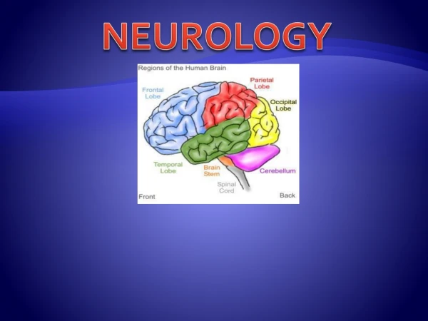
Neurology
Neurology. What not to miss in the ER Danielle Pirrie CCPA Toronto East General Hospital dpirr@tegh.on.ca. Objectives. Review the less common S/S of stroke/TIA Discuss need for testing (echo, Holter , carotid dopplers ) Review CNS infection S/S. Case # 1.

Neurology
E N D
Presentation Transcript
Neurology What not to miss in the ER Danielle Pirrie CCPA Toronto East General Hospital dpirr@tegh.on.ca
Objectives • Review the less common S/S of stroke/TIA • Discuss need for testing (echo, Holter, carotid dopplers) • Review CNS infection S/S
Case # 1 • 78yo male, minimal English, from a rehab hospital (for minor) deconditioning, 2 day hx of being confused, telling translator that he is in his village in Serbia, being chased by bandits in masks. • PMHx: • HTN, • previous left MCA stroke 7 yrs ago left with minor right arm weakness, • high cholesterol
Case #1 • By the next day, his speech (when talking with family) was like word salad, not making any sense. • But he could tell me in English that he was fine and “want to go home”
Case #1 • P/E: • VS: T 36.7, HR 86, BP 154/92, RR 18 SpO2 94% RA • Neuro exam: CN II-XII normal, no focal weakness, no dysarthria, upgoing toes bilat • DDx • Infection • Stroke • Encephalopathy
Stroke CT scan showed a left parietal stroke relating to Wernicke’s area
Stroke • Typical anterior circulation stroke S/S • Unilateral weakness • Slurred speech • Decreased LOC • Other anterior circulation stroke S/S • Cognitive impairment • Difficulty with speech, word finding difficulty • Weakness or clumsiness • Changes of sensation • Visual losses – hemianopia
Stroke • Posterior circulation stroke S/S • Acute vision loss • Confusion • Dizziness • Nausea • Memory loss
Stroke/TIA • Dizziness • Usually associated with other brainstem S/S such as double vision, dysarthria, ataxia, dysphasia. • DDx: benign paroxysmal positional vertigo, migraine, Meniere’s, low BP, vestibular neuronitis, acoustic tumours, medications, anxiety, etc.
Stroke/TIA • Aphagia/dysphagia • Can be completely non-verbal or simply word finding difficulty • Damage to frontal lobe results in problems speaking (expressive) • Damage to temporal lobe results in problems understanding (receptive)
Stroke/TIA • Decrease LOC • Most likely to be caused by a brain stem stroke or hemorrhagic stroke • Brain stem stroke difficult to diagnose on CT scan
Stroke workup • CT scan • Carotid dopplers • If 70-99% stenosis and TIA or nondisabling stroke, may be candidate for surgery or stenting. • Echocardiogram • Holter monitoring N Engl J Med July 1, 2010
Stroke Summary • If TIA, ensure pt has followup for stroke workup to reduce future risk of stroke • Posterior circulation strokes have many mimics
Case #2 • 27yo female comes into ER with fever, headache, fatigue and loss of appetite, • After a few hours of waiting in the waiting room, her boyfriend notices that she is trying to use a pop can as a cell phone, that she is speaking gibberish and not making any sense. She is then brought into a room and examined.
Case #2 • P/E • temp of 39.8oC, HR 110, BP 114/72, RR 28, SpO2 98% RA • CN: PERLA 3+, left visual field defect, no facial asymmetry • Motor: no focal deficits, no neck stiffness • Labs • CBC: WBC 10.4, Hb 140, Plt 247 • Normal electrolytes, LFT, RFT
Case #2 • DDx • Bacterial meningitis • Viral meningitis • Herpes simplex encephalitis • Stroke
Case #2 • Anytime there is HA, mental status changes and fever, need to do LP • CSF analysis: • Glucose: 2.7 (normal) • Protein: 0.4 (normal) • Culture did not grow anything • CT scan head normal
CNS Infections • Herpes Simplex Encephalitis • Typically HSV-1 • S/S: fever, headache, psychiatric or mental changes, seizure, vomiting, focal weakness, memory loss. • CSF: mononuclear lymphocytes, high RBC, protein normal or high, glucose normal or low, send for viral cultures and PCR • CT may be negative • Need MRI to diagnose definitively
HSV on MRI (T2) • Hyperintesity in right temporal lobe • Treatment with acyclovir IV
CNS Infections • Meningitis • May be bacterial, viral, tubercular, or fungal • Bacterial meningitis: children under 2. • s/s: evolve over hours, starts with URTI s/s then develop fever, lethargy, N/V, stiff neck, photophobia • CFS: high polymorphonuclear leukocytes, high protein, low sugar • Urgent management is vital as severe cortical damage can result from any delay in treatment
CNS Infections • Abscesses • Severe HA • Mental status changes • Unilateral weakness/paralyisis • Fever
CNS Infection Summary • Low threshold for LP in pts with fever and mental status changes • Treat empirically for HSV-1 to ensure no irreversible brain damage • Abscesses are usually seen on CT
Case #3 • 73yo male, sudden onset of L HA while at home • Pt took 2 ASA for pain but it did not resolve so he took 2 more ASA 2 hours later • Approx 1 hr after, he suddenly noticed not being able to read the computer screen and having decreased vision on the right side
Case #3 • PMHx: • A-fib for which he takes ASA • HTN • Dyslipidemia • Prior small right occipital lobar bleed in 2007 • ETOH approx 3 drinks/day • Smokes a pipe • Son is a neurologist in NY state
Case #3 • PE: • VS normal except for irregular pulse • CN mostly normal except for right visual field defect • No motor, sensation, coordination deficits • Normal verbal • Visual acuity
Case #3 • This came out as “beautiful story run April” • When he tried to spell “road” it was P-F-G-O
Intracranial bleed • CT head showed a lobar hemorrhage.
Intracranial bleeds • Intra-axial bleeds • Within the brain itself (as in previous case) • Hemorrhagic stroke intraparenchymal intraventricular
Intracranial bleed • Causes: • HTN • Trauma • Aneurysm • AV malformation • Tumour • Amyloid angiopathy
Intracranial Bleed • Extra-axial bleeds
Intracranial bleed • All bleeds require discussion with neurosurgery. • Blood in brain can increase ICP • At risk for seizures