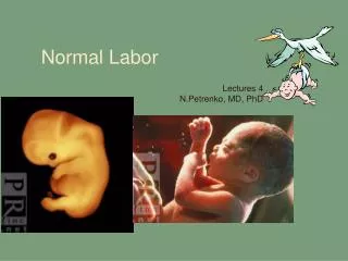
Normal Labor
Normal Labor. Lectures 4 N.Petrenko, MD, PhD. 1 Birth Passage. 1 Birth Passage. 1 Birth Passage. 1 Birth Passage. 1 Birth Passage. Four different types of pelvises, but frequently mixed types. anthrapoid. android. gynaecoid. platypelloid. Pelvic inlets. Platypoid. Gynecoid.

Normal Labor
E N D
Presentation Transcript
Normal Labor Lectures 4 N.Petrenko, MD, PhD
1 Birth Passage • Four different types of pelvises, but frequently mixed types anthrapoid android gynaecoid platypelloid
Pelvic inlets Platypoid Gynecoid Anthropoid Android
1 Birth Passage Asymmetrical pelvises • Abnormality of lower limb • Abnormality of pelvic girdle • Abnormality of vertebral column Osteomalacic pelvis Robert’s pelvis Scoliotic pelvis Coxalgic pelvis Split pelvis • Naegele’s pelvis
1 Birth Passage Measurement of AP conjugates •Diagonal conjugate ~12.0 cm •True conjugate ~11.0 cm •AP outlet ~12.5 cm
1 Birth Passage • Assess shape of sacrum
2 Fetus Lambdoidal suture • Sutures: • Frontal • Sagittal • Coronal • Lambdoidal Sagittal suture Coronal suture Frontal suture Note: sutures are actually membranous spaces that meet at fontanels
Foetal skull 1. Bones: 2 parietals, 2 frontals, 2 temporals, occipital 2. Sutures: sagital, frontal, lamboidal, coronal, temporal 3. Fontanelles: anterior, posterior, 2 anterior temporals, 2 posterior temporals.
Fetal skull 1 4 5 2 1. Suboccipitobregamatic: ~9.5 cc Vertex 2. Suboccipitofrontal: ~10.0 cm Sinciput 3. Occipitofrontal:~11.24 cm persistent OP 4. Mentovertical: ~13.8 cm brow 5. Submentobregmatic: ~9.5 cm Face 6. Submentovertical: ~11.25 cm incompletely extended face 7. Biparietal diameter: ~9.5 cm 6 3 7
Fetus • ☺Fontanelles: intersection of sutures, allows for molding, helps identify position of head • Anterior (bregma) • Diamond shaped • Approx 2-3 cm • Ossifies in ~12-18 months • Posterior • Triangle shaped • Smaller • Closes in 8-12 weeks
Fetus Fetal lie Longitudinal Transverse
Fetus Fetal lie Cephalic • Breech Shoulder
Fetus • Fetal presentation: Cephalic • ☺Vertex presentation • Most common • Head completely flexed on chest • Suboccipitobregmatic (Smallest diameter) • Occiput in presenting part
Fetus • Fetal presentation: Cephalic • Military presentation • Fetal head neither flexed nor extended • Occipitofrontal diameter presents • Top of the head is presenting part
Fetus • Fetal presentation: Cephalic • Brow presentation • Fetal head partially extended • Occipitomental diameter presents • Sinciput is presenting part
Fetus • Fetal presentation: Cephalic • Face presentation • Head hyperextended • Submentobregmatic diameter presents • Face is presenting part
Fetus • Fetal presentation: Breech • Sacrum is the landmark • Complete breech • Knees and hips are flexed, thighs on abdomen (“fetal position”) • Buttocks and feet are presenting parts
Fetus • Fetal presentation: Breech • Sacrum is the landmark • Frank breech • Hips flexed, knees extended • Buttocks is presenting part
Fetus • Fetal presentation: Breech • Sacrum is the landmark • Footling breech • Hips and legs extended • Feet are presenting parts (single vs double)
Fetus • Fetal presentation: Shoulder • Acromion process of shoulder is presenting part
Station In Gynaecoid & Android pelvis distance between ischial spine to brim is ~5 cm. In Anthropoid pelvis distance is ~7 cm In Platypelloid pelvis distance is ~3 cm Station of the head in relation to ischial spines
Partogram Normal dilatation Alert line Abnormal dilatation Acton line